Week in BioE (February 2, 2018)

Broccoli + Yogurt = Cancer Prevention?

broccoli
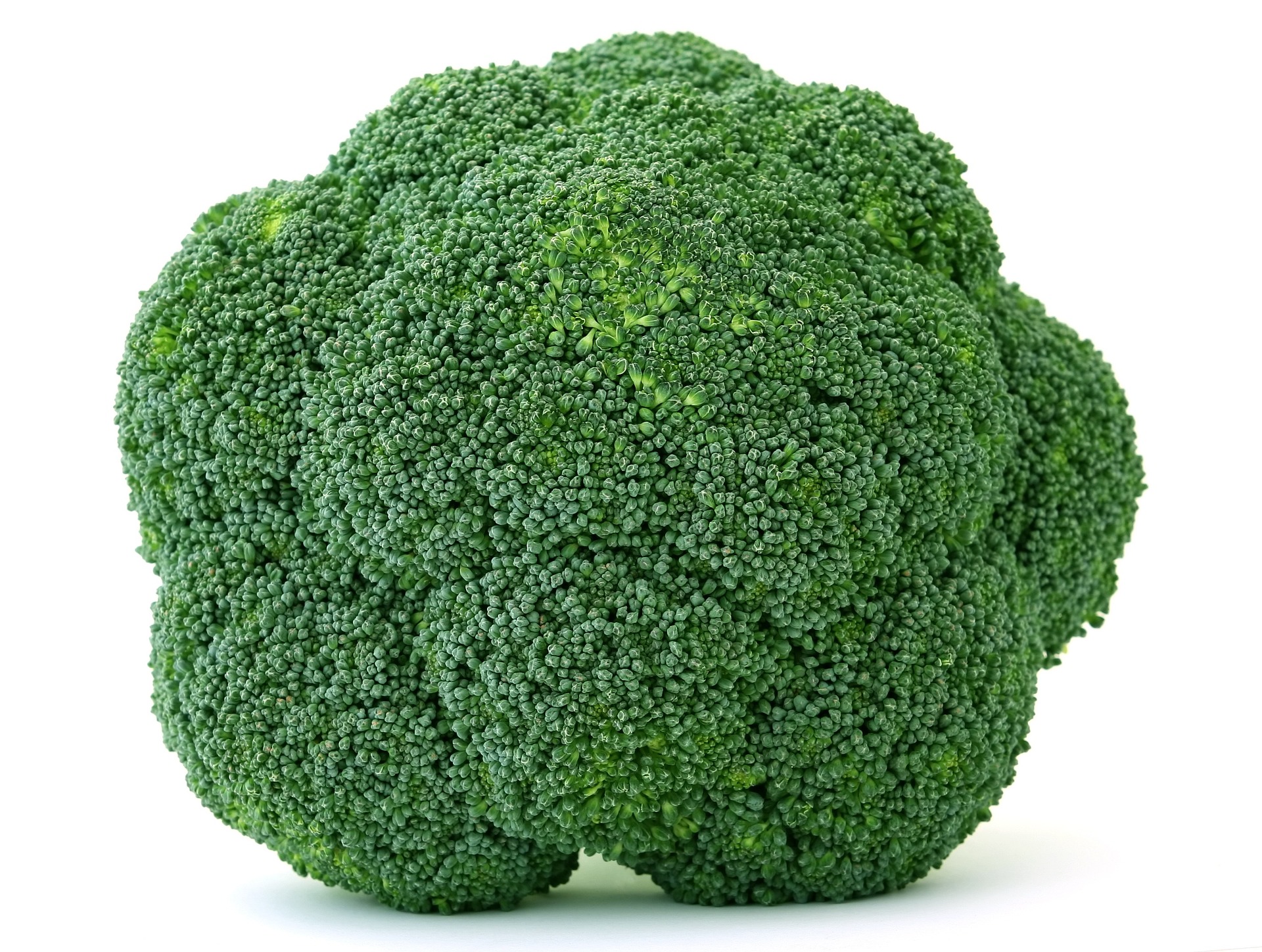
George H.W. Bush refused to eat it, but maybe he should start. It turns out that broccoli, combined with bioengineered yogurt, could provide effect cancer prevention. We’ve known for some time that compounds in certain fresh vegetables can increase chemoprevention, but the levels are usually too low to be effective, or they can’t be assimilated optimally by the body.  However, scientists in Singapore found that engineered bacteria, when ingested by mice with colorectal cancer, had anticancer effects. The bacteria caused the secretion of an enzyme by the cancer cells that transformed glucosinolates — compounds found in vegetables — into molecules with anticancer efficacy. The scientists report their findings in Nature Biomedical Engineering.

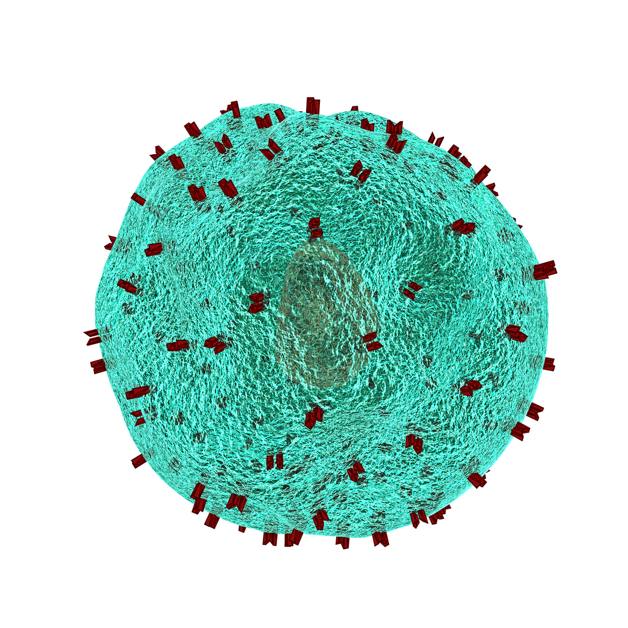
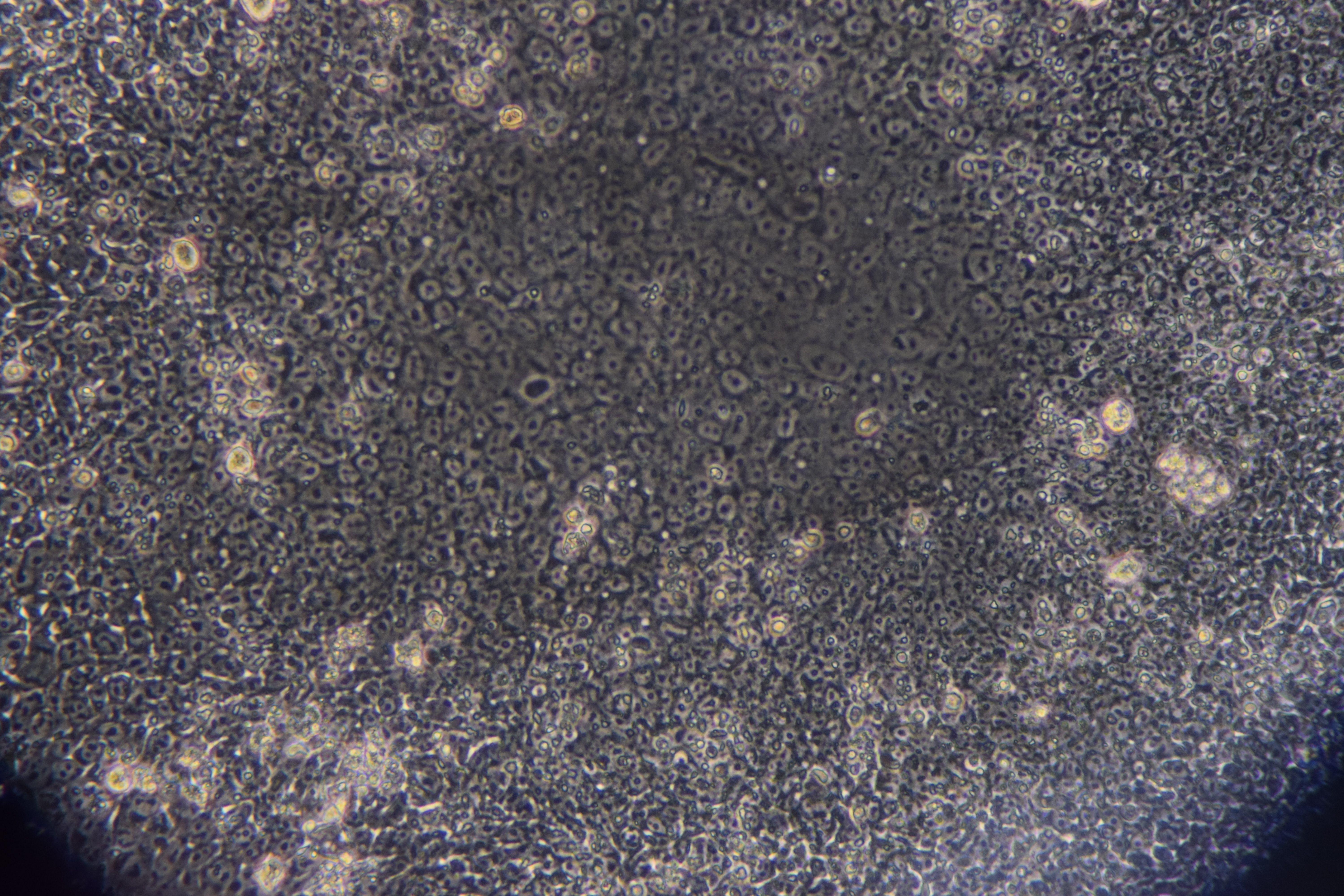
The authors programmed an E. coli cell line to bind to heparan sulfate proteoglycan, a cell surface protein that occurs in colorectal cancer cells. Once the engineered bacteria bound to the cancer cells, the bacteria secreted myrosinase, an enzyme that commonly occurs in many plants to defend them against aphids. In the cell model employed by the authors, myrosinase caused the conversion of glucosinolates into sulforaphane, which in turn could inhibit cancer cell growth.

The scientists then applied their system in a mouse model of colorectal cancer, feeding the mice yogurt infused with the engineered bacteria. They found that the mice fed broccoli plus the yogurt developed fewer and smaller tumors than mice fed broccoli alone. Additional testing is necessary, of course, but the study authors believe that their engineered bacteria could be used both as a preventive tool in high-risk patients and as a supplement for cancer patients after surgery to remove their tumors.

The Gates of CRISPR

About two years ago, software giant Microsoft unveiled Azimuth, a gene-editing tool for CRISPR/Casa9 that it had developed in collaboration with scientists at the Broad Institute. Now, in response to concerns that CRIPR may edit more of the genome than a bioengineer wants, the team has introduced a tool called Elevation. A new article in Nature Biomedical Engineering discusses the new tool.

In the article, the team, co-led by John C. Doench, Ph.D., Institute Scientist at the Broad Institute, describes how it developed Azimuth and Elevation, both of which are machine learning models, and deployed the tools to compare their ability to predict off-target editing with the ability of other approaches. The Elevation model outperformed the other methods. In addition, the team has implemented a cloud-based service for end-to-end RNA design, which should alleviate some of the time and resource handicaps that scientists face in using CRISPR.

Reducing Infant Mortality With an App

Among the challenges still faced in the developing world with regard to health care is high infant mortality, with the most common cause being perinatal asphyxia, or lack of oxygen reaching the infant during delivery. In response, Nigerian graduate student Charles C. Onu, a Master’s student in the computer science lab of Doina Precup, Ph.D., at McGill University in Montreal, founded a company called Ubenwa, an Igbo word that means “baby’s cry.”

With Ubenwa and scientists from McGill, Onu developed a smartphone app and a wearable that apply machine learning to instantly diagnose birth asphyxia based on the sound of a baby’s cry. In initial testing, the device performed well, with sensitivity of more than 86% and specificity of more than 89%. You can read more about the development and testing of Ubenwa at Arxiv.

People and Places

Several universities have announced that they are introducing new centers for research in bioengineering. Purdue University secured $27 million in funding from Semiconductor Research Corp. for its Center for Brain-inspired Computing Enabling Autonomous Intelligence, or C-BRIC, which opened last month. The center will develop, among other technologies, robotics that can operate without human intervention.

In Atlanta, Emory University received a $400 million pledge from the Robert W. Woodruff Foundation for two new centers — the Winship Cancer Institute Tower and a new Health Sciences Research Building. The latter will host five research teams, including one specializing in biomedical engineering. Further north in Richmond, Virginia Commonwealth University announced that it will begin construction on a new $92 million Engineering Research Building in the fall.  The uppermost floors of the new building will include labs for the college’s Department of Biomedical Engineering.

Finally, North Carolina’s Elon College will introduce a bachelor’s degree program in engineering in the fall. The program will offer concentrations in biomedical engineering and computer engineering. Sirena Hargrove-Leak, Ph.D., is director of the program.

Week in BioE (January 24, 2018)

Using AI to Better Understand Cancer Immunology

ImmunoMap
A T cell with receptors highlighted in red

T lymphocytes in the immune system play a vital role in the body to recognize invasion by an outside element. When foreign bacteria enter the body, receptors on the T cell surface detect antigens associated with the bacteria and send a signal deploying phagocytes to attack and defeat the invading bacteria. While evolution and vaccination make the immune system very efficient, the inability of T cell receptors (TCRs) to detect cancer makes normal T cells relatively ineffective in resisting cancer. One of the ways to overcome this limitation of the immune system is to better understand how the TCRs respond to antigens. Analyses of the proteins involved in TSR responses are useful but limited by several factors, including the dizzying amount of data involved. Data analysis techniques have been helpful but have offered little information about the general reactions of TSRs, rather than how they react to specific antigens.  

A possible solution to this obstacle is ImmunoMap, developed by scientists collaborating between Johns Hopkins University and Memorial Sloan Kettering Cancer Center. In a study recently published in Cancer Immunology Research, the authors, led by Jonathan P. Schneck, M.D., Ph.D., a professor of pathology at Johns Hopkins associated with the university’s Institute for Cell Engineering and Institute for Nanobiotechnology, describe their creation and deployment of ImmunoMap, a group of artificial intelligence algorithms that use machine learning to process large amounts of sequencing data and compare data from different antigens with each other.

The authors trained ImmunoMap initially using data from a mouse model of melanoma, in which the algorithm demonstrated significantly better performance than traditional methods. Subsequently, ImmunoMap was applied to patient response data from a melanoma clinical trial of the chemotherapy agent nivolumab. The algorithm discovered a new group of patients that would respond positively to nivolumab treatment — a finding missed by popular past methods. More testing of ImmunoMap is necessary, but the technology could make significant contributions to the monitoring of cancer patients receiving chemotherapy. In addition, it could to help to better predict response in patients before they begin specific chemotherapy regimes.

Wearables Improving Health

Among the most troubling health disparities related to global wealth inequality is the higher rate of mortality among children suffering from cancer. Fever is a common symptom of children undergoing cancer treatment, and this symptom may indicate more serious health issues that require the attention of a doctor. However, continuously monitoring skin temperature in children from low resource settings is difficult. Seeking to help remedy this problem, undergraduate engineering students at Harvard collaborated with the Dana-Farber/Boston Children’s Cancer & Blood Disorders Center’s Global Health Initiative to develop tools for earlier fever detection and treatment.

In a course taught by David Mooney, Ph.D., Robert P. Pinkas Family Professor of Bioengineering at Harvard, students developed a wearable device that sounds an alarm when the wearer needs medical help. The app can send patients’ recorded messages to their doctors, who can then review the temperature data and messages from the children before responding. Fashioned like a wristwatch, the extra-durable and waterproof device will next move into pilot testing among a larger patient population.

Meanwhile, at Northwestern, John A. Rogers, Ph.D., the Louis Simpson and Kimberly Querrey Professor of Materials Science and Engineering, Biomedical Engineering, and Neurological Surgery in Northwestern’s McCormick School of Engineering, has partnered with cosmetics giant L’Oréal to create the world’s smallest wearable. The device, which is smaller than an adult fingernail, measures UV sun exposure for the wearer and can tell when they should go back inside instead of risking overexposure. Unsurprisingly, it’s solar powered, and it was demonstrated a couple of weeks ago at a consumer electronics show in Las Vegas.


Growing Hydrogels Like Human Tissue

Scientists at Carnegie Mellon University and Nanyang Technological University in Singapore have collaborated in a process to create polyacrylamide gels that grow in a manner resembling natural tissue. K. Jimmy Hsia, Ph.D., Professor of Biomedical Engineering at Carnegie Mellon, is co-lead author of a new study in PNAS describing this new growth mode.

In the study, Dr. Hsia and his coauthors report that, in the same way that growth factors secreted by a living organism affect the generation of new tissue, oxygen can be modulated to control how hydrogels grow. Moreover, while growth is under way, the process could be continued to efficiently manage the mass transfer of nutrients from cell to cell. Finally, the authors detail the mechanical processes that help to shape the final product. With this new process, the ability to design and create materials for applications such as robotics and tissue engineering comes a step closer to resembling living tissue as closely as possible.

People and Places

Engineers at Virginia Tech have been awarded a $1.1 million grant from the Virginia Research Investment Committee to develop a device that uses low-energy electric fields for the treatment of brain tumors. Rafael Davalos, Ph.D., L. Preston Wade Professor of Biomedical Engineering and Mechanics, is the chief investigator on the grant.

The Department of Biomedical Engineering has announced the appointment of Kam W. Leong, Ph.D., as the Samuel Y. Sheng Professor of Biomedical Engineering. Dr. Leong earned his Ph.D. in chemical engineering from the University of Pennsylvania and taught at Duke and Johns Hopkins before arriving at Columbia in 2006. He was previously the James B. Duke Professor of Biomedical Engineering at Duke. Congratulations to Dr. Leong!

Week in BioE (December 15, 2017)

A New Model of the Small Intestine

small intestine
Small intestinal mucosa infested with Giardia lamblia parasites

Diseases of the small intestine, including Crohn’s disease and microbial infections, impose a huge burden on health. However, finding treatments for these diseases is challenged by the lack of optimal models for studying  disease. Animal models are only so close to human disease states, and laboratory models using cell lines do not completely mimic the environment inside the gut.

However, these limitations might be overcome soon thanks to the research of scientists at Tufts University.  In an article recently published in PLOS ONE, a team led by David L. Kaplan, Ph.D., of the Tufts Department of Biomedical Engineering, describes how they used donor stem cells and a compartmentalized biomimetic scaffold to model and generate small intestine cells that could differentiate into the broad variety of cell types common to that organ.

The study team tested the response of its cell model to E. coli, a common pathogen. At the genetic level, the model matched the reaction of the human small intestine when exposed to this bacterium. The success of the model could translate into its use in the near future to better understand the digestive system’s response to infection, as well as to test individualized treatments for inflammatory bowel diseases such as Crohn’s.

Saving Battle-wounded Eyes

The increase in combat survival rates has led to a higher incidence of veterans with permanent vision loss due to catastrophic damage to the eye. Globe injuries will recover of some vision, if caught in time. However, combat care for eye injuries often occurs hundreds or thousands of miles away from emergency rooms with attending ophthalmologists. With this unavoidable delay in treatment, people with globe injuries suffer blindness and often enucleation.

However, battle medics might soon have something in their arsenals to prevent such blinding injuries immediately in the combat theater. As reported recently in Science Translational Medicine, engineers at the University of Southern California (USC) and ophthalmologists from USC’s Roski Eye Institute have collaborated in creating a new material for temporary sealing of globe injuries. The study authors, led by John J. Whalen, III, Ph.D., used a gel called poly(N-isopropylacrylamide) (PNIPAM), already under investigation for treating retinal injuries. PNIPAM is a thermoresponsive sealant, meaning it is a liquid at cooler temperature but an adhesive gel at warmer temperature. These interesting properties mean PNIPAM can be applied as a liquid and then solidifies quickly on the eye. The authors manipulated PNIPAM chemically to make it more stable at body temperature. As envisioned, the gel, when used with globe injuries, could be applied by medics and then removed with cold water just before the eye is treated.

The study team has tested the gel in rabbits, where it showed statistically significant improvement in wound sealing and no negative effects on the eyes or overall health of the rabbits. The authors believe the material will be ready for human testing in 2019.

Predicting Seizures in Epilepsy

Epilepsy is a central nervous system disorder characterized by seizure activity that can range in severity from mild to debilitating. Many patients with epilepsy experience adequate control of seizures with medications; however, about a third of epileptic patients have intractable cases requiring surgery or other invasive procedures.

In what could be a breakthrough in the treatment of refractive epilepsy, scientists from Australia in collaboration with IBM Research-Australia have used big data from epilepsy patients to develop a computer model that can predict when seizures will occur. So far, the technology predicts 69% of seizures in patients. While it’s still short of a range of accuracy making it feasible for use in patients outside of experimental settings, the acquisition of ever-increasing amounts of data will render the model more accurately.

The Art of Genetic Engineering

Among the techniques used in genetic engineering is protein folding, which is one of the naturally occurring processes that DNA undergoes as it takes on three dimensions. Among the major developments in genetic engineering was the discovery of the ability to fold DNA strands artificially, in a process called DNA origami.

Now, as suggested by the name “origami,” some people have begun using the process in quasi-artistic fashion. In an article recently published in Nature, bioengineers at CalTech led by Lulu Qian, Ph.D., assistant professor of bioengineering, showed they were able to produce a variety of shapes and designs using DNA origami, including a nanoscale replica of Leonardo da Vinci’s Mona Lisa.

DNA now also has another unique artistic application — tattoos, although people’s opinions of whether tattooing constitutes art might vary. Edith Mathiowitz, Ph.D., of Brown University’s Center for Biomedical Engineering, is among the patenters of Everence, a technology that takes DNA provided by a customer and incorporates it into tattoo ink. Potential tattooees can now have the DNA of loved ones incorporated into their bodies permanently, if they should so wish.

People and Places

The University of Washington has launched its new Institute for Nano-engineered Systems, cutting the ribbon on the building on December 4. The center will house facilities dedicated to scalable nanomanufacturing and integrated photonics, among others. Meanwhile, at the University of Chicago, Rama Ranganathan, M.D., Ph.D., a professor in the Department of Biochemistry and Molecular Biology and the Institute for Molecular Engineering, will lead that college’s new Center for Physics of Evolving Systems. Congratulations!

Week in BioE (December 8, 2017)

Brain Implant Advance to Cure Dementia

Alzheimer's
Neurofibrillary tangles in the hippocampus of a patient with Alzheimer’s disease.

The dramatic increase in life expectancy over the past couple of generations has one unfavorable consequence: an increase in the incidence of age-related dementias that include Alzheimer’s disease. Drugs like donepezil, which inhibits hydrolization of acetylcholine and thus increases its presence at the neural synapses, is one treatment that can slow the progression of these diseases, but there is currently no cure. 

An alternative technology that directly stimulates the brain with an implantable chip holds promise to reverse the effects of Alzheimer’s. At the annual meeting of the Society for Neuroscience, held last month in Washington, D.C., Dong Song, Ph.D., Research Associate Professor of Biomedical Engineering at USC’s Viterbi School of Engineering, gave a lecture on his lab’s device, which uses an array of implantable electrodes to improve human memory.

Dr. Song tested his device in epilepsy patients, who often receive implants designed to control their seizures in intractable cases. Twenty such patients volunteered to receive Dr. Song’s implant, and data from these patients showed that short-term memory increased by 15% and working memory by 25%. While additional testing is needed on more patients, it might not be long before implants like Dr. Song’s become the standard of care in treatment dementias.

Genetic Variation in the Human Microbiome

The human body is host to a veritable universe of microbes that play important roles in the organ systems and other bodily processes. E. coli, for example, is present in the large intestine and it participates in the breaking down of food for energy. Like all other forms of life, these microbes evolve. creating variations in genetic information and, ultimately, new bacteria species. Within any given species of bacteria, the number of differences in the genome sequences can vary broadly; with E. coli, some areas of the genome can vary radically between strains and cannot be explained by DNA copying errors.
   
To determine why the genome of E. coli subject to such variation, scientists at the University of Illinois, Urbana-Champaign (UIUC), led by Sergei Maslov, Ph.D., professor of bioengineering and physics at UIUC, investigated the issue by developing computational models using Multi Locus Sequence Typing (MLST). In their findings, published in Genetics, they concluded that the variation can be ascribed to the process of recombination, by which different sequences from different sources are combined into the same chromosome. When such events are frequent, they result in a sort of genetic stability in which variation in genetic information increases without speciation.

The study provides an important contribution to basic science in helping to better explain how different strains of bacteria develop, including virulent and drug-resistant strains. In addition, it sheds further light on the mechanisms underlying evolution.

A Step Closer to Water-efficient Agriculture

Drought and famine are closely related phenomena. Some plants are more resistant to drought than others, but few of these plants are fit for human consumption. Determining how plants resist drought could provide a key to engineering crops to become drought-resistant.
Investigating this topic, scientists at the Oak Ridge National Laboratory of the U.S. Department of Energy sought to understand better the process of crassulacean acid metabolism (CAM), by which drought-resistant plants keep their stomata, or pores, closed during sunlight hours to retain water and open them at night. The team reports in Nature Communications that they compared the genomes of three drought-resistant plants — orchid, pineapple, and Kalanchoë fedtschenkoi, a species of plant native to Madagascar.  Among the authors’ discoveries was a variation in a gene encoding phosphoenolpyruvate carboxylase, an enzyme that plays a role in CAM.

With this increased knowledge of the evolutionary development of drought resistance, we come a step closer to being able to expedite the evolution of plants that are typically not resistant to drought to developing the CAM mechanism and developing this resistance.

Computer Model Can Mimic Heart Attack

Heart disease remains the leading cause of death in developed countries. A major obstacle in reducing the deaths due to cardiac arrest is the inability to determine the precise mechanics unfolding in the heart when it stops suddenly. Abnormal heart rhythms (arrythmias) are a major cause of death, but the reasons how arrhythmias occur at the cellular level is poorly understood.

In a recent study published in PLOS Computational Biology,  Raimond L. Winslow, Ph.D. who is Raj and Neera Singh Professor in the Department of Biomedical Engineering at Johns Hopkins University, and his colleagues developed a computer model of calcium dynamics in cardiac cells. The model predicted a new mechanism for arrythmia that would occur when cardiac cells expelled calcium, creating an electrical charge outside the cell that could evoke an arrhythmia.

The authors believe that their research will facilitate the development of drugs to prevent cardiac arrhythmias and treatments for sudden cardiac arrest. In addition, the work shows that it could be easier to predict the statistical relationship between arrhythmias and cardiac arrest on the basis of far less data.

People and Places

Stevens Institute of Technology in Hoboken, N.J., has announced plans to divide its Department of Biomedical Engineering, Chemistry and Biological Sciences (BCB) into two new departments: the Department of Biomedical Engineering and the Department of Chemistry and Chemical Biology. Hongjun Wang, Ph.D., associate professor in the BCB department, will be the new chair of BME. Congratulations Hongjun!

Week in BioE (December 1, 2017)

Pretreatment Determination of Cancer Therapy Efficacy

multiple myeloma
Micrography showing malignant plasma cells with Russell bodies (eosinophilic uniformly staining membrane bound bodies containing immunoglobulin).

Multiple myeloma is a type of blood cell cancer affecting the blood plasma cells. Although significant advances have improved the treatment of multiple myeloma, the 5-year survival rate remains only 50%. Among the obstacles to increasing survival is that some patients do not respond to drugs for the disease. Similarly, there are only limited ways to predict whether a patient will respond to any given drug.

However, that limitation might be a thing of the past. In a study published recently in Nature Communications, MIT engineers and scientists showed that the mass accumulation rate (MAR) of cancer cells predicted the likelihood of cancerous cells responding to specific drugs. The lead author of the study, Scott R. Manalis of MIT’s Department of Biological Engineering, coauthored the paper with scientists from the Koch Institute for Integrative Cancer Research at MIT, the Dana-Farber Cancer Institute in Boston, and Harvard Medical School.

Dr. Manalis and his colleagues found in previous studies that the MAR, which is the rate at which single cells increase in mass, was predictive of drug sensitivity. The authors used a cleverly designed device called a suspended microchannel resonator to measure the cells’ MAR — itself an impressive feat of engineering given the microscopic size of the myeloma cells. The device was used to analyze multiple myeloma cells obtained from nine patients with the disease.  The authors concluded that the MAR could predict the cells’ sensitivity to standard treatment, as well as combination therapies and investigational drugs. If this technology proves effective in larger cohorts, it could significantly increase survival rates for patients with this disease.

Glaucoma Treatment Implant Could Replace Eye Drops

Glaucoma is a common eye disease in which there are abnormal increases in intraocular pressure (IOP), which is a causal predictor for damage to the optic nerve and a precursor to permanent vision loss. Luckily, there are many available treatments for this disease, many of which involve the use of eye drops. However, given the correlation between advanced age and glaucoma, ophthalmologists find that many patients are unable to administer eye drops on their own. Unless they have someone who can administer the drops for them, the patients will lose their vision.

Scientists at the University of California, San Francisco (UCSF), have made a significant advance in solving this problem. The UCSF team, led by Tejal A. Desai, Ph.D., Professor and Chair of the Department of Bioengineering and Therapeutic Sciences, developed a long-term implant for glaucoma patients using polycaprolactone, a type of biodegradable polyester, to eliminate the need for eye drops to treat glaucoma. The authors report in the Journal of Controlled Release that the device could effectively administer a glaucoma drug in rabbits over a six month period. The authors will continue testing, first in larger animals and ultimately, if all goes well, in humans.

Using Deep Learning to Develop Better Microscopes

Artificial neural networks are one type of technology used by scientists to develop machine learning — the process by which computers are designed to learn on their own without being programmed beforehand. In deep learning, a subtype of machine learning, computers process raw data to determine the characteristics they need to know, rather than being “taught.”

The applications of deep learning are potentially limitless. In one application, researchers from the Bioengineering Department at the University of California, Los Angeles (UCLA), are using deep learning to develop more accurate microscopes. Aydogan Ozcan, Ph.D., Chancellor’s Professor and HHMI Professor at UCLA, is lead author of a paper published in Optica describing how he and his colleagues created a deep neural network trained to increase resolution based on visual information. Using images obtained with a regular microscope as their initial data, their network produced significantly higher-quality images that resembled images obtained with higher-magnification lenses. Their findings show that deep learning could improve the quality of low resolution microscopy images, which could significantly enhance point of care applications.

Keeping Bioengineering Ethical

If you’re a frequent reader of this blog, you know we’ve begun producing podcasts. However, a recent podcast produced by Russ Altman, MD, PhD, Professor of Bioengineering, Genetics, Medicine and Biomedical Data Science at Stanford, caught our interest. In the podcast, Dr. Altman interviews Dr. Megan J. Palmer, a Senior Research Scholar at the Center for International Security and Cooperation at Stanford, and they discuss the security challenges faced by scientists involved in biotechnology. Enjoy!

People and Places

Two institutions announced new centers recently for areas related to bioengineering. First, the Texas Medical Center in Houston has opened its Center for Device Innovation — a collaboration between TMC and Johnson & Johnson to facilitate the development of new devices from idea to marketing. In addition, Saint Vincent College near Pittsburgh dedicated a new engineering and biomedical sciences building, the $6 million James F. Will Engineering and Biomedical Science Hall, which will house the college’s biomedical science program.

Week in BioE (November 24, 2017)

Hear Ye, Hear Ye

auditory cortext
Electron microscope image of the auditory cortex

Last week, we reported on researchers at Purdue University studying how the brain processes visual data. A recent report from biomedical engineers at Washington University-St. Louis studies another intriguing aspect of brain function: how we detect and interpret sound. The popular perception is that that neurons in the brain’s auditory cortex first identify that a sound is present (introductory reaction) and then determine the sound content (secondary reaction). Dennis Barbour, MD, Ph.D., associate professor of biomedical engineering at WashU and lead author on this study, tested whether the accuracy of the information encoded during the first process was less accurate than that recorded during the second process. While animals were exposed to auditory stimuli, the activity of neurons in their auditory cortices was measured and recorded using event-related potentials and functional MRI.

Dr. Barbour refuted the popular assumption of less accuracy earlier in sound processing. The group’s data showed that neurons were equally accurate in communicating sound information regardless of whether it was an introductory or secondary reaction. Therefore, it is likely that these two reactions serve a different purpose than initially suspected. Whether this model of neuron reaction to stimuli pertains to the other sensory organs remains to be seen.

Stem Cells Regenerate Damaged Arteries

Peripheral artery disease (PAD) is one complication of diabetes, characterized by a narrowing of blood vessels in the peripheral circulation. PAD can lead to poor oxygenation of tissue in the limbs, and in the most severe cases, it can lead to limb amputation. Therefore, there is a great unmet clinical need to reverse the poor circulation caused in PAD. In a recent issue of Theranosticsmesenchymal stem cells (MSCs) were used to regrow blood vessels damaged by PAD. Led by Wawrzyniec Lawrence Dobrucki, Ph.D., professor of bioengineering and of medicine and head of the Experimental Molecular Imaging Laboratory at the Beckman Institute for Advanced Science and Technology at the University of Illinois, Urbana-Champaign, this report showed the development of new blood vessels (angiogenesis) could be accelerated by injecting MSCs into mice following limb ischemia. The authors found that angiogenesis was 80% greater than the angiogenesis in untreated animals. These changes in the blood vessel network were also matched with functional improvement, as blood perfusion increased by 42% and muscle strength by 70% in animals treated with MSCs.

The study provides additional evidence for the multiple medical applications of stem cells. Dr. Dobrucki believes the technology tested in this study could eventually be applied not only to regenerate damaged vascular tissue but also to diagnose diseases like PAD.

Saliva Test for Lupus

Blood testing provides a simple and effective way to diagnose many diseases. But what can healthcare professionals do if obtaining a blood sample isn’t possible? Children and patients who fear needles pose the biggest problems here, but collecting blood can also be difficult for patients in remote areas. To both reduce the discomfort and increase patient accessibility to diagnostic tests, there is a great interest in replacing blood-based diagnostic tests with tests using other fluids like saliva and urine.  Using a grant from the National Institutes of Health,  Chandra Mohan, Ph.D., Hugh Roy and Lillie Cranz Cullen Endowed Professor of biomedical engineering at the University of Houston, intends to address this issue by developing a saliva-based test for lupus, an autoimmune disorder that affects approximately 1.5 million Americans. Based on the discovery that anti-double stranded DNA antibodies appear in the blood and saliva of lupus patients, Dr. Mohan will develop and then test the new diagnostic method to evaluate the potential of replacing blood-based detection with saliva samples.

Engineering Better Plastics

Along with concerns about climate change, environmental concerns regarding pollution have been an emphasis of scientists and activists for decades. Garbage poses a particular problem because most of the plastic in garbage is not biodegradable.

In response to this environmental concern, a team of engineering students at the University of Iowa have used genetic engineering to develop sensors for biodegradable plastics. Bacteria already produce a biodegradable plastic – 3-hydroxypropionate (3HP) – that could be a replacement for the non-degradable plastics that are used in the market today. However, manufacturing 3HP is more expensive, and new production methods would be more efficient if there were a sensor available to determine 3HP amounts during the manufacturing process. The Iowa team engineered bacteria that emit light based on the 3HP present in the microenvironment. By monitoring the emitted light during the manufacture of 3HP, we could control and optimize the production of 3HP and eventually make it an affordable alternative to non-degradable plastics. The team presented its research last week at the Giant Jamboree sponsored by the International Genetically Engineered Machine Foundation in Boston.

People and Places

The University of California, Santa Barbara, opened its new bioengineering building recently. The building will house at least a dozen faculty and their research groups and both the Center for Bioengineering and the Institute for Collaborative Biotechnologies. At the University of Southern California, officials announced the creation of a new center: the USC Michelson Center for Convergent Bioscience, designed to take advantage of collaborative research teams to tackle major health problems, including cancer, infection and drug development. The center will be run by chemistry faculty member Valery Fokin and by Peter Kuhn, Ph.D., Professor of Aerospace and Mechanical Engineering & Biomedical Engineering at USC. Finally, last week, Tulane University’s Department of Biomedical Engineering celebrated its 40th anniversary. Happy anniversary, Tulane!

Week in BioE (November 10, 2017)

Building Muscle at the Cellular Level

mitochondria
Cells with the mitochondria in green.

We’ve known for many years that exercise is good for you, but it was less clear how muscle strength and stamina were assembled at the molecular level. Based the principle that the health of mitochondria – a key organelle within the muscle cell – regulates muscle health, recent work identifies some of the key signaling pathways in vivo that can switch a cell between degrading damaged mitochondria or creating new mitochondria. Zhen Yan, M.D., Ph.D., of the University of Virginia, used a fluorescent reporter gene (MitoTimer) to “report back” the information for individual mitochondria in muscle cells prior to and following exercise. The results reported in a recent issue of Nature Communications show very clearly that mitochondria can switch a muscle cell’s fate. Dr. Yan’s research team identified a new signaling pathway within skeletal muscle that is essential to mitophagy. Knowledge of this pathway could help to develop a variety of therapies for diseases of the muscles or damage to the muscles due to injury.

Understanding How the Brain Processes Visual Data

As a model for how the brain “computes” the information surrounding all of us, researchers have studied how visual information is processed by the brain. One method for investigating this question is the use of artificial neural networks to recognize visual information that they have previously “seen.” A recent article in Cerebral Cortex details how a team at Purdue University, led by Zhongming Liu, Ph.D., Assistant Professor of Electrical and Computer Engineering and Biomedical Engineering, used an artificial neural network to predict and decode information obtained with functional magnetic resonance imaging (fMRI). By collecting fMRI brain activation data when people watch movies, the artificial neural network could generate feature maps that strongly resembled the objects depicted by the initial stimuli. Available now in open access format, the team at Purdue intends to repeat these experiments with more complex networks and more detailed imaging modalities

Preventing Prosthesis-related Infection

Prostheses have improved by leaps and bounds over the years, with the development of osseointegrated prostheses — which are fused directly to the existing bones — a major step in this evolution. However, these prostheses can lead to severe infections that would require the removal of the prosthesis. These problems have been seen more commonly over the last decade or so in the military, where wounded soldiers have received prostheses but suffered subsequent infections.

In a major step forward to address this issue, Mark Ehrensberger, PhD, assistant professor of biomedical engineering at SUNY Buffalo, is the principal investigator on a two-year $1.1 million grant from the Office of Naval Research in the U.S. Department of Defense, awarded for the purpose of investigating implant-related infections. Initial research by Dr. Ehrensberger, who shares the grant award with scientists from the departments of orthopaedics and microbiology and immunology, showed that delivering electrical stimulation to the site of the prosthesis could be effective. One method the team will investigate is using titanium from within the implants themselves to conduct the current to the site.

Success with this grant could mean that patients receiving prostheses show better recovery rates and much lower rates of rejection. It could also reduce the antibiotics used by such patients, which would be a welcome outcome given the increasing rates of antibiotic resistance in health care.

Bioengineering Treatments for Depression

Depression is a largely invisible illness, but it brings with it a massive burden on both the patient and society, with health care costs exceeding $200 billion per year in the U.S. alone. Different drugs are used to treat depression, but all have significant side effects. Psychotherapy also has some effectiveness, but not all patients are helped with therapy.

One promising alternative to treat depression uses transcranial magnetic stimulation, but the devices used in this treatment are often cumbersome. In response to calls to develop more accessible forms of therapy for depression, a startup company in Sweden called Flow Neuroscience has developed a wearable device that uses transcranial direct-current stimulation targeted at the left frontal lobe. The device is noninvasive and is smaller than a sun visor, and the company claims it will be relatively inexpensive (estimated at $750). Flow Neuroscience is in the process of applying for regulatory approval in the European Union.

People and Places

United Kingdom Chancellor of the Exchequer Philip Hammond  has announced that the British government will provide £7 million (approximately $9.2 million) in funding to create the UK Centre for Engineering Biology, Metrology and Standards. The government is collaborating with the the Francis Crick Institute in London, with the goal of supporting startup companies in Great Britain dedicated to using engineering and the biological sciences to develop new products.

Closer to home, the Universities of Shady Grove — a partnership of nine Maryland public universities where each university provides its most heavily demanded program — have begun construction on a $162 million biomedical sciences building. The building is slated for completion in 2019 and is expected to nearly double the enrollment at Shady Grove.

Here at Penn, Adam Pardes, a current Ph.D. candidate in our own Department of Engineering, is one of the cofounders of NeuroFlow, a company developing a mobile platform to track and record biometric information obtained from wearables.  NeuroFlow recently received $1.25 million in investments to continue developing its technology and ultimately bring it to market. Congratulations, Adam!

Finally, California State University, Long Beach, is our newest national BME program this fall. Burkhard Englert, Ph.D., professor and chair of the Department of Computer Engineering and Computer Science at CSULB, heads the new program as interim chair until a permanent chair is hired.

Penn Bioengineering at BMES 2017

BMES 2017

The annual meeting of the Biomedical Engineering Society (BMES) was held in Phoenix on October 11-14. The professional society for bioengineers and biomedical engineers this year played host not only to faculty from Penn’s Bioengineering Department but also to several undergraduate and graduate students, as well as staff

As previously mentioned here, three of the undergraduate students from the Center for Engineering MechanoBiology (CEMB) presented their work at the BMES meeting. The three students – Kimberly DeLuca from New Jersey Institute of Technology; John Durel from the University of Virginia; and Olivia Leavitt from Worcester Polytechnic Institute – spent 10 weeks over the summer at Penn working on individual research projects in the labs of Penn faculty.

Olivia worked in the laboratory of Beth Winkelstein, Ph.D., Professor of Bioengineering and Vice Provost for Education at Penn. Olivia’s project studied how matrix proteases influence the nerve impulses, but not the structure, of connective tissue. Jacob’s project, developed with Professor Jason Burdick, Ph.D., generated new insights into how single stem cells sense the mechanical environment and ‘make decisions’ about which type of cell they will become.  Kimberly’s work was done in the lab of Robert Mauck, Ph.D., Professor of Orthopaedic Surgery at Penn’s Perelman School of Medicine, and it studies how to make materials with unique mechanical properties that could eventually find use in tissue engineering applications.

“I am very pleased to have been a part of the CEMB’s first round of undergraduate summer interns, and while there are certainly some small kinks to be worked out around the edges, the CEMB offered an invaluable experience. If I had to go back and decide again whether or not to chose this internship versus others, I would do it again in a heart-beat,” John Durel said.

BMES 2017
(left to right) Bioengineering Department Chair David Meaney, BMES Co-president Olivia Teter, and GABE board members Meagan Ita and Varsha Viswanath.

Also attending BMES were officers of the undergraduate chapter of BMES at Penn. As we previously reported, the chapter won the Student Outreach Achievement Award for the year, repeating its win from 2015. Penn’s contingent from the BMES chapter, as well as from the Graduate Association of Bioengineers (GABE), were on hand to receive awards and recognition (see photo above).

BMES 2017
Sevile Mannickarottu

Finally, Sevile Mannickarottu, instructional laboratories director for the Bioengineering Department, presented a paper at one of the conference sessions. Alongside presenters from MIT, Johns Hopkins, Berkeley, UCSD, UIUC, and Stanford, Sevile (see photo right) participated in a special sessions on curricular innovation held on Friday, October 13. Sevile did a great job explaining the innovations introduced to Penn’s undergraduate lab over the course of the last few years, and the presentation was very well received.

Next year’s BMES conference will be held in Atlanta on October 17-20, followed by the 2019 meeting in Philadelphia, to be co-chaired by Penn BE’s Jason Burdick.

Week in BioE (October 30, 2017)

Using Stem Cells to Repair Damaged Tissue

CCND2
Induced pluripotent stem cells

Repairing heart tissue after a heart attack is a major focus of tissue engineering. A key challenge here is keeping grafted cardiomyocytes in place within the tissue to promote repair. As we reported a couple of weeks ago, using tissue spheroids and nanowires is one approach to overcome this challenge. Another approach involves manipulating the cell cycle — the process by which normal cells reproduce, grow, and eventually die.

In the latest advance in cellular engineering for this purpose, Jianyi Zhang, M.D., Ph.D., chair of the Department of Biomedical Engineering at the University of Alabama, Birmingham (UAB) and T. Michael and Gillian Goodrich Endowed Chair of Engineering Leadership, published an article in Circulation Research showing how to control key cell-cycle activators to improve the success rate of cardiomyocyte transplants. Dr. Zhang and his coauthors, using a mouse model of myocardial infarction, engineered the transplanted cells so that they expressed much higher levels of cyclin d2, a protein that plays a key role in cell division. Cardiac function improved significantly, and infarct size decreased in mice receiving these engineered the cells. The authors plan to test their discovery next in larger animal models.

Use of stem cells in tissue regeneration isn’t limited to the heart, of course. Stephanie Willerth, Ph.D., Canada Research Chair in Biomedical Engineering at the University of Victoria in Canada, is one of two recipients from that school of an Ignite Award from the British Columbia Innovation Council. Dr. Willerth will use her award to create “bioink” for three-dimensional printers. The bioink will convert skin cells into pluripotent stem cells using technology developed by Aspect Biosystems, a biotech company in Vancouver. Once induced, the pluripotent stem cells can be converted again into a number of different cell types. Dr. Willerth’s specific focus is building brain tissue with this technology.

Making Music

Prosthetic limbs have been a standard of care for amputees and people with underdeveloped arms or legs. Many current prostheses are designed to resemble actual limbs and use myoelectrical interfaces to re-create normal movements. Alternatively, other prostheses designed for specific purposes, such as the Flex-Foot Cheetah prosthetic foot for running, do not resemble the human limb but are optimized for a specific prosthetic function.

Now, a group of undergraduate bioengineering students at George Mason University (GMU) produced a prosthetic arm to play the violin. The students, who were instructed by Laurence Bray, Ph.D., associate chair of the Department of Bioengineering at GMU, were connected with a local fifth grader from nearby Alexandria, Va., named Isabella Nicola. Nicola was born without a left hand and only part of her left arm, and she had been learning violin using a prosthesis designed for her by her music teacher. The teacher, a GMU alumnus, reached out the department for help.

The design team used a three-dimensional printer to create a prosthetic arm for Isabella. The prosthesis is made of durable, lightweight plastic and includes a built-in bow, which Isabella can use to play her instrument. The prosthesis is hot pink — the color of Isabella’s choosing. She can now play the violin much more easily than before. Whether a symphony chair is in her future is up to her.

People and Places

The University of New Hampshire will use a five-year Center of Biomedical Research Excellence grant by the National Institutes of Health to create the Center of Integrated Biomedical and Bioengineering Research. The center will unite several colleges under the rubric of bioengineering and biomedical engineering. Similarly, the University of Iowa will use a $1.4 million grant from the Roy J. Carver Charitable Trust, an Iowa-based charity, to add a biomedical engineering laboratory for its College of Engineering.

Finally, congratulations to University of Minnesota Ph.D. BME student Lizzy Crist, who has been named the NCAA’s Woman of the Year, for her undergraduate record as a scholar-athlete (soccer) at Washington University in St. Louis.  She joins last year’s winner, MIT biological engineering student Margaret Guo, a swimmer who is now an M.D./Ph.D. student at Stanford.

Week in BioE (October 20, 2017)

spina bifida
Computed tomography showing spina bifida occulta (arrow).

Fetal Repair Without Surgery?

Spina bifida is a fairly common type of birth defect caused by incomplete closure of the backbone and tissue surrounding the spinal cord. Fetal surgery can repair the defect before delivery, but this invasive surgery can lead to high risk preterm delivery.

A new material may dramatically reduce the invasiveness of surgery needed to correct spina bifida. In a new article in Macromolecular Bioscience, surgeons and bioengineers from the University of Colorado report on one of these alternatives. One of the lead authors was Daewon Park, Ph.D., assistant professor of bioengineering. Dr. Park and his colleagues developed a reverse thermal gel, which is an injectable liquid that forms a gel at higher temperatures when injected into the body. Ultimately a gel like this one could be injected at or near the spine, where it would cover the defect in a spina bifida patient, harden into a gel, and ultimately repair the defect by deploying stem cells or engineered tissue.

The research team’s most recent study indicates that their gel retained its stability in amniotic fluid and was compatible with neural tube cells. They also tested the gel in two animal models, with successful results. The gel is still far from being used in actual fetal surgery cases, but the authors will continue to test the gel under conditions increasingly similar to the human amniotic sac.

Building Better Brains

UCLA scientists have developed an improved system for generating brain structures from stem cells. The team of scientists, led by Bennett G. Novitch, Ph.D., professor of neurobiology at UCLA, report their findings in Cell Reports.  Importantly, the methods used by Dr. Novitch and his colleagues fine-tuned and simplified earlier efforts in this area, developing a method that did not require any specific reactors to generate the tissue. They were also able to generate tissue resembling the basal ganglia for the first time, indicating promise for using these tissues to model diseases affecting that part of the brain, including Parkinson’s disease.

Next, the authors demonstrated the usefulness of these “organoids” in modeling damage due to Zika virus. After exposing the generated organoids to Zika, the authors measured the cellular responses of the tissue, demonstrating the ability to use these tissues to model the disease.  Given the recent epidemic of Zika virus in the Western Hemisphere, which focused attention on the virus’s effects on the human brain, in addition to microcephaly and other birth defects when the disease is transmitted from pregnant mothers to their children, understanding how Zika affects the developing brain is key to determining how to prevent the damage it causes and possibly repairing it. Reliable models of brain development are necessary, and the UCLA team’s findings seem to indicate that they’ve found one.

Rebuilding Brain Circuits After Injury

Among the issues in the prevention and treatment of head injury is that we still lack complete information about the mechanism underlying these injuries.  However, a key piece of basic research recently published by a team at the University of North Carolina, Chapel Hill, demonstrates that a key aspect of this mechanism occurs in the axons, which are the stalks that grow from neurons to signal to each other. In an article published in Nature Communications, the team, led by Anne Marion Taylor, Ph.D., assistant professor of biomedical engineering at UNC, reports on using microfluidics technology to determine how neurons react when axons are severed. The authors found that damage to axons causes a compensatory loss of collateral connections to neighboring neurons. This loss in connectivity could be reversed by adding a protein, netrin-1, into the solution surrounding the neurons. Although netrin-1 was already known for its importance in rebuilding damaged axons, this work showed that netrin-1 has more widespread effects in rebuilding neural circuits after trauma.

Seeing Inside the Body

One of the key problems with minimally invasive surgical procedures is difficulty in shining sufficient light in the target region to see and manipulate tissue during surgery. Glass filaments are currently used, but they pose a health risk because they can break off in the body. A new citrate polymer fiber invented by scientists at Penn State University represents a much safer alternative to glass filaments. Reporting in Biomaterials, the scientists, led by Jian Yiang, Ph.D., associate professor of biomedical engineering at Penn State, describe how they developed the fiber and show how much less likely this polymer filament is to break when manipulated. If biocompatibility tests show that this polymer does not affect tissue health, it could eventually appear in surgical microscopes and make glass filaments a thing of the past.
Creating better illumination tools is one way of seeing inside the body. An elegant device developed by a team including MIT biomedical engineer Giovanni Traverso, Ph.D., consists of a proof-of-concept ingestable sensor that attaches to the stomach lining and can provide upper gastrointestinal system information for two days. The team reports on the device in a recent issue of Nature Biomedical Engineering. Unlike earlier ingestable diagnostic devices, the sensor developed by Dr. Traverso and his colleagues uses the contraction of the gut to power the device. It also surpassed pill-sized ingestable cameras by providing data from a longer time period.

Implants That Grow With Chilren

Using implants to treat medical problems in children is difficult for one simple reason: children can quickly outgrow their devices. The problem has been particularly acute among pediatric cardiac surgeons, for whom implants are commonly used devices. Now, thanks to collaboration between surgeons and a team including Jeffrey Karp, Ph.D., a Harvard biomedical engineer, valve implants that grow with patients could be here soon. Inspired by the clever design of a Chinese finger trap, the collaborators developed an implant that grows longer but thinner over time. They report on their device in Nature Biomedical Engineering, including its proof-of-principle testing in growing piglets. Based on these data, the study authors will continue to adapt and develop the device.

Say What?

Humans are especially good at listening to many voices at once or focusing on one voice in a crowded room. However, we really don’t know how we do this so well. Scientists at Imperial College London (ICL) solved part of this mystery. Reporting in eLife, the ICL authors developed a mathematical method to measure how the response to speech in a person’s brainstem changed when that person’s attention moved from one person to another. Perceptible changes in brainstem activity occurred when a person was intently listening to someone, and it disappeared when the person ignored speech. In addition to supplementing what we already know about how the brain stem participates in sensory tasks, the ICL team’s findings mean that damage to the brainstem – common to several neurological disorders – can easily affect how we process speech and interact with each other.