Week in BioE (June 28, 2019)

by Sophie Burkholder

Innovations in Vascularization Could Lead to a New Future in Bioprinting

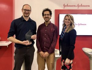
We may be one step closer to 3D-printing organs for transplants thanks to innovations in vascularization from researchers at Rice University and Washington University. Jordan Miller, Ph.D., a Penn Bioengineering alumnus, now an assistant professor of bioengineering at Rice, worked with his colleague Kelly Stevens, Ph.D., an assistant professor of the bioengineering department at Washington, to develop 3D-printed networks that mimicked the vascularized pathways for the transport of blood, lymph, and other fluids in the body. Their work appeared on a recent cover of Science, featuring a visual representation of the 3D-printed vessels in vasculature meant to mirror that of the human lung.

Relying heavily on open source 3D-printing, Miller and Stevens, along with collaborators from a handful of other institutions and start-ups, found ways to model dynamic vasculature systems similar to heart valves, airways systems, and bile ducts to keep 3D-printed tissue viable. The video below demonstrates the way the team successfully modeled vasculature in a small portion of the lung by designing a net-like structure around a sack of air. But Miller, a long-time supporter of open source printing and bioprinting, hopes that this is merely one step closer to what he sees as the ultimate goal of allowing for all organs to be bioprinted. Having that sort of power would reduce the complex issues that come with organ transplants, from organ availability to compatibility, and bring an end to a health issue that affects the over 100,000 people on the organ transplant waiting list.

A Combination of Protein Synthesis and Spectrometry Improve Cell Engineering

One goal of modern medicine is to create individualized therapeutics by figuring out a way to control cell function to perform specific tasks for the body without disrupting normal cell function. Balancing these two goals often proves to be one of the greatest difficulties of this endeavor in the lab, but researchers at Northwestern University found a way to combine the two functions at once in methods they’re calling cell-free protein synthesis and self-assembled monolayer desorption ionization (SAMDI) mass spectrometry. This innovation in the combination of the two methods accelerates the trial and error process that comes with engineering cells for a specific need, allowing researchers to cover a lot more ground in determining what works best in a smaller amount of time.

Leading the study are Milan Mrksich, Ph.D., a Henry Wade Rogers Professor of Biomedical Engineering at Northwestern, and Michael Jewett, Ph.D., a Charles Deering McCormick Professor of Teaching Excellence and co-director of the Center for Synthetic Biology at Northwestern. Together, they hope to continue to take advantage of the factory-like qualities of cell operations in order to use cells from any organisms to our advantage as needed. By helping to reduce the amount of time spent on trial and error, this study brings us one step closer to a world of efficient and individualized medicine.

Non-Invasive Sensory Stimulation as New Way of Treating Alzheimer’s

What if we could reduce the effects of Alzheimer’s disease with a non-invasive therapy comprised of only sensory inputs of light and sound? A recent study between Georgia Tech and MIT tries to make that possible. Alzheimer’s patients often have a larger than normal number of amyloid plaques in their brains, which is a naturally occurring protein that in excess can disrupt neurological function. The treatment —  designed in part by Abigail Paulson, a graduate student in the lab of Annabelle Singer, Ph.D., assistant professor of Biomedical Engineering at Georgia Tech and Emory University — uses a combination of light and sound to induce gamma oscillations in brain waves of mice with high amounts of these amyloid plaques. Another lead author of the study is Anthony Martorell, a graduate student in the Tsai Lab at MIT, where Singer was a postdoctoral researcher.

This new approach is different from other non-invasive brain therapies for memory improvement, as tests demonstrated that it had the power to not only reach the visual cortex, but that it also had an effect on the memory centers in the hippocampus. An innovation like this could bring about a more widespread form of treatment for Alzheimer’s patients, as the lack of a need for surgery makes it far more accessible. Singer hopes to continue the project in the future by looking at how these sensory stimulations affect the brain throughout a variety of processes, and more importantly, if the therapy can be successfully applied to human patients.

NIH Grant Awarded to Marquette Biomedical Engineering Professor for Metal Artifact Reduction Techniques in CT Scans

Taly Gilat-Shmidt, Ph.D., an associate professor of biomedical engineering at Marquette University, recently received a $1.4 million grant from the National Institute of Health to improve methods for radiation treatment through metal artifact reduction techniques. When patients have some sort of metal that can’t be removed, such as an orthopaedic implant like a hip or knee replacement, it can interfere with the imaging process for CT scans and lead to inaccuracies by obscuring some tissue in the final images. These inaccuracies can lead to difficulty in devising treatment plans for patients who require radiation, as CT scans are often used to assess patients and determine which line of treatment is most appropriate. Gilat-Schmidt hopes to use the grant to implement tested algorithms to help reduce this variability in imaging that comes from metal implants.

People and Places

Activities for Community Education in Science (ACES), founded by Penn chemistry graduate students in 2014, aims to inspire interest and provide a positive outlook in STEM for kids and their families. The biannual event provides students grades 3–8 with an afternoon of demonstrations, experiments, and hands-on activities focused on physics and chemistry.

After an explosive opening demonstration, more than 70 students made their way between experiments in small groups, each participating in different experiments based on their age.

Read the rest of this story on Penn Today.

The Society of Women Engineers (SWE) is a non-profit organization serving as one of the world’s largest advocates for women in engineering and technology over the past six decades. With a mission to empower women to become the next leading engineers of the world, SWE is just one of many agents hoping to bring more diversity to the field. Our chapter of SWE at Penn focuses particularly on professional development, local educational outreach, and social activities across all general body members. In a new article from SWE Magazine, the organization collected social media responses from the public on the women engineers we should all know. With a diverse list of engineers from both the past and present, the article helps bring to light just how much even a handful of women contributed to the field of engineering already.


Chip Diagnostics receives the JPOD @ Philadelphia QuickFire Challenge Award

By Erica K. Brockmeier

Chip Diagnostics is the awardee of the JPOD @ Philadelphia QuickFire Challenge sponsored by Johnson & Johnson Innovation — JLABS. The Challenge was designed to accelerate healthcare innovation and commercialization within the greater Philadelphia area.

David Issadore (center) was announced as the awardee of the JPOD @ Philadelphia QuickFire Challenge by Katherine Merton (right), head of JLABS New York City, Boston, and Philadelphia, at last week’s BIO 2019 International convention. (Photo: Johnson & Johnson Innovation)

Chip Diagnostics is a Philadelphia-based device company founded in 2016 based on research from the lab of David Issadore, Assistant Professor of Bioengineering and Electrical and Systems Engineering in the School of Engineering and Applied Science. The startup combines microelectronics, microfluidics, and nanomaterials with the aim to better diagnose cancer. The company is developing technologies and digital assays for minimally-invasive early cancer detection and screening that can be done using mobile devices.

There has been a long interest in diagnosing cancer using blood tests by looking for proteins, cells, or DNA molecules shed by tumors, but these tests have not worked well for many cancers since the molecules shed tend to be either nonspecific or very rare.

Issadore’s group aims to target different particles called exosomes: Tiny particles shed by cells that contain similar proteins and RNA as the parent cancer cell. The problem, explains Issadore, is that because of the small size of the exosomes, conventional methods such as microscopy and flow cytometry wouldn’t work. “As an engineering lab, we saw an opportunity to build devices on a nanoscale that could specifically sort the cancer exosomes versus the background exosomes of other cells,” he explains.

After Issadore was approached by the IP group at PCI Ventures in the early stages of their research, Chip Diagnostics has since made huge strides as a company. Now, as the awardee of the JPOD @ Philadelphia QuickFire Challenge, Chip Diagnostics will receive $30,000 in grant funding to further develop the first-in-class, ultra-high-definition exosomal-based cancer diagnostic. The award also includes one year of residency at Pennovation Works as well as access to educational programs and mentoring provided by Johnson & Johnson Family of Companies global network of experts.

Originally posted on the Penn Engineering Medium Blog. Continue reading at Penn Today.

Week in BioE (June 14, 2019)

by Sophie Burkholder

Bio-inspiration Informs New Football Helmet Design from IUPUI Students

Art, design, biology, and engineering all interact with each other in a recent design for a football helmet from two students one of media arts and the other of engineering at the Indiana University – Purdue University Indianapolis. Directed by Lecturer in Media Arts and Science Zebulun Wood, M.S., and Associate Professor of Mechanical and Energy Engineering and Assistant Professor of Biomedical Engineering Andres Tovar, Ph.D., the students found inspiration in biological structures like a pomelo peel, nautilus shell, and woodpecker skull to create energy-absorbing helmet liners. The resulting design took these natural concussion-reducing structures and created compliant mechanism lattice-based liners the replace the foam traditionally placed in between two harder shells of a typical helmet. Their work not only exemplifies the benefits of bio-inspiration, but demonstrates the way that several different domains of study can overlap in the innovation of a new product.

Study of Mechanical Properties of Hyaluronic Acid Could Help Inform Current Debates Over Treatment Regulation for Osteoarthritis

Arthritis is an extremely common condition, especially in older patients, in which inflammation of the joints can cause high amounts of stiffness and pain. Osteoarthritis in particular is the result of the degradation of flexible tissue between the bones of a joint, which increases friction in joint motion. A common treatment of this form of arthritis is the injection of hyaluronic acid, which is meant to provide joint lubrication, and decreases this friction between bones. Recently, however, there has been a debate over hyaluronic acid’s classification by the FDA and whether it should remain based on the knowledge of the mechanical actions of the acid in treatment for osteoarthritis or if potential chemical action of the acid should be considered as well.

Because of limited ways of testing the mechanical properties of the acid, many researchers felt that there could be more to hyaluronic acid’s role in pain relief for arthritic patients. But Lawrence Bonassar, Ph.D., the Daljit S. and Elaine Sarkaria Professor in Biomedical Engineering at the Meinig School of Bioengineering of Cornell University, had another idea. With his lab, he created a custom-made tribometer to measure the coefficient of friction of a given lubricant by rubbing a piece of cartilage back and forth across a smooth glass plate. The research demonstrated that hyaluronic acid’s ability to reduce the coefficient of friction aligned with patients’ pain relief. Bonassar and his team hope that these results will demonstrate the heavy contribution of mechanical action that hyaluronic acid has in osteoarthritis treatment, and help bring an end to the debate over its FDA classification.

A New Way of Mapping the Heart Could Lead to Better Understanding of Contractile Activity

Though reduced contractions in certain regions of the heart can be an indicator of a certain condition, there is currently no way to directly measure contractile activity. This is why Cristian Linte, Ph.D., an Associate Professor of Biomedical Engineering in the Kate Gleason College of Engineering at the Rochester Institute of Technology (RIT), hopes to create a map of the heart that can quantify contraction power. In collaboration with Niels Otani, Ph.D., an Associate Professor in the School of Mathematics at RIT, Linte plans to use an $850,000 grant from the National Science Foundation to achieve a more comprehensive understanding of the heart through both medical imaging and mechanical modeling. The group hopes that their approach will lead to not only a better way to diagnose certain heart conditions and diseases, but also open up understanding of active contraction, passive motion, and the stresses within the heart walls that underlie each.

Celebrity Cat Lil Bub Helps Penn and German Researchers Draw Public Attention to Genetics

Lil Bub’s unique appearance has garnered millions of online fans, and now, an avenue for researchers to talk about genetics. (Photo Courtesy of Mike Bridavsky)

In 2015, a group of curious researchers set out to sequence the genome of a celebrity cat named Lil Bub. They were hoping to understand the genetics behind Lil Bub’s extra toes and unique skeletal structure, which contribute to her heart-warming, kitten-like appearance. However, an equally important goal of their “LilBUBome” project was to invite the general public into the world of genetics.

Orsolya “Uschi” Symmons, a postdoctoral researcher at Penn in Associate Professor of Bioengineering Arjun Raj’s lab, led the research team along with Darío Lupiáñez at the Max-Delbrück Center for Molecular Medicine in Berlin, and Daniel Ibrahim at the Max Planck Institute for Molecular Geneticsin Berlin. Lil Bub’s owner, Mike Bridavsky, also contributed to the project.

Because of Lil Bub’s online fame, the project garnered attention from her fans and the media, all hoping to discover the secret to Lil Bub’s charm. As early as 2015, Gizmodo’s Kiona Smith-Strickland reported on the team’s intentions to sequence Lil Bub’s genome, and, since then, many have been awaiting the results of the LilBUBome.

To read more of this story, visit Penn Engineering’s Medium Blog.

People and Places

The Alfred P. Sloan Foundation awarded a six-year grant to Barnard College and Columbia University’s School of Engineering and Applied Science to support graduate education for women in engineering. The funding will go towards a new five-year program that enables Barnard students to attain both a B.A. and M.S. in one year after their traditional four years of undergraduate education. The program will offer M.S. degrees in chemical engineering, biomedical engineering, and industrial engineering and operations research, and is one of the first of its kind for women’s colleges.

We would like to congratulate Jean Paul Allain, Ph.D., on being named the first head of the new Ken and Mary Alice Lindquist Department of Nuclear Engineering at Penn State. Allain, who is currently a Professor and head of graduate programs in the University of Illinois at Urbana-Champaign’s Department of Nuclear, Plasma, and Radiological Engineering, conducts research in models of particle-surface interactions. In addition to being head of the new department at Penn State, Allain will also hold a position as a Professor of Biomedical Engineering at the university.

We would also like to congratulate Andrew Douglas, Ph.D., on his appointment as the Vice Provost for Faculty Affairs at Johns Hopkins University. Douglas currently holds the position of Vice Dean for Faculty at the Whiting School of Engineering, and has joint appointments in Mechanical and Biomedical Engineering. Douglas’s research at Hopkins focuses on mechanical properties and responses of compliant biological tissue and on the nonlinear mechanics of solids, with a focus on soft tissues and organs like the heart and tongue.

César de la Fuente Named One of GEN’s ‘Top 10 Under 40’

Cesar de la Fuente-Nunez, PhD

César de la Fuente, assistant professor in the Departments of Psychiatry and Microbiology in the Perelman School of Medicine and the Department of Bioengineering in the School of Engineering and Applied Science, has been listed as one of the top 10 emerging professionals in his field under the age of 40 by GEN, a publication that covers genetic engineering and biotechnology news. The list recognizes up-and-coming leaders in the field of life sciences, both in industry and academia.

De la Fuente, who started at Penn earlier this year, was recognized because he “is pioneering the computerization of biological systems for the development of transformative biotechnologies designed to solve societal grand challenges such as antibiotic resistance.”

Read the full story at the Penn Engineering Medium blog.

Week in BioE (May 31, 2019)

by Sophie Burkholder

Vector Flow Imaging Helps Visualize Blood Flow in Pediatric Hearts

A group of biomedical engineers at the University of Arkansas used a new ultrasound-based imaging technique called vector flow imaging to help improve the diagnosis of congenital heart disease in pediatric patients. The study, led by associate professor of biomedical engineering Morten Jensen, Ph.D., collaborated with cardiologists at the local Children’s Hospital in Little Rock to produce images of the heart in infants to help potentially diagnose congenital heart defects. Though the use of vector flow imaging has yet to be developed for adult patients, this type of imaging could possibly provide more detail about the direction of blood flow through the heart than traditional techniques like echocardiography do. In the future, the use of both techniques could provide information about both the causes and larger effects of heart defects in patients.

Using Stem Cells to Improve Fertility in Leukemia Survivors

One of the more common side effects of leukemia treatment in female patients is infertility, but researchers at the University of Michigan want to change that. Led by associate professor of biomedical engineering Ariella Shikanov, Ph.D., researchers in her lab found ways of increasing ovarian follicle productivity in mice, which directly relates to the development of mature eggs. The project involves the use of adipose-derived stem cells, that can be found in human fat tissue, to surround the follicles in an ovary-like, three-dimensional scaffold.  Because the radiation treatments for leukemia and some other cancers are harmful to follicles, increasing their survival rate with this stem cell method could reduce the rate of infertility in patients undergoing these treatments. Furthermore, this new approach is innovative in its use of a three-dimensional scaffold as opposed to a two-dimensional one, as it stimulates follicle growth in all directions and thus helps to increase the follicle survival rate.

Penn Engineers Look at How Stretching & Alignment of Collagen Fibers Help Cancer Cells Spread

Cancer has such a massive impact on people’s lives that it might be easy to forget that the disease originates at the cellular level. To spread and cause significant damage, individual cancer cells must navigate the fibrous extracellular environment that cells live in, an environment that Penn Engineer Vivek Shenoy has been investigating for years.

Shenoy is the Eduardo D. Glandt President’s Distinguished Professor with appointments in Materials Science and Engineering, Mechanical Engineering and Applied Mechanics, and Bioengineering. He is also the Director of the Center for Engineering MechanoBiology (CEMB), one of the NSF’s twelve Science and Technology Centers.

Shenoy’s most recent study on cancer’s mechanical environment was led by a postdoctoral researcher in his lab, Ehsan Ban. Paul Janmey, professor in Physiology and Bioengineering, and colleagues at Stanford University also contributed to the study. Shenoy also received the Heilmeier Award this March and delivered the Heilmeier Award Lecture in April.

Read the rest of this story on Penn Engineering’s Medium Blog.

Controlled Electrical Stimulation Can Prevent Joint Replacement Infections

Joint replacements are one of the most common kinds of surgery today, but they still require intense post-operative therapy and have a risk of infection from the replacement implant. These infections are usually due to the inflammatory response that the body has to any foreign object, and can become serious and life-threatening if left untreated. Researchers at the University of Buffalo Jacobs School of Medicine and Biomedical Sciences hope to offer a solution to preventing infections through the use of controlled electrical stimulation. Led by Mark Ehrensberger, Ph.D., Kenneth A. Krackow, M.D., and Anthony A. Campagnari, Ph.D., the treatment system uses the electrical signal to create an antibacterial environment at the interface of the body and the implant. While the signal does not prevent infections completely, these antibacterial properties will prevent infections from worsening to a more serious level. Patented as the Biofilm Disruption Device TM, the final product uses two electrode skin patches and a minimally invasive probe that delivers the electrical signal directly to the joint-body interface. The researchers behind the design hope that it can help create a more standard way of effectively treating joint replacement infections.

People and Places

TBx: Gabriel Koo, Ethan Zhao, Daphne Cheung, and Shelly Teng

For their senior design project, four bioengineering seniors Gabriel Koo, Ethan Zhao, Daphne Cheung, and Shelly Teng created a low-cost tuberculosis diagnostic that they called TBx. Using their knowledge of the photoacoustic effect of certain dyes, the platform the group created can detect the presence of lipoarabinomannan in patient urine. The four seniors presented TBx at the Rice360 Design Competition in Houston, Texas this spring, which annually features student-designed low-cost global health technologies.

Dan Huh’s Space-based Organ-on-a-Chip Experiments Featured in WIRED

By Lauren Salig

SpaceX launched its 17th resupply mission to the International Space Station on May 4, with bioengineering professor Dan Huh’s organ-on-a-chip experiments in tow.

Dan Huh, the Wilf Family Term Assistant Professor in the Department of Bioengineering, researches human organs and the diseases that infect them by engineering devices made of living cells that act as stand-ins for organs. Huh’s lab has developed imitations of many organs, including the placenta and the eye, but it’s his lung-on-a-chip and his bone-marrow-on-a-chip that are reaching unprecedented heights as part of a new experiment taking place at the International Space Station (ISS).

On May 4, SpaceX launched a ISS-bound cargo capsule carrying Huh’s organ-on-a-chip experiments, which will remain in space for a month. Once back on Earth, the chips that spent time in space will be compared to control chips from Huh’s lab that are being monitored in parallel. Huh’s team is looking to see how being in space affects bacterial infections in lungs and white blood cell behavior in bone marrow. The researchers’ hope is that their studies will reveal important information about how human organs function both in space and on Earth.

Daniel Oberhaus of WIRED wrote an article describing the multiple organs-on-a-chip experiments being conducted at the ISS, including the two experiments headed by Huh:

Dan Huh is a bioengineer at the University of Pennsylvania and the lead researcher on the lung tissue chip headed to the ISS. This lung chip models a human airway and will be infected with Pseudomonas aeruginosa, a species of bacteria that had previously been found on the ISS. On Earth this bacteria is usually associated with respiratory infections, which are one of the leading types of illness on long-duration missions to the ISS.

Huh says scientists still know very little about why astronauts’ immune response seems to become suppressed in orbit, and the tissue chips are aimed at building a better understanding of the phenomenon.


Originally posted at the Penn Engineering Medium Blog.

Read the entire article at WIRED.

Penn Engineers: Cells Require Gene Expression Feedback to Keep Moving

By Lauren Salig

When cells move throughout the body, they do so by dragging themselves, using molecular “arms” to pull themselves closer to where they need to be while unlatching themselves from the area they’re moving away from. In a recent study, Penn Engineers looked at a few mechanobiological factors that help regulate cells’ migration towards their destination, providing new insight into the gene expression feedback loops that keep them from getting stuck.

Joel Boerckel and Devon Mason

The research was led by Joel Boerckel, Assistant Professor of Orthopaedic Surgery in the Perelman School of Medicine and in Bioengineering in Penn Engineering, and bioengineering graduate student Devon Mason. Co-authors include bioengineering graduate student Joseph Collins and researchers from the University of Notre Dame, Indiana University and Purdue University.

The study was published in the Journal of Cell Biology.

Read the full story at the Penn Engineering Medium Blog.

Week in BioE: April 19, 2019

by Sophie Burkholder

New Vascularized Patches Could Help Patient Recovery from Heart Attacks

Heart attacks are the result of a stoppage of blood flow to the heart – an interruption to normal function that can result in severe tissue damage, or even tissue death. This loss of healthy tissue function is one of the biggest challenges in treating patients that undergo heart attacks, as the damaged tissue increases their risk of having future attacks. One of the main solutions to this issue right now is the creation of cardiac tissue scaffolds using stem cells to create a platform for new and healthy tissue to grow in vivo. A group of biomedical engineers at Michigan Technological University hopes to expand on this basis by focusing not just on cellular alignment in the scaffold but on that of microvessels too. Led by Feng Zhao, Ph.D., Associate Professor of Biomedical Engineering, the team hopes that this new attention on microvessel organization will improve the vasculature of the scaffolds, and thus improve the success of the scaffolds in vivo, allowing for a better recovery from heart attacks.

Some Stem Cells May Be More Fit Than Others

Stem cells are one of the hottest research areas in the field of bioengineering today. Widely known as the cells in the human embryo that have the ability to eventually transform into specific cells for the brain, lung, and every other organ, stem cells are also of recent interest because researchers found ways to reverse this process, transforming organ-specific cells back to the pluripotent stem cell level. This achievement however, is mostly applicable to individual stem cells, and doesn’t fully encapsulate the way this process might work on a larger population level. So Peter Zandstra, Ph. D., a bioengineering faculty member at the University of British Columbia, decided to research just that.

Using mouse embryonic fibroblasts (MEFs), Zandstra and his lab attempted to track the cells throughout their reprogramming, to more clearly trace each back to its respective parent population. Surprisingly, they found that after only one week of reprogramming, nearly 80% of the original cell population had been removed, meaning that most of the parent generation was not “fit” enough to undergo the process of reprogramming, indicating that perhaps some stem cells will have a better chance of survival in this process than others. This research may suggest that not all cells have the capacity to undergo reprogramming, as many researchers originally thought.

A New Microdevice Will Help Model Bronchial Spasms

The difficulty in breathing associated with asthma is the result of bronchial spasms, which are a kind of muscle contraction in the airways. But little was known about just how these spasms occurred in patients, so Andre Levchenko, Ph.D., Professor of Biomedical Engineering at Johns Hopkins, and his lab created a microdevice to model them. Calling the device a “bronchi on a chip,” Levchenko and his team used a microphysiological model to look at some of the biochemical and mechanical signals associated with these kinds of muscle contractions. They found that the contractions operate in a positive feedback system, so that those caused by disturbance from allergens will subsequently cause even more contractions to occur. But surprisingly, they also found that a second contraction, if triggered at the right time during the initial contraction, could actually stop the process and allow the muscles to relax. Because asthma is a notoriously difficult disease to translate from animal to human models, this new device opens the door to understanding different mechanisms of asthma before taking research to clinical trials.

New CHOP Research Center to Focus Research on Pediatric Airway Disorders

A new bioengineering lab at the Children’s Hospital of Philadelphia called the Center for Pediatric Airway Disorders will specialize in a variety of airway procedures for pediatric patients such as tracheal reconstruction and recurrent laryngeal nerve reinnervation. This new lab will be one of the first to give a unique focus to the application of bioengineering to pediatric laryngology. The interdisciplinary center brings together students and researchers from all different fields, including materials science and microbiology, to find new ways of repairing tissue and regenerating organs related to respiratory disorders. Specific areas of research will involve the modeling of children’s vocal cords, understanding the mechanisms of fibrosis, and improving surgical procedures.

Deeper Understanding of Sickle Cell Anemia Could Lead to New Treatments

Though sickle cell anemia is a common and well-known disease, a new study of its causes at the nanoscale level might reveal previously unknown information about the assembly of hemoglobin fibers. Using microscopes with the ability to visualize these molecules at such a small level, researchers at the University of Minnesota found that the beginning organizations that lead to sickle cell anemia are much less ordered than originally thought. Led by Associate Professor of Biomedical Engineering David Wood, Ph.D., the team of researchers used this higher level of microscopy to find that hemoglobin self-assembly process, which was originally thought to be 96% efficient, is actually only 4% efficient. Wood hopes that this new knowledge will help allow for the development of new and better treatments for patients with sickle cell anemia, as there are currently only two FDA-approved ones on the market.

People & Places

Penn Today asked five Penn researchers about the women in STEM who have been a source of inspiration and encouragement throughout their own careers. Their responses include active researchers who have paved the way for better inclusion in STEM and famous female scientists from the past who broke boundaries as they made strides with their research.

Dr. Danielle Bassett, the Eduardo D. Glandt Faculty Fellow and associate professor of bioengineering and electrical and systems engineering in the School of Engineering and Applied Science, has two heroes: “Ingrid Daubechies for her work on wavelets, or “little waves,” which are beautiful mathematical objects that can be used to extract hidden structure in complex data. “Also, Maryam Mirzakhani for inspiring a child to believe that mathematics is simply painting. Would that we all could see the world just that bit differently.”

Read the full story on Penn Today.

Joel Boerckel, Ph.D, Assistant Professor of Orthopaedic Surgery and Bioengineering

This week, we want to congratulate Joel Boerckel, Ph.D., Assistant Professor of Orthopaedic Surgery and Bioengineering, and his lab on receiving a second R01 Grant from the National Institute of Arthritis and and Musculoskeletal Skin Diseases for their work on defining the roles of YAP and TAZ in embryonic bone morphogenesis and mechanoregulation of fracture repair. Dr. Boerckel is a member of the McKay Orthopaedic Research Laboratory.

We would also like to congratulate Christopher Yip, Ph. D., on being appointed as the new dean of the University of Toronto’s Faculty of Applied Science and Engineering. A professor in both the Department of Chemical Engineering and Applied Chemistry the Institute of Biomaterials and Biomedical Engineering, Dr. Yip’s research involves the use of molecular imaging to understand the self-assembly of proteins.

Week in BioE: April 5, 2019

by Sophie Burkholder

Tulane Researchers Use Cancer Imaging Technique to Help Detect Preeclampsia

Preeclampsia is potentially life-threatening pregnancy disorder that typically occurs in about 200,000 expectant mothers every year. With symptoms of high blood pressure, swelling of the hands and feet, and protein presence in urine, preeclampsia is usually treatable if diagnosed early enough. However, current methods for diagnosis involve invasive procedures like cordocentesis, a procedure which takes a sample of fetal blood.

Researchers at Tulane School of Medicine led by assistant professor of bioengineering Carolyn Bayer, Ph.D., hope to improve diagnostics for preeclampsia with the use of spectral photoacoustic imaging. Using this technique, Bayer’s team noticed a nearly 12 percent decrease in placental oxygenation in rats with induced preeclampsia when compared to normal pregnant rats after only two days. If success in using this imaging technology continues at the clinical level, Bayer plans to find more applications of it in the detection and diagnosis of breast and ovarian cancers as well.

New CRISPR-powered device detects genetic mutations in minutes 

Two groups of researchers from the University of California, Berkeley and the Keck Graduate Institute of the Claremont Colleges recently collaborated to design what they call a “CRISPR-Chip” –  a combination of the CRISPR-Cas9 System with a graphene transistor to sequence DNA for the purpose of genetic mutation diagnosis. While companies like 23andMe made genetic testing and analysis more common and accessible for the general public in recent years, the CRISPR-Chip looks to streamline the technology even more.

This new chip eliminates the long and expensive amplification process involved in the typical polymerase chain reaction (PCR) used to read DNA sequences. In doing so, the CRISPR-Chip is much more of a point-of-care diagnostic, having the ability to quickly detect a given mutation or sequence when given a pure DNA sample. Led by Kiana Aran, Ph.D., the research team behind the CRISPR-Chip hopes that this new combination of nanoelectronics and modern biology will allow for a new world of possibilities in personalized medicine.

New Method of Brain Stimulation Might Alleviate Symptoms of Depression

Depression is one of the most common mental health disorders in the United States, with nearly 3 million cases every year. For most patients suffering from depression, treatment involves prolonged psychotherapy, antidepressant medication, or even electroconvulsive therapy in extreme cases. Now, scientists at the University of North Carolina School of Medicine study the use of transcranial alternating current stimulation (tACS) to alleviate symptoms of depression.

Led by Flavio Frohlich, Ph.D., who has an adjunct appointment in biomedical engineering, this team of researchers based this new solution on information from each patient’s specific alpha oscillations, which are a kind of wave that can be detected by an electroencephalogram (EEG). Those who suffer from depression tend to have imbalanced alpha oscillations, so Frohlich and his team sought to use tACS to restore this balance in those patients. After seeing positive results from data collected two weeks after patients in a clinical trial receives the tACS treatment, Frohlich hopes that future applications will include treatment for even more mental health disorders and psychiatric illnesses.

University of Utah Researchers Receive Grant to Improve Hearing Devices for Deaf Patients

Engineers at the University of Utah are part of team that recently received a $9.7 million grant from the National Institute of Health (NIH) to design new implantable hearing devices for deaf patients, with the hope to improve beyond the sound quality of existing devices. The work will build upon a previous project at the University of Utah called the Utah Electrode Array, a brain-computer interface originally developed by Richard Normann, Ph.D., that can send and receive neural impulses to and from the brain. This new device will differ from a typical cochlear implant because the Utah Electrode Array assembly will be attached directly to the auditory nerve instead of the cochlea, providing the patient with a much higher resolution of sound.

People & Places

Vivek Shenoy, Eduardo D. Glandt President’s Distinguished Scholar in the Department of Materials Science and Engineering and Secondary Faculty in Bioengineering, has been named the recipient of the 2018–19 George H. Heilmeier Faculty Award for Excellence in Research for “for pioneering multi-scale models of nanomaterials and biological systems.”

The Heilmeier Award honors a Penn Engineering faculty member whose work is scientifically meritorious and has high technological impact and visibility. It is named for George H. Heilmeier, a Penn Engineering alumnus and advisor whose technological contributions include the development of liquid crystal displays and whose honors include the National Medal of Science and Kyoto Prize.

Read the rest of the story on Penn Engineering’s Medium blog.

We would also like to congratulate Jay Goldberg, Ph.D., from Marquette University on his election as a fellow to the National Academy of Inventors. Nominated largely for his six patents involving medical devices, Goldberg also brings this innovation to his courses. One in particular called Clinical Issues in Biomedical Engineering Design allows junior and senior undergraduates to observe the use of technology in clinical settings like the operating room, in an effort to get students thinking about how to improve the use of medical devices in these areas.


Week in BioE: March 29, 2019

by Sophie Burkholder

New Studies in Mechanobiology Could Open Doors for Cellular Disease Treatment

When we think of treatments at the cellular level, we most often think of biochemical applications. But what if we began to consider more biomechanical-oriented approaches in the regulation of cellular life and death? Under a grant from the National Science Foundation (NSF),Worcester Polytechnic Institute’s (WPI) Head of the Department of Biomedical Engineering Kristen Billiar, Ph.D., performs research that looks at the way mechanical stimuli can affect and trigger programmed cell death.

Billiar, who received his M.S.E. and Ph.D. from Penn, began his research by first noticing the way that cells typically respond to the mechanical stimuli in their everyday environment, such as pressure or stretching, with behaviors like migration, proliferation, or contraction. He and his research team hope to find a way to eventually predict and control cellular responses to their environment, which they hope could open doors to more forms of treatment for disorders like heart disease or cancer, where cellular behavior is directly linked to the cause of the disease.

Self-Learning Algorithm Could Help Improve Robotic Leg Functionality

Obviously, one of the biggest challenges in the field of prosthetics is the extreme difficulty in creating a device that perfectly mimics whatever the device replaces for its user. Particularly with more complex designs that involve user-controlled motion for joints in the limbs or hands, the electrical circuits implemented are by no means a perfect replacement of the neural connections in the human body from brain to muscle. But recently at the University of Southern California Viterbi School of Engineering, a team of researchers led by Francisco J. Valero-Cuevas, Ph. D.,  developed an algorithm with the ability to learn new walking tasks and adapt to others without any additional programming.

The algorithm will hopefully help to speed the progress of robotic interactions with the world, and thus allow for more adaptive technology in prosthetics, that responds to and learns with their users. The algorithm Valero-Cuevas and his team created takes inspiration from the cognition involved with babies and toddlers as they slowly learn how to walk, first through random free play and then from pulling on relevant prior experience. In a prosthetic leg, the algorithm could help the device adjust to its user’s habits and gait preferences, more closely mimicking the behavior of an actual human leg.

Neurofeedback Can Improve Behavioral Performance in High-Stress Situations

We’re all familiar with the concept of being “in the zone,” or the feeling of extraordinary focus that we can sometimes have in situations of high-stress. But how can we understand this shift in mindset on a neuroengineering level? Using the principal of the Yerkes-Dodson law, which says that there is a state of brain arousal that is optimal for behavioral performance, a team of biomedical engineering researchers at Columbia University hope to find ways of applying neurofeedback to improving this performance in demanding high-stress tasks.

Led by Paul Sajda, Ph.D., who received his doctoral degree from Penn, the researchers used a brain-computer interface to collect electroencephalography (EEG) signals from users immersed in virtual reality aerial navigation tasks of varying difficulty levels. In doing so, they were able to make connections between stressful situations and brain activity as transmitted through the EEG data, adding to the understanding of how the Yerkes-Dodson law actually operates in the human body and eventually demonstrating that the use of neurofeedback reduced the neural state of arousal in patients. The hope is that neurofeedback may be used in the future to help treat emotional conditions like post-traumatic stress disorder (PTSD).

Ultrasound Stimulation Could Lead to New Treatments for Inflammatory Arthritis

Arthritis, an autoimmune disease that causes painful inflammation in the joints, is one of the more common diseases among older patients, with more than 3 million diagnosed cases in the United States every year. Though extreme measures like joint replacement surgery are one solution, most patients simply treat the pain with nonsteroidal anti-inflammatory drugs or the adoption of gentle exercise routines like yoga. Recently however, researchers at the University of Minnesota led by Daniel Zachs, M.S.E., in the Sensory Optimization and Neural Implant Coding Lab used ultrasound stimulation treatment as a way to reduce arthritic pain in mice. In collaboration with Medtronic, Zachs and his team found that this noninvasive ultrasound stimulation greatly decreased joint swelling in mice who received the treatment as opposed to those that did not. They hope that in the future, similar methods of noninvasive treatment will be able to be used for arthritic patients, who otherwise have to rely on surgical remedies for serious pain.

People and Places

Leadership and Inspiration: EDAB’s Blueprint for Engineering Student Life

To undergraduates at a large university, the administration can seem like a mysterious, all-powerful entity, creating policy that affects their lives but doesn’t always take into account the reality of their day-to-day experience. The Engineering Deans’ Advisory Board (EDAB) was designed to bridge that gap and give students a platform to communicate with key decision makers.

The 13-member board meets once per week for 60 to 90 minutes. The executive board, comprised of four members, also meets weekly to plan out action items and brainstorm. Throughout his interactions with the group, board president Jonathan Chen, (ENG ‘19, W ‘19), has found a real kinship with his fellow board members, who he says work hard and enjoy one another’s company in equal measure.

Bioengineering major Daphne Cheung (ENG’19) joined the board as a first-year student because she saw an opportunity to develop professional skills outside of the classroom. “For me, it was about trying to build a different kind of aptitude in areas such as project management, and learning how to work with different kinds of people, including students and faculty, and of course, the deans,” she says.

Read the full story on Penn Engineering’s Medium Blog. Media contact Evan Lerner.

Purdue University College of Engineering and Indiana University School of Medicine Team Up in New Engineering-Medicine Partnership

The Purdue University College of Engineering and the Indiana University School of Medicine recently announced a new Engineering-Medicine partnership, that seeks to formalize ongoing and future collaborations in research between the two schools. One highlight of the partnership is the establishment of a new M.D./M.S. degree program in biomedical engineering that will allow medical students at Indiana University to receive M.S.-level training in engineering technologies as they apply to clinical practice. The goal of this new level of collaboration is to further involve Purdue’s engineering program in the medical field, and to exhibit the benefits that developing an engineering mindset can have for medical students. The leadership of this new partnership includes