Week in BioE (March 15, 2019)

by Sophie Burkholder

Synthetic Spinal Discs from a Penn Research Team Might Be the Solution to Chronic Back Pain

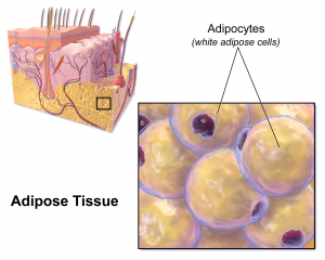
Spinal discs, the concentric circles of collagen fiber found between each vertebra of the spine, can be the source of immense back pain when ruptured. Especially for truck and bus drivers, veterans, and cigarette smokers, there is an increased risk in spinal disc rupture due to overuse or deterioration over time. But these patients aren’t alone. In fact, spinal discs erode over time for almost everyone, and are one of the sources of back pain in older patients, especially when the discs erode so much that they allow direct bone-to-bone contact between two or more vertebrae.

Robert Mauck, Ph.D.

Robert Mauck, Ph.D., who is the director of the McKay Orthopaedic Research Laboratory here at Penn and a member of the Bioengineering Graduate Group Faculty, led a research team in creating artificial spinal discs, with an outer layer made from biodegradable polymer and an inner layer made with a sugar-like gel. Their findings appear in Science Translational Medicine. These synthetic discs are also seeded with stem cells that produce collagen over time, meant to replace the polymer as it degrades in vivo over time. Though Mauck and his time are still far from human clinical trials for the discs, they’ve shown some success in goat models so far. If successful, these biodegradable discs could lead to a solution for back pain that integrates itself into the human body over time, potentially eliminating the need of multiple invasive procedures that current solutions require. Mauck’s work was recently featured in Philly.com.

An Untethered, Light-Activated Electrode for Innovations in Neurostimulation

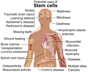
Neurostimulation, a process by which nervous system activity can be purposefully modulated, is a common treatment for patients with some form of paralysis or neurological disorders like Parkinson’s disease. This procedure is typically invasive, and because of the brain’s extreme sensitivity, even the slightest involuntary movement of the cables, electrodes, and other components involved can lead to further brain damage through inflammation and scarring. In an effort to solve this common problem, researchers from the B.I.O.N.I.C. Lab run by Takashi D.Y. Kozai, Ph. D., at the University of Pittsburgh replaced long cables with long wavelength light and a formerly tethered electrode with a smaller, untethered one.

The research team, which includes Pitt senior bioengineering and computer engineering student Kaylene Stocking, centered the device on the principle of the photoelectric effect – a concept first described in a publication by Einstein as the local change in electric potential for an object when hit with a photon. Their design, which includes a 7-8 micron diameter carbon fiber implant, is now patent pending, and Kozai hopes that it will lead to safer and more precise advancements in neurostimulation for patients in the future.

A New Microfluidic Chip Can Detect Cancer in a Drop of Blood

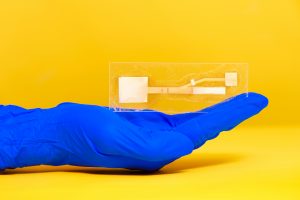
Many forms of cancer cannot be detected until the disease has progressed past the point of optimum treatment time, increasing the risk for patients who receive late diagnoses of these kinds of cancer. But what if the diagnostic process could be simplified and made more efficient so that even a single drop of blood could be enough input to detect the presence of cancer in a patient? Yong Zeng, Ph.D., and his team of researchers at the University of Kansas in Lawrence sought to answer that question.

They designed a self-assembled 3D-nanopatterned microfluidic chip to increase typical microfluidic chip sensitivity so that it can now detect lower levels of tumor-associated exosomes in patient blood plasma. This is in large part due to the nanopatterns in the structure of the chip, which promote mass transfer and increase surface area, which in turn promotes surface-particle interactions in the device. The team applied the device to their studies of ovarian cancer, one of the notoriously more difficult kinds of cancer to detect early on in patients.

A Wearable Respiration Monitor Made from Shrinky Dinks

Michelle Khine, Ph. D., a professor of biomedical engineering at the University of California, Irvine incorporates Shrinky Dinks into her research. After using them once before in a medical device involving microfluidics, her lab recently worked to incorporate them into a wearable respiration monitor – a device that would be useful for patients with asthma, cystic fibrosis, and other chronic pulmonary diseases. The device has the capability to track the rate and volume of its user’s respiration based on measurements of the strain at the locations where the device makes contact with the user’s abdomen.

Paired with Bluetooth technology, this sensor can feed live readings to a smartphone app, giving constant updates to users and doctors, as opposed to the typical pulmonary function test, which only provides information from the time at which the test takes a reading. Though Khine and her team have only tested the device on healthy patients so far, they look forward to testing with patients who have pulmonary disorders, in hopes that the device will provide more comprehensive and accessible data on their respiration.

People and Places

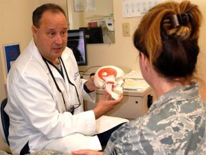
Ashley Kimbel, a high school senior from Grissom High School in Huntsville, Alabama, designed a lightweight prosthetic leg for local Marine, Kendall Bane, after an attack in Afghanistan led him to amputate one of his legs below the knee. Bane, who likes to keep as active as possible, said the new lighter design is more ideal for activities like hiking and mountain biking, especially as any added weight makes balance during these activities more difficult. Kimbel used a CAD-modeling software produced by Siemens called Solid Edge, which the company hopes to continue improving in accessibility so that more students can start projects like Kimbel’s.

This week, we would like to congratulate Angela Belcher, Ph.D., on being named the new head of the Department of Biological Engineering at the Massachusetts Institute of Technology (MIT). With her appointment to this role, now half of the MIT engineering department heads are women. Belcher’s research is in the overlap of materials science and biological engineering, with a particular focus on creating nanostructures based on the evolution of ancient organisms for applications in medical diagnostics, batteries, solar cells, and more.

We would also like to congratulate Eva Dyer, Ph.D., and Chethan Pandarinath, Ph.D., both of whom are faculty members at the Walter H. Coulter Department of Biomedical Engineering at Georgia Tech and Emory University, on receiving research fellowships from the Alfred P. Sloan Foundation. Dr. Dyer, who formerly worked with Penn bioengineering faculty member Dr. Konrad Kording while he was at Northwestern University, leads research in the field of using data analysis methods to quantify neuroanatomy. Dr. Pandarinth leads the Emory and Georgia Tech Systems Neural Engineering Lab, where he works with a team of researchers to use properties of artificial intelligence and machine learning to better understand large neural networks in the brain.


Week in BioE (February 28, 2019)

by Sophie Burkholder

Louisiana Tech Sends First All-Female Team to RockOn

A team of faculty and students from Louisiana Tech University will participate in RockOn, a NASA-sponsored workshop on rocketry and engineering. Mechanical Engineering Lecturer Krystal Corbett, Ph.D., and Assistant Professor of bioengineering Mary Caldorera-Moore, Ph.D., will work together to lead the university’s first team of three all-female students at the event. At the program, they will have the chance to work on projects involving components of spacecraft systems, increasing students’ experience in hands-on activities and real-world engineering.

Refining Autism Treatments Using Big Data

Though treatments like therapy and medication exist for patients with autism, one of the biggest challenges that those caring for these patients face is in measuring their effects over time. Many of the markers of progress are qualitative, and based on a given professional’s opinion on a case-by-case basis. But now, a team of researchers from Rensselaer Polytechnic Institute (RPI) hopes to change that with the use of big data.

Juergen Hahn, Ph. D., and his lab recently published a paper in Frontiers in Cellular Neuroscience discussing their findings in connecting metabolic changes with behavioral improvements in autistic patients. Their analysis looks for multiple chemical and medical markers simultaneously in data from three distinct clinical trials involving metabolic treatment for patients. Being able to quantitatively describe the effects of current autism treatments would revolutionize clinical trials in the field, and lead to overall better patient care.

Penn Engineers Can Detect Ultra Rare Proteins in Blood Using a Cellphone Camera

One of the frontiers of medical diagnostics is the race for more sensitive blood tests. The ability to detect extremely rare proteins could make a life-saving difference for many conditions, such as the early detection of certain cancers or the diagnosis of traumatic brain injury, where the relevant biomarkers only appear in vanishingly small quantities. Commercial approaches to ultrasensitive protein detection are starting to become available, but they are based on expensive optics and fluid handlers, which make them relatively bulky and expensive and constrain their use to laboratory settings.

Knowing that having this sort of diagnostic system available as a point-of-care device would be critical for many conditions — especially traumatic brain injury — a team of engineers led by Assistant Professor in the Department of Bioengineering, David Issadore, Ph.D., at the University of Pennsylvania have developed a test that uses off-the-shelf components and can detect single proteins with results in a matter of minutes, compared to the traditional workflow, which can take days.

Read the full story on Penn Engineering’s Medium blog.

Treating Cerebral Palsy with Battery-Powered Exoskeletons

Cerebral palsy is one of the most common movement disorders in the United States. The disorder affects a patient’s control over even basic movements like walking, so treatments for cerebral palsy often involve the use of assistive devices in an effort to give patients better command over their muscles. Zach Lerner, Ph.D., is an Assistant Professor of Mechanical Engineering and faculty in Northern Arizona University’s Center for Bioengineering Innovation whose research looks to improve these kinds of assistive devices through the use of battery-powered exoskeletons.

Lerner and his lab recently received three grants, one each from the National Institute of Health (NIH), the National Science Foundation (NSF), and the Arabidopsis Biological Resource Center, to continue their research in developing these exoskeletons. Their goal is to create devices with powered assistance at joints like the ankle or knee to help improve patient gait patterns in rehabilitating the neuromuscular systems associated with walking. The team hopes that their work under these new grants will help further advance treatment for children with cerebral palsy, and improve overall patient care.

People & Places

David Aguilar, a 19-year-old bioengineering student at Universitat Internacional de Catalunya made headlines recently for a robotic prosthetic arm that he built for himself using Lego pieces. Due to a rare genetic condition, Aguilar was born without a right forearm, a disability that inspired him to play with the idea of creating his own prosthetic arm from age nine. His design includes a working elbow joint and grabber that functions like a hand. In the future, Aguilar hopes to continue improving his own prosthetic designs, and to help create similar versions of affordable devices for other patients who need them.

This week, we would like to congratulate two recipients of the National Science Foundation’s Career Awards, given to junior faculty that exemplify the role of teacher-scholars in their research. The first recipient we’d like to acknowledge is the University of Arkansas’ Kyle Quinn, Ph.D., who received the award for his work in developing new image analysis methods and models using the fluorescence of two metabolic cofactors. Dr. Quinn completed his Ph.D. here at Penn in Dr. Beth Winkelstein’s lab, and received the Solomon R. Pollack Award for Excellence in Graduate Bioengineering Dissertation Research for his work.

The second recipient of the award we wish to congratulate is Reuben Kraft, Ph.D., who is an Assistant Professor in Mechanical and Biomedical Engineering at Penn State. Dr. Kraft’s research centers around developing computational models of the brain through linking neuroimaging and biomechanical assessments. Dr. Kraft also collaborates with Kacy Cullen, Ph.D., who is a secondary faculty member in Penn’s bioengineering department and a member of the BE Graduate Group faculty.

Finally, we’d like to congratulate Dawn Elliott, Ph.D., on being awarded the Orthopaedic Research Society’s Adele L. Boskey, PhD Award, awarded annually to a member of the Society with a commitment to both mentorship and innovative research. Dr. Elliott’s spent 12 years here at Penn as a member of the orthopaedic surgery and bioengineering faculty before joining the University of Delaware in 2011 to become the founding director of the bioengineering department there. Her research focuses primarily on the biomechanics of fibrous tissue in tendons and the spine.

Week in BioE (February 21, 2019)

by Sophie Burkholder

Detecting Infectious Diseases with Paper-Based Devices

Dr. Linnes’ paper device. Image used courtesy of Erin Easterling, Purdue College of Engineering.

Despite great advancements in diagnostics technology over the past few decades, patient accessibility to these technologies remains one of the biggest challenges of the field today. Particularly in low-resource areas, even simple processes can end up taking weeks or months to return results from tests that are normally completed in days. But what if these tests could be simplified to smaller, at-home tests based on properties of microfluidics – something like a pregnancy test but for infectious diseases like HIV?

Jacqueline Linnes, Ph.D., and her team of researchers at Purdue University are working towards finding a way to do just that by creating paper-based devices that use microfluidics to help carry out the necessary diagnostic tests. Specifically, her lab designed such a paper-based system that can detect HIV nucleic acids within 90 minutes of receiving a drop of patient blood. The success of this design shows promise for producing devices for diseases whose diagnostics process involve similar pathways of pathogen detection, opening the door to more applications of at-home tests based in the properties of paper microfluidics.

Here at Penn, undergraduate bioengineering students enrolled in the two-semester laboratory course Bioengineering Modeling, Analysis, and Design (BE 309 & BE 310) have the chance to create their own models of paper microfluidics delivery systems based on given time constraints in a multi-step process. Though the students’ challenge only involves water as a substrate, Linnes’ research demonstrates the later implications of studying fluid flow through a medium as cheap and accessible as paper.

Watch the video below demonstrating Dr. Linnes’ device:

Funding for Cancer Research in Tumor Mimicry and Imaging

Two of the deadliest forms of cancer today are breast cancer and pancreatic cancer, with the latter having a five-year survival rate of only about 8%. Because cancer treatments are often adjusted according to a unique patient-to-patient basis, learning how to improve predictions of tumor behavior could help determine proper therapies sooner.

Chien-Chi Lin, Ph.D., an associate professor of biomedical engineering at Indiana University – Purdue University Indianapolis, recently received a grant from the National Institute of Health to advance his research in pancreatic cancer treatment. His project under the grant involves the development of bio-inspired, responsive, and viscoelastic (BRAVE) cell-laden hydrogels to help understand cell interactions in pancreatic ductal adenocarcinoma, which is the most common form of malignancy in the pancreas. These hydrogels mimic tumor tissue, as well as model tumor development over time, helping to eventually find better ways of treating pancreatic cancer.

In other news surrounding cancer-related research, a team of researchers led by Kenneth Tichauer, Ph.D., at the Illinois Institute of Technology won the university’s Nayar Prize for their development of the Agent-Dependent Early Photon Tomography (ADEPT) Cancer Imager, a machine designed to find early tumor development in the lymph nodes of breast cancer patients. Through the use of a special dyeing process that now dyes the entire lymph node, providing a sharper image that allows for a quicker discovery of smaller tumors.

Penn’s Women in Computer Science (WiCS) Hosts FemmeHacks

Penn President Amy Gutmann and Penn Engineering Dean Vijay Kumar stopped by FemmeHacks at the Pennovation Center Feb. 9. The annual event is a beginner-friendly collegiate hackathon for women-identifying people with an interest in computer programming, and featured a day of all-levels workshops Feb. 8. The event is sponsored by Penn’s Women in Computer Science student organization.

Though the event is not specifically tailored towards applications in bioengineering, skills relating to coding and software development are increasingly important for those interested in pursuing a career in medical device design. In fact, in the evaluation of new medical devices, the FDA often focuses more on software over hardware, as the former is associated with more security liabilities, due to its relative novelty.

Read the full story and see pictures on FemmeHacks on Penn Today.

People & Places

In December, the department of biomedical engineering at the University of Alabama at Birmingham received the Ruth L. Kirschstein National Research Service Awards Institutional Research Training Grant, which will support predoctoral students enrolled in the university’s biomedical engineering graduate programs. The department plans to use the grant for research in cardiovascular tissue engineering.

Case Western Reserve University and Cleveland Clinic announced the launch of an alliance last year with the goal of creating better synergy across the two renowned institutions, hoping to provide more opportunities for students with interest in medicine at all levels, from high school to postdoctoral education. Though researchers from both institutions frequently partner on projects, this new alliance will create a more structured platform for future collaborations.

We would like to commend Steven George, M.D./Ph.D., on his new position as the chair of the Department of Biomedical Engineering at the University of California at Davis. His research involves the development of “organ-on-a-chip” technologies using stem cells and microfluidics to mimic human organ functions of vascularized cardiac, tumor, and pancreatic tissues.

Finally, we want to congratulate Paul Yock, M.D., on his being chosen to receive the National Academy of Engineering’s 2019 Fritz J. and Dolores H. Russ Prize. The prize honors two of Dr. Yock’s inventions from his research in interventional cardiology, one of which is Rapid Exchange, which is a kind of stenting and balloon angioplasty system. Dr. Yock is the Martha Meier Weiland Professor in the School of Medicine and Professor of Bioengineering.


Week in BioE (February 13, 2019)

by Sophie Burkholder

Bioengineers Tackle Heart Disease

Heart disease is currently the leading cause of death in the United States, resulting in about 630,000 deaths every year according to the Center for Disease Control. One of the most common side effects of heart disease is damage to blood vessels and cardiac tissue, which can ultimately lead to conditions like high blood pressure, arrhythmia, and even cardiac arrest. In serious cases of irreversible heart damage, often the only option for patients is a full heart transplant, and efforts to engineer vascularized cardiac tissue grafts have proved challenging in research so far.

But researchers Ying Zheng, Ph.D., and Charles Murry, M.D., Ph.D., both of whom have joint appointments in Bioengineering at the University of Washington, have found success in using human microvascular grafts to create working blood vessels in vitro to treat infarcted rat hearts. The new heart muscle, developed from human embryonic stem cell-derived endothelial cells in petri dishes, was grown with a focus on not only being able to easily integrate it in vivo, but also in creating a patch of vasculature that closely mirrored that of the heart. In concentrating more on the mechanical aspects of the blood vessel network, Zheng and Murry were able to better restore normal blood flow to the damaged rat hearts after integration of the grafts. The study appears in a recent edition of Nature Communications.

Another team of bioengineers, led by Michael Sacks, Ph.D. at the University of Texas at Austin, recently invented a software-based method for repairing mitral valves in the heart. Their work, published in the International Journal for Numerical Methods in Biomedical Engineering, uses computational modeling techniques to create a noninvasive way of simulating repairs to the mitral valve, which will allow for a better prediction of surgical procedures and postoperative side effects on a more patient-specific basis. This ability to know which treatment plan may be best-suited for a given patient is important especially for valve repair, as heart valves are notoriously difficult to model or image due to the complexity of their functions. But through the use of advanced technology in 3D echocardiography, Sacks and his team say that their new model is accurate enough to rely on in clinical settings.

Virtual Reality Assists in the Evaluation of Surgery

Any form of surgery is always a high risk procedure, as it is subject to a wide variety of sources of human error and irregularity, even with the best surgeons. Certainly, there should be a system in place to not only continually assess the knowledge of surgeons throughout their careers, but also to evaluate their practices and techniques during operation. Such an evaluation, however, would put patients at risk during the assessment of the surgeon.

But now a team of researchers from Rensselaer Polytechnic Institute has developed a way of simulating colorectal surgical procedures using virtual reality technology. Suvranu De, Sc.D. — the J. Erik Jonsson ‘22 Distinguished Professor of Engineering and Head of the Department of Mechanical, Aerospace and Nuclear Engineering with joint appointments in Biomedical Engineering and Information Technology and Web Science —leads the project which incorporates both visual and tactile feedback for users to employ as a tool for both training and evaluating colorectal surgeons. While virtual reality simulators have been used for similar applications related to procedures like the colonoscopy, they have yet to be fully developed for open surgical procedures, because of the difficulties in creating a fully engaged and immersive environment. Nonetheless, De and his team hope that their work will lead to the creation of the first “Virtual Intelligent Preceptor,” which will allow for more advanced technological innovations in aspects of surgical education that have so far been difficult to standardize. Support for the project comes from the National Institute of Biomedical Imaging and Bioengineering (NBIB).

Penn BE’s Dr. Bassett on Understanding Knowledge Networks in the Brain

Dr. Danielle Bassett, Ph.D., Eduardo D. Glandt Faculty Fellow and Associate Professor of Bioengineering

As a network neuroscientist, Danielle Bassett, Ph.D., Eduardo D. Glandt Faculty Fellow and Associate Professor in the Department Bioengineering, brings together insights from a variety of fields to understand how the brain’s connections form and change: mathematics, physics, electrical engineering and developmental biology, to name a few. Bassett’s recent work on the learning process also draws from linguistics, educational theory and other domains even further afield.

The intersection and interaction of knowledge from multiple sources doesn’t just describe Bassett’s methodology; it’s at the heart of her research itself. At the Society for Industrial and Applied MathematicsAnnual Meeting last year, Bassett provided an address on how the structure of knowledge networks can influence what our brains can do when it comes to learning new things.

Read the full story on Penn Engineering’s Medium blog.

People & Places:

Tammy Dorsey, a graduate student at Wichita State University, created a non-invasive in utero tool to help read the oxygen levels of unborn babies as part of her senior design project. Dorsey says the inspiration for the project came from complications during the birth of her middle child, who despite having a normal heart rate throughout the entire pregnancy, was born blue. The device Dorsey created uses measurements of the baby’s pH to read fetal oxygen levels. She hopes that the design will help doctors better detect when a fetus is in distress during pregnancy and childbirth.

The field of bioengineering is constantly growing, and new programs are always in development. Boise State University has announced the launch of a new doctoral program in bioengineering that will begin in the fall of 2019. Developed through the collaboration of the university’s College of Health Sciences, College of Engineering, Graduate College, and College of Arts and Sciences, this new opportunity to do research in the field of bioengineering will have three study tracks available in biomechanics, mechanobiology, and human performance.

The new biomedical engineering department at the University of Massachusetts Amherst has announced the department’s first faculty appointments. The founding department head will be Professor Tammy L. Haut Donahue, Ph.D., whose research focus is on the biomechanics of the musculoskeletal system. Another professor joining the department’s new faculty is Seth W. Donahue, Ph.D., who has also done research in the field of biomechanics, and specifically how it pertains to tissue regeneration.

Since we last posted, there have also been several significant academic appointments in the field of Bioengineering. This week, we would like to congratulate Bruce Tromberg, Ph.D., on his appointment as the director of the National Institute of Biomedical Imaging and Bioengineering (NIBIB). Dr. Tromberg is currently a Professor with appointments in Biomedical Engineering and Surgery at the University of California at Irvine, where he leads research in bioimaging and biophotonics. He has also served on the External Advisory Board of NIH P41 Center for Magnetic Resonance and Optical Imaging here at Penn since 2009, and has also given several lectures here on his work in bioimaging.

Secondly, we congratulate the University of Toronto’s Professor Warren Chan, Ph.D., who was recently named as a Tier 1 Canada Research Chair in Nanobioengineering. Professor Chan, who is also the director of the Institute of Biomaterials and Biomedical Engineering at the University of Toronto, conducts research in the field of nanotechnology for applications in the treatment and diagnosis of cancer and viral diseases.

And finally, we also want to congratulate Frank Pintar, Ph.D., on his appointment as the Founding Chair of the Marquette University and Medical College of Wisconsin. Dr. Pintar’s research in bioengineering involves the study of the biomechanics involved with brain and spinal cord injury, with a focus on motor vehicle crash trauma.

Michael Mitchell Receives NIH Director’s New Innovator Award

Michael Mitchell, Skirkanich Assistant Professor of Innovation in Penn Engineering’s Department of Bioengineering, is drawing on a variety of fields — biomaterials engineering, data science, gene therapy and machine learning — to tailor the next generation of drug delivery vehicles with this level of precision.

His work in this field has earned him a $2.4 million NIH Director’s New Innovator Award, which is part of the NIH Common Fund’s High-Risk, High-Reward Research program. The High-Risk, High-Reward Research program supports innovative research proposals that might not prove successful in the conventional peer-review process despite their potential to advance medicine.

Read the full story at Penn Medium.

Week in BioE (August 16, 2018)

Microscopic Magnets Reduce Pain

A new approach uses “mechanoceuticals” to treat pain.

Drugs are commonly injected directly into an injury site to speed healing. For chronic pain, clinicians can inject drugs to reduce inflammation in painful joints, or can inject nerve blockers to block the nerve signals that cause pain. In a recent study, a group from UCLA developed a technique to deform a material surrounding nerve fibers to trigger a response in the fibers that would relieve pain. The combination of mechanics and treatment – i.e., ‘mechanoceuticals’ – is a clever way to trick fibers and reverse painful symptoms. Done without any injections and simply controlling magnetic fields outside the body, this approach can be reused as necessary.

The design of this mechanoceutical was completed by Dino Di Carlo, PhD, Professor of Bioengineering, and his team at UCLA’s Sameuli School of Engineering. By encasing tiny, magnetic nanoparticles within a biocompatible hydrogel, the group used magnetic force to stimulate nerve fibers and cause a corresponding decrease in pain signals. This promising development opens up a new approach to pain management, one which can be created with different biomaterials to suit different conditions, and delivered “on demand” without worrying about injections or, for that matter, any prescription drugs.

Understanding the Adolescent Brain

It’s no surprise that adults and adolescents often struggle to understand one another, but the work of neurologists and other researchers provides a possible physical reason for why that might be. Magnetic resonance elastrography (MRE) is a tool used in biomedical imaging to estimate the mechanical properties, or stiffness, of tissue throughout the body. Unexpectedly, a recent study suggests that brain stiffness correlates with cognitive ability, suggesting MRE may provide insight into patients’ behavior, psychology, and psychiatric state.

A new paper in Developmental Cognitive Neuroscience published the results of a study using MRE to track the relative “stiffness” vs. “softness” of adult and adolescent brains. The University of Delaware team, led by Biomedical Engineering Assistant Professor Curtis Johnson, PhD, and his doctoral student Grace McIlvain, sampled 40 living subjects (aged 12-14) and compared the properties to healthy adult brains.

The study found that children and adolescent brains are softer than those of adults, correlating to the overall malleability of childhood development. The team hopes to continue their studies with younger and older children, looking to demonstrate exactly when and how the change from softness to stiffness takes place, and how these properties correspond to individual qualities such as risk-taking or the onset of puberty. Eventually, establishing a larger database of measurements in the pediatric brain will help further studies into neurological and cognitive disorders in children, helping to understand conditions such as multiple sclerosis, autism, and cerebral palsy.

Can Nanoparticles Replace Stents?

Researchers and clinicians have made amazing advances in heart surgery. Stents, in particular, have become quite sophisticated: they are used to both prop open clogged arteries as well as deliver blood-thinning medication slowly over days to weeks in the area of the stent. However, the risk of blood clotting increases with stents and the blood vessels can constrict over time after the stent is placed in the vessel.

A recent NIH grant will support the design of a stent-free solution to unclog blood vessels. Led by Shaoqin Gong, PhD, Vilas Distinguished Professor of Biomedical Engineering at UW-Madison, the team used nanoparticles (or nanoclusters) to directly target the affected blood vessels and prevent regrowth of the cells post-surgery, eliminating the need for a stent to keep the pathways open. These nanoclusters are injected through an intravenous line, further reducing the risks introduced by the presence of the stent. As heart disease affects millions of people worldwide, this new material has far-reaching consequences. Their study is published in the September edition of Biomaterials.

NIST Grant Supports

The National Institute of Standards and Technology (NIST) awarded a $30 million grant to Johns Hopkins University, Binghamton University, and Morgan State University as part of their Professional Research Experience Program (PREP). Over five years, this award will support the collaboration of academics from all levels (faculty, postdoc, graduate, and undergraduate) across the three universities, enabling them to conduct research and attend NIST conferences.

The principal investigator for Binghamton U. is Professor and Chair of the Biomedical Engineering Department, Kaiming Ye, PhD. Dr. Ye is also the Director of the Center of Biomanufacturing for Regenerative Medicine (CBRM), which will participate in this collaborative new enterprise. Dr. Ye hopes that this grant will create opportunities for academics and researchers to network with each other as well as to more precisely define the standards for the fields of regenerative medicine and biomaterial manufacturing.

People and Places

The A. James Clark Scholars Program has been established in the School of Engineering and Applied Science at the University of Pennsylvania with an extraordinary $15 million gift from the A. James & Alice B. Clark Foundation. It is the largest one-time gift to undergraduate support in the University’s history. The Clark Scholars Program will provide financial aid and create a new academic program for undergraduate engineering students.

The gift honors the late A. James Clark, former CEO of Clark Enterprises and Clark Construction Group LLC, one of the country’s largest privately-held general building contractors. It is designed to prepare future engineering and business leaders, with an emphasis on low income families and first-generation college students. Clark never forgot that his business successes began with an engineering scholarship. This has guided the Clark family’s longstanding investments in engineering education and reflects its commitment to ensure college remains accessible and affordable to high-potential students with financial need.

Read the full story at Penn Today. Media contact Evan Lerner and Ali Sundermier.

We are proud to say that three incoming Clark Scholars from the Freshman Class of 2022 will be part of the Bioengineering Department here at Penn.

And finally, our congratulations to the new Dean of the School of Engineering at the University of Mississippi: David A. Puleo, PhD. Dr. Puleo earned his bachelor’s degree and doctorate in Biomedical Engineering from Rensselaer Polytechnic Institute. Most recently he served as Professor of Biomedical Engineering and Associate Dean for Research and Graduate Studies at the University of Kentucky’s College of Engineering. Building on his research in regenerative biomaterials, he also founded Regenera Materials, LLC in 2014. Over the course of his career so far, Dr. Puleo received multiple teaching awards and oversaw much departmental growth within his previous institution, and looks poised to do the same for “Ole Miss.”

Week in BioE (August 9, 2018)

Converting Fat to Fight Obesity

White fat stories calories and provides the body with insulation.

There are two types of fat in the human body: brown and white. Brown fat, the “good” fat, is rich in mitochondria, which gives it its brown appearance. Whereas white fat stores calories and acts as an insulator, mitochondria-rich brown fat burns energy to produce heat throughout the body and maintains body temperature. White fat, conversely, uses its stored energy to insulate the body and keep its temperature level. While all fat serves a purpose in the body, an excess of white fat cells causes obesity, a condition affecting one in three adults in the U.S. and the root cause of many potential health problems. Finding ways to convert white fat to brown opens a possibility of treating this problem naturally.

A new study in Scientific Reports proposes a clever way to convert fat types. Professor of Biomedical Engineering Samuel Sia, PhD, of the Columbia University School of Engineering and Applied Science, led a team which developed a method of converting white fat into brown using a tissue-grafting technique. After extracting and converting the fat, it can then be transplanted back into the patient. White fat is hard-wired to convert to brown under certain conditions, such as exposure to cold temperatures, so the trick for Dr. Sia’s team was finding a way to make the conversion last for long periods. The studies conducted with mice suggested that using these methods, newly-converted fat stayed brown for a period of two months.

Dr. Sia’s team will proceed to conduct further tests, especially on the subjects’ metabolism and overall weight after undergoing the procedure, and they hope that eventual clinical trials will result in new methods to treat or even prevent obesity in humans.

Cremins Lab Student Appointed Blavatnik Fellow

Linda Zhou is currently pursuing her MD/PhD in Genomics and Computational Biology under the supervision of Dr. Jennifer Phillips-Cremins.

The Perelman School of Medicine named Linda Zhou, a student in BE’s Cremins Laboratory, a Blavatnik Fellow for the 2018-2019 academic year. The selection process for this award is highly competitive, and Linda’s selection speaks to the excellent quality of her scholarship and academic performance. The fellows will be honored in a special ceremony at the Museum of Natural History in New York City.

Linda received her B.S. in Biophysics and Biochemistry from Yale University and is currently pursuing her M.D./Ph.D. in the Genomics and Computational Biology Program at Penn. “I am honored to be named a Blavatnik Fellow and am extremely excited to continue my graduate studies investigating neurological disorders and the 3D genome,” she said. “This support will be integral to achieving my long term goal of driving scientific discovery that will help treat human disease.”

Linda’s research is overseen by Penn Bioengineering Assistant Professor Jennifer Phillips-Cremins, PhD. “Linda is an outstanding graduate student,” said Dr. Cremins. “It is a true delight to work with her. She is hard working, intelligent, kind, and has extraordinary leadership ability. Her unrelenting search for ground-state truth makes her a shining star.”

The Blavatnik Family Fellowship in Biomedical Research is a new award announced by the Perelman School of Medicine in May of this year. This generous gift from the Blavatnik Family Foundation awards $2 million to six recipients in the Biomedical Graduate Studies Program at Penn for each of the next four years.

Growing Lungs in a Lab

As the demand for lung transplants continues to rise, so does the need for safe and effective transplanted lungs. Bioengineered lungs grown or created in labs are one way of meeting this demand. The problem – as is ever the case with transplants – is the high rate of rejection. The results of success are always better when cells from the patient herself (or autologous cells) are used in the transplanted organ.

Recently Joan Nichols, PhD, Professor of Internal Medicine, and Microbiology and Immunology, at the University of Texas Medical Branch at Galveston, successfully bioengineered the first human lung. Her latest study published in Science Translational Medicine describes the next milestone for Dr. Nichols’ lab: successfully transplanting a bioengineered lung into a pig.

These advances are possible due to Dr. Nichols’ work with autologous cells, continuing the trend of “on demand” medicine (i.e. medicine tailor for a specific patient) which we track on this blog. Dr. Nichols’ particular method is to build the structure of a lung (using the harvested organs of dead pigs in this case), de-cellularize the tissue, and then repopulate it with autologous cells from the intended recipient. This way, the host body recognizes the cells as friendly and the likelihood of acceptance increases. While further study is needed before clinical trials can begin, Dr. Nichols and her team see the results as extremely promising and believe that we are on the way to bioengineered human lungs.

Nanoparticles Combat Dental Plaque

Combine a diet high in sugar with poor oral hygiene habits and dental cavities likely result. The sugar triggers the formation of an acidic biofilm (plaque) on the teeth, eroding the surface. Early childhood dental cavities affect one in every four children in the United States and hundreds of millions more globally. It’s a particularly severe problem in underprivileged populations.

In a study published in Nature Communications this week, researchers led by Hyun (Michel) Koo of the University of Pennsylvania School of Dental Medicine in collaboration with David Cormode of Penn’s Perelman School of Medicine and School of Engineering and Applied Science used FDA-approved nanoparticles to effectively disrupt biofilms and prevent tooth decay in both an experimental human-plaque-like biofilm and in an animal model that mimics early-childhood caries.

Dr. David Cormode is Assistant Professor of Radiology and Secondary Faculty in Bioengineering at Penn. His research includes Bioengineering Therapeutics, Devices and Drug Delivery and Biomaterials.

Read the full story at Penn Today. Media contact Katherine Unger Baillie.

Stopping the Flu from Catching On

The flu virus is notoriously contagious, but there may be a way to stop it before it starts. In order for the influenza virus to successfully transport itself into the cells of a human host, it needs a certain protein called hemagglutinin which mediates its entry. By interfering with this vital ingredient, researchers can effectively kill the virus.

A new study in the Proceedings of the National Academy of Sciences discusses a method of disrupting the process by which this protein causes the virus to infect its host cells. This discovery could lead to more effective flu vaccines that target the flu virus at its root, rather than current ones which have to keep up with the ongoing changes and mutations of the virus itself. Indeed, the need for different vaccines to address various “strains” of the flu is moot if a vaccine can stop the virus from infecting people in the first place.

This breakthrough results from grants provided by the NSF, the Welch Foundation, and the NIH to Rice University and Baylor College of Medicine. Lead researchers José Onuchic, PhD, Harry C. and Olga K. Wiess Chair of Physics and Professor of Chemistry and BioSciences at Rice University; Jianpeng Ma, PhD, Professor of Bioengineering at Rice University and Lodwick T. Bolin Professor of Biochemistry at Baylor College of Medicine; and Qinghua Wang, PhD, Assistant Professor of Biochemistry at Baylor College of Medicine. Their team will continue to study the important role proteins play in how the flu virus operates.

People and Places

This week, we congratulate a few new leadership appointments in bioengineering. First, the Georgia Institute of Technology appointed Penn BE alumnus Andréas García, PhD, the new Executive Director of the Parker H. Petit Institute for Bioengineering and Bioscience. In addition to his new role, Dr. García is also the George W. Woodruff School of Mechanical Engineering Regents Professor. He conducts research in biomolecular, cellular, and tissue engineering and collaborates with a number of research centers across Georgia Tech. Dr. García graduated with both his M.S.E. and Ph.D. from the University of Pennsylvania’s Department of Bioengineering.

Secondly, the University of Minnesota Institute for Engineering in Medicine (IEM) named the Distinguished McKnight University Professor John Bischof, PhD, their new director. This follows Dr. Bischof’s recent position as interim director for the IEM. Dr. Bischof earned his Ph.D. in Mechanical Engineering at the University of California at Berkeley, and is currently a faculty member in both the Mechanical Engineering and Biomedical Engineering Departments at the University of Minnesota. Dr. Bischof holds the Carl and Janet Kuhrmeyer Chair in Mechanical Engineering.

At an earlier, but no less impressive, point in his academic career, Tanishq Abraham became the youngest person to graduate with a degree in biomedical engineering. The fifteen year old recently graduated summa cum laude from the University of California, Davis. As part of his graduating research, Abraham – a first-generation Indian-American – designed a device to measure the heart rates of burn victims. Abraham has already been accepted by U.C. Davis for his Ph.D. and plans to continue on to his M.D.

Finally, the work continues to create affordable and well-fitted prosthetics, especially for remote, rural, and underfunded areas both in the U.S. and abroad. Unfortunately, recent studies published by the Centre for Biomedical Engineering at the India Institute of Technology Delhi (IIT) demonstrate the uphill nature of this battle; stating that India alone contains over half a million upper limb amputees. To address this explosive population, researchers and entrepreneurs are using new bioengineering technologies such as digital manufacturing, 3D scanning and printing, and more. The best innovations are those that save time, resources, and money, without sacrificing quality in the prosthetic or patient comfort. Penn Engineering’s Global Biomedical Service (GBS) program similarly responds to this need, as each year students follow an academically rigorous course with a two-week immersive trip to China, where they learn how to create and fit prosthetic limbs for local children in conjunction with Hong Kong Polytechnic University.

Week in BioE (July 31, 2018)

New Data Analysis Methods

Like many other fields, biomedical research is experiencing a data explosion. Some estimates suggest that the amount of data generated from the health sciences is now doubling every eighteen months, and experts expect it to double every seventy-three days by 2020.  One challenge that researchers face is how to meaningfully analyze this data tsunami.

The challenge of interpreting data occurs at all scales, and a recent collaboration shows how new approaches can allow us to handle the volumes of data emerging at the level of individual cells to infer more about how biology “works” at this level.  Wharton Statistics Department researchers Mo Huang and Jingshu Wang (PhD Student and Postdoctoral Researcher, respectively) collaborated with Arjun Raj’s lab in Bioengineering and published their findings in recent issues of Nature Methods and Proceedings of the National Academy of Sciences.  One study focused on a de-noising technique called SAVER to provide more precise data from single cell experiments and significantly improves the ability to detect trends in a dataset, similar to how increasing sample size helps improve the ability to determine differences between experimental groups.  The second method, termed DESCEND, creates more accurate characterization of gene expression that occur in individual cells. Together these two methods will improve data collection for biologists and medical professionals working  to diagnose, monitor, and treat diseased cells.

Dr. Raj’s team contributed data to the cause and acted as consultants on the biological aspects of this research. Further collaboration involved Mingyao Li, PhD, Professor of Biostatistics at the Perelman School of Medicine, and Nancy Zhang, PD, Professor Statistics at the Wharton School. “We are so happy to have had the chance to work with Nancy and Mingyao on analyzing single cell data,” said Dr. Raj. “The things they were able to do with our data are pretty amazing and important for the field.”

Advancements in Biomaterials

This blog features many new biomaterials techniques and substances, and there are several exciting new developments to report this week. First, the journal of Nature Biomedical Engineering published a study announcing a new therapy to treat or even eliminate lung infections, such as those acquired while in hospital or as the result of cystic fibrosis, which are highly common and dangerous. Researchers identified and designed viruses to target and kill the bacteria which causes these infections, but the difficulty of administering them to patients has proven prohibitive. This new therapy, developed by researchers at the Georgia Institute of Technology, is administered as a dry powder directly to the lungs and bypasses many of the delivery problems appearing in past treatments. Further research on the safety of this method is required before clinical trials can begin.

A team at Harvard University published another recent study in Nature Biomedical Engineering announcing their creation of a tissue-engineered scale model of the left human heart ventricle. This model is made from degradable fibers that simulate the natural fibers of heart tissue. Lead investigator Professor Kevin Kit Parker, PhD, and his team eventually hope to build specific models culled from patient stem cells to replicate the features of that patient’s heart, complete with the patient’s unique DNA and any heart defects or diseases. This replica would allow researchers and clinicians to study and test various treatments before applying them to a specific patient.

Lastly, researchers at the Tufts University School of Engineering published in the Proceedings of the National Academy of Sciences on their creation of flexible magnetic composites that respond to light. This material is capable of macroscale motion and is extremely flexible, allowing its adaptation into a variety of substances such as sponges, film, and hydrogels. Author and graduate student Meg Li explained that this material differs from similar substances in its complexity; for example, in the ability for engineers to dictate specific movements, such as toward or away from the light source. Co-author Fiorenzo Omenetto, PhD, suggests that with further research, these movements could be controlled at even more specific and detailed levels.

CFPS: Getting Closer to “On Demand” Medicine

A recent and growing trend in medicine is the move towards personalized or “on demand” medicine, allowing for treatment customized to specific patients’ needs and situations. One leading method is Cell-Free Protein Synthesis (CFPS), a way of engineering cellular biology without using actual cells. CFPS is used to make substances such as medicine, vaccines, and chemicals in a sustainable and portable way. One instance if the rapid manufacture of insulin to treat diabetic patients. Given that many clinics most in need of such substances are found in remote and under-served locations far away from well-equipped hospitals and urban infrastructure, the ability to safely and quickly create and transport these vital substances to patients is vitally important.

The biggest limiting factor to CFPS is difficulty of replicating Glycosylation, a complex modification that most proteins undergo. Glycosylation is important for proteins to exert their biological function, and is very difficult to synthetically duplicate. Previously, achieving successful Glycosylation was a key barrier in CFPS. Fortunately, Matthew DeLisa, PhD, the Williams L. Lewis Professor of Engineering at Cornell University and Michael Jewett, PD, Associate Professor of Chemical and Biological Engineering at Northwestern University, have created a “single-pot” glycoprotein biosynthesis that allows them to make these critical molecules very quickly. The full study was recently published in Nature Communications. With this new method, medicine is one step closer to being fully “on demand.”

People and Places

The Institute of Electrical and Electronics Engineers (IEEE) interviewed our own Penn faculty member Danielle Bassett, PhD, the Edwardo D. Glandt Faculty Fellow and Associate Professor in Bioengineering, for their website. Dr. Bassett, who shares a joint appointment with Electrical Systems Engineering (ESE) at Penn, has published groundbreaking research in Network Neuroscience, Complex Systems, and more. In the video interview (below), Dr. Bassett discusses current research trends in neuroscience and their applications in medicine.

Finally, a new partnership between Case Western Reserve University and Cleveland Clinic seeks to promote education and research in biomedical engineering in the Cleveland area. Cleveland Clinic Lerner Research Institute‘s Chair of Biomedical Engineering, Geoff Vince, PhD, sees this as an opportunity to capitalize on the renown of both institutions, building on the region’s already stellar reputation in the field of BME. Dozens of researchers from both institutions will have the opportunity to collaborate in a variety of disciplines and projects. In addition to professional academics and medical doctors, the leaders of this new partnership hope to create an atmosphere that can benefit all levels of education, all the way down to high school students.

Week in BioE (July 25, 2018)

Advances in Stem Cell Research

Stem cell therapy has been used to treat a variety of conditions.

A paper published this month in Scientific Reports announced a new a strategy for the treatment of segmental bone defects. The new technique, called Segmental Additive Tissue Engineering (or SATE) comes from a team of researchers with the New York Stem Cell Foundation Research Institute (NYSCF). A press release from the NYSCF and an accompanying short video (below) describe the breakthrough technique, which will “[allow] researchers to combine segments of bone engineered from stem cells to create large scale, personalized grafts that will enhance treatment for those suffering from bone disease or injury through regenerative medicine.”

Segmental Additive Tissue Engineering from NYSCF on Vimeo.

Ralph Lauren Senior Investigator Guiseppe Maria de Peppo, PhD, and first author Martina Sladkova, PhD, express their hope that this new procedure will help address some of the limitations of bone grafts, such as immune system rejection, the need for growing bones in pediatric patients, and the difficulty of creating larger bone grafts made from patient stem cells.

Elsewhere in stem cell research, the Spanish Agency for Medicines and Medical Devices has given the company Viscofan BioEngineering approval to start clinical trials for stem cell therapy to treat heart failure. Already a world leader in the market for medical collagen, Viscofan is now turning its research toward using collagen (a protein found in the connective tissue of mammals) to strengthen the weakened heart muscle of those with ischemic cardiomyopathy, a type of heart failure and the leading cause of death in the world. This new “Cardiomesh” project includes collaborators from industry, academia, and hospitals to create this elastic and biodegradable product. Viscofan expects to start clinical trials after the summer of this year, and the full details can be found in Viscofan’s press release.

Federal Grant Supports International Bioengineering Research

The Canadian government awarded a $1.65 million federal grant to two top Canadian universities to develop a center based on engineering RNA. The University of Lethbridge and the Université de Sherbrooke will team up with international collaborators from the United States, Germany, France, Australia, and more and to found and develop the Ribonucleic Acid Bioengineering and Innovation Network Collaborative Research and Training Experience over the next six years. This comes as part of the Canadian government’s CREATE initiative, which awards grants to research teams across the country to support research, innovation, and jobs-creation in the sciences. These two universities are national leaders in the field of RNA research, a diverse and interdisciplinary field. This new network will focus on training of both young academics transitioning to industry and entrepreneurs looking to develop new technologies. This project is led by Hans-Joachim Wieden, PhD, Chemistry and Biochemistry faculty at the University of Lethbridge and an Alberta Innovates Strategic Chair in RNA Bioengineering.

Lehigh University Awarded Grant in Ebola Research

Close to Philadelphia in Allentown, PA, researchers at Lehigh University received a National Science Foundation (NSF) grant to support their research into one of the deadliest of modern diseases, the Ebola virus, which is highly infectious and currently without vaccine or cure. Entitled “TIM Protein-Mediated Ebola Virus-Host Cell Adhesion: Experiments and Models,” the goal of this research is to create a “predictive and quantitative model of the Ebola Virus (EBOV)-host cell interactions at the molecular through single-virus levels.” Building on past research, the investigators ultimately hope to provide the first quantitative study of this type of cell interaction. By studying how EBOV enters the body through healthy cells, the aim is to understand how it works and ultimately develop a technique to stop its entry. The lead investigator, Anand Jagota, PhD, is the current Professor and Founding Chair of Lehigh University’s Bioengineering program.

New Research in Brain Tumor Removal

The National Institute of Biomedical Imaging and Bioengineering (NIBIB) awarded a grant to Fake (Frank) Lu, PhD, Assistant Professor of Biomedical Engineering at the State University of New York (SUNY) at Binghamton in support of his research to design more accurate techniques for the removal of brain tumors. His technique, called Stimulated Raman Scattering or SRS, is a mode of identifying molecules during surgery which can be used to create a highly detailed and accurate image. Dr. Lu’s SRS techniques will improve both the speed of the surgery and the accuracy of the tissue removal. With this grant support, Dr. Lu’s team will collaborate with local universities and hospitals on collecting more data as their next step before making the technology more widely available.

People and Places

Undergraduate students at our neighbor Drexel University received the Robert Noyce Scholarship, an NSF program that supports students seeking their teacher certification in science and math at the middle school level. The co-investigators and undergraduates are from a variety of disciplines and programs across the university, including co-investigator Donald L. McEachron, PhD, Teaching Professor of Biomedical Engineering, Science and Health Systems. The students’ curriculum in the DragonsTeach Middle Years program will combine rigorous preparation for teaching STEM subjects and the foundational knowledge to work with under-served schools.

Another group of students, this time from California State University, Long Beach, used their victory in the university’s annual Innovation Challenge as an opportunity to launch a startup called Artemus Labs. Their first product, “Python,” uses body heat other physical sensations to regulate a prosthetic liner, useful in making sure prosthetic limbs are comfortable for the wearer. The students explained that their idea was driven by need, as few prosthetic manufacturers prioritize such factors as temperature or sweat regulation in the creation of their products.

Finally, the University of Southern California Viterbi School of Engineering has a new Chair of Biomedical Engineering: Professor K. Kirk Shung, PhD. Dr. Shung obtained his doctorate from the University of Washington and joined USC in 2002. With a background in electrical engineering, Dr. Shung’s research focuses on high frequency ultrasonic imaging and transducer development, and has been supported by a NIH grant as well as won multiple awards from the American Institute of Ultrasound in Medicine and the Institute of Electrical and Electronics Engineers (IEEE), among others.

Week in BioE (July 9, 2018)

A New Treatment for Joint Dysfunction

TMD is a common condition affecting movement of the jaw

Medical researchers have long been baffled by the need to find safe and effective treatment for a common condition called temporomandibular joint dysfunction (TMD). Affecting around twenty-five percent of the adult population worldwide, TMD appears overwhelmingly in adolescent, premenopausal women. Many different factors such as injury, arthritis, or grinding of the teeth can lead to the disintegration of or damage to the temporomandibular joint (TMJ), which leads to TMD, although the root cause is not always clear. A type of temporomandibular disorder,  TMD can result in chronic pain in the jaw and ears, create difficulty eating and talking, and even cause occasional locking of the joint, making it difficult to open or close one’s mouth.  Surgery is often considered a last resort because the results are often short-lasting or even dangerous.

The state of TMD treatment may change with the publication of a study in Science Translational Medicine. With contributions from researchers at the University of California, Irvine (UCI), UC Davis, and the University of Texas School of Dentistry at Houston, this new study has successfully implanted engineered discs made from rib cartilage cells into a TMJ model. The biological properties of the discs are similar enough to native TMJ cells to more fully reduce further degeneration of the joint as well as potentially pave the way for regeneration of joints with TMD.

Senior author Kyriacos Athanasiou, PhD, Distinguished Professor of Biomedical Engineering at UCI, states the next steps for the team of researchers include a long-term study to ensure ongoing effectiveness and safety of the implants followed by eventual clinical trials. In the long run, this technique may also prove useful and relevant to the treatment of other types of arthritis and joint dysfunction.

Advances in Autism Research

Currently, diagnosis of autism spectrum disorders (ASD) has been limited entirely to clinical observation and examination by medical professionals. This makes the early identification and treatment of ASD difficult as most children cannot be accurately diagnosed until around the age of four, delaying the treatment they might receive. A recent study published in the journal of Bioengineering & Translational Medicine, however, suggests that new blood tests may be able to identify ASD with a high level of accuracy, increasing the early identification that is key to helping autistic children and their families. The researchers, led by Juergen Hahn, PhD, Professor and Department Head of Biomedical Engineering at the Rensselaer Polytechnic Institute, hope that after clinical trials this blood test will become commercially available.

In addition to work that shows methods to detect autism earlier, the most recent issue of Nature Biomedical Engineering includes a study to understand the possible causes of autism and, in turn, develop treatments for the disease. The breakthrough technology of Cas9 enzymes allowed researchers to edit the genome, correcting for symptoms that appeared in mice which resembled autism, including exaggerated and repetitive behaviors. This advance comes from a team at the University of California, Berkeley, which developed the gene-editing technique known as CRISPR-Gold to treat symptoms of ASD by injecting the Cas9 enzyme into the brain without the need for viral delivery. The UC Berkeley researchers suggest in the article’s abstract that these safe gene-editing technologies “may revolutionize the treatment of neurological diseases and the understanding of brain function.” These treatments may have practical benefits for the understanding and treatment of such diverse conditions as addiction and epilepsy as well as ASD.

Penn Professor’s Groundbreaking Bioengineering Technology

Our own D. Kacy Cullen, PhD, was recently featured in Penn Today for his groundbreaking research which has led to the first implantable tissue-engineered brain pathways. This technology could lead to the reversal of certain neurodegenerative disorders, such as Parkinson’s disease.

With three patents, at least eight published papers, $3.3 million in funding, and a productive go with the Penn Center for Innovation’s I-Corps program this past fall, Dr. Cullen is ready to take this project’s findings to the next level with the creation of a brand new startup company: Innervace. “It’s really surreal to think that I’ve been working on this project, this approach, for 10 years now,” he says. “It really was doggedness to just keep pushing in the lab, despite the challenges in getting extramural funding, despite the skepticism of peer reviewers. But we’ve shown that we’re able to do it, and that this is a viable technology.” Several Penn bioengineering students are involved in the research conducted in Dr. Cullen’s lab, including doctoral candidate Laura Struzyna and recent graduate Kate Panzer, who worked in the lab all four years of her undergraduate career.

In addition to his appointment as a Research Associate Professor of Neurosurgery at the Perelman School of Medicine at the University of Pennsylvania, Dr. Cullen also serves as a member of Penn’s Department of Bioengineering Graduate Group Faculty, and will teach the graduate course BE 502 (From Lab to Market Place) for the BE Department this fall 2018 semester. He also serves as the director for the Center of Neurotrauma, Neurodegeneration, and Restoration at the VA Medical Center.

New Prosthetics Will Have the Ability to Feel Pain

New research from the Department of Biomedical Engineering at Johns Hopkins University (JHU) has found a way to address one of the difficult aspects of amputation: the inability for prosthetic limbs to feel. This innovative electronic dermis is worn over the prosthetic, and can detect sensations (such as pain or even a light touch), which are conveyed to the user’s nervous system, closing mimicking skin. The findings of this study were recently published in the journal Science Robotics.

While one might wonder at the value of feeling pain, both researchers and amputees verify that physical sensory reception is important both for the desired realism of the prosthetic or bionic limb, and also to alert the wearer of any potential harm or damage, the same way that heat can remind a person to remove her hand from a hot surface, preventing a potential burn. Professor Nitish Thakor, PhD, and his team hope to make this exciting new technology readily available to amputees.

People and Places

Women are still vastly outnumbered in STEM, making up only twenty percent of the field, and given the need for diversification, researchers, educators, and companies are brainstorming ways to proactively solve this problem by promoting STEM subjects to young women. One current initiative has been spearheaded by GE Healthcare and Milwaukee School of Engineering University (MSOE) who are partnering to give middle school girls access to programs in engineering during their summer break at the MSOE Summer STEM Camp, hoping to reduce the stigma of these subjects for young women. GE Girls also hosts STEM programs with a number of institutions across the U.S.

The National Science Policy Network (NSPN) “works to provide a collaborative resource portal for early-career scientists and engineers involved in science policy, diplomacy, and advocacy.” The NSPN offers platforms and support including grant funding, internships, and competitions. Chaired and led by emerging researchers and professors from around the country, including biomedical engineering PhD student Michaela Rikard of the University of Virginia, the NSPN seeks to provide a network for young scientists in the current political climate in which scientific issues and the very importance of the sciences as a whole are hotly contested and debated by politicians and the public. The NSPN looks to provide a way for scientists to have a voice in policy-making. This new initiative was recently featured in the Scientific American.

Upon its original founding in 2000, the Bill and Melinda Gates Foundation has included the eradication of malaria as part of its mission, pledging around $2 billion to the cause in the years since. One of its most recent initiatives is the funding of a bioengineering project which targets the type of mosquitoes which carry the deadly disease. Engineered mosquitoes (so-called “Friendly Mosquitoes”) would mate in the wild, passing on a mosquito-killing gene to their female offspring (only females bite humans) before they reach maturity. While previous versions of “Friendly Mosquitoes” have been met with success, concerns have been raised about the potential long-term ecological effects to the mosquito population. UK-based partner Oxitec expects to have the new group ready for trials in two years.