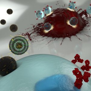
An artist’s illustration of nanoparticles transporting mRNA into a T cell (blue), allowing the latter to express surface receptors that recognize cancer cells (red). (Credit: Ryan Allen, Second Bay Studios)
New cancer immunotherapies involve extracting a patient’s T cells and genetically engineering them so they will recognize and attack tumors. This type of therapy is not without challenges, however. Engineering a patient’s T cells is laborious and expensive. And when successful, the alterations to the immune system immediately make patients very sick for a short period of time, with symptoms including fever, nausea and neurological effects.
Now, Penn researchers have demonstrated a new engineering technique that, because it is less toxic to the T cells, could enable a different mechanism for altering the way they recognize cancer, and could have fewer side effects for patients.
The technique involves ferrying messenger RNA (mRNA) across the T cell’s membrane via a lipid-based nanoparticle, rather than using a modified HIV virus to rewrite the cell’s DNA. Using the former approach would be preferable, as it only confers a temporary change to the patient’s immune system, but the current standard method for getting mRNA past the cell membrane can be too toxic to use on the limited number of T cells that can be extracted from a patient.
Michael Mitchell, Margaret Billingsley, and Carl June
The researchers demonstrated their technique in a study published in the journal Nano Letters. It was led by Michael Mitchell, Skirkanich Assistant Professor of Innovation of bioengineering in the School of Engineering and Applied Science, and Margaret Billingsley, a graduate student in his lab.
They collaborated with one of the pioneers of CAR T therapy: Carl June, the Richard W. Vague Professor in Immunotherapy and director of the Center for Cellular Immunotherapies in the Abramson Cancer Center and the director of the Parker Institute for Cancer Immunotherapy at the Perelman School of Medicine.
University of Washington Researchers Engineer a New Way to Study Circulatory Obstruction
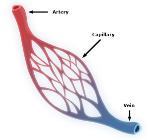
Capillaries are one of the most important forms of vasculature in our body, as they allow our blood to transfer nutrients to other parts of our body. But for how much effect capillary functionality can have on our health, their small size makes them extremely difficult to engineer into models for a variety of diseases. Now, researchers at the University of Washington led by Ying Zheng, Ph. D., engineered a three-dimensional microvessel model with living cells to study the mechanisms of microcirculatory obstruction involved with malaria.
Rather than just achieving a physical model of capillaries, these researchers created a model that allowed them to study typical flow and motion through capillaries, before comparing it to deficiencies in this behavior involved with diseases like malaria. The shape of the engineered model is similar to that of an hourglass, allowing the researchers to study instances where red blood cell transit may encounter bottlenecks between the capillaries and other vessels. Using multiphoton technology, Zheng and her team created 100mm capillary models with etched-in channels and a collagen base, to closely model the typical size and rigidity of the vessels. Tested with malaria-infected blood cells, the model showed similar circulatory obstructive behavior to that which occurs in patients, giving hope that this model can be transferred to other diseases involving such obstruction, like sickle cell anemia, diabetes, and cardiovascular conditions.
Understanding a Cell Membrane Protein Could Be the Key to New Cancer Treatments
Almost every cell in the body has integrins, a form of proteins, on its membrane, allowing cells to sense biological information from beyond their membranes while also using this feedback information to initiate signals within cells themselves. Bioengineers at the Imperial College of London recently looked at the way another membrane protein, called syndecan-4, interacts with integrins as a potential form of future cancer treatment. Referred to as “cellular hands” by lead researcher of the study Armando del Rio Hernandez, Ph.D., syndecan-4 sometimes controls the development of diseases or conditions like cancer and fibrosis. Hernandez and his team specifically studied the ties of syndecan-4 to yes-associated protein (YAP) and enzyme called P13K, both of which are affiliated with qualities of cancer progression like halted apoptosis or cell stiffening. Knowing this, Hernandez and his team hope to continue research into understanding the mechanisms of syndecan-4 throughout the cell, in search of new mechanisms and targets to focus on with future developments of cancer treatments.
A New Medical Device Could Improve Nerve Functionality After Severe Damage
Serious nerve damage remains difficult to repair surgically, often involving the stretching of nerves for localized damage, or the transfer of healthy nerve cells from another part of the body to fill larger gaps in nerve damage. But these imperfect solutions limit the return of full nerve function and movement to the damaged part of the body, and in more serious cases with large areas of nerve damage, can also risk damage in other areas of the body that healthy nerves are borrowed from for treatment. A new study from the University of Pittsburgh published in Science Translational Medicine led by Kacey Marra, Ph. D., has successfully repaired nerve damage in mice and monkeys using a biodegradable tube that releases growth factors called glial-cell-derived neurotrophic factors over time.
Marra and her team showed that this new device restored nerve function up to 80% in nonhuman primates, where current methods of nerve replacement often only achieve 50-60% functionality restoration. The device might have an easier time getting FDA-approval, since it doesn’t involve the use of stem cells in its repair mechanisms. Hoping to start human clinical trials in 2021, Marra and her team hope that the device will help both injured veterans and typical patients with nerve damage, and see potential future applications in facial nerve damage as well.
A New Computational Model Could Improve Treatments for Cancer, HIV, and Autoimmune Diseases
With cancer, HIV, and other autoimmune diseases, the best treatment options for patients are often determined with trial-and-error methods, leading to prolonged instances of ineffective approaches and sometimes unnecessary side effects. A group of researchers led by Wesley Errington, Ph.D., at the University of Minnesota decided to take a computational approach this problem, in an effort to more quickly and efficiently determine the most appropriate treatment for a given patient. Based on parameters controlling interactions between molecules with multiple binding sites, the team’s new model looks primarily at binding strength, linkage rigidity, and size of linkage arrays. Because diseases can often involve issues in molecular binding, the model aimed to model the 78 unique binding configurations for cases of when interacting molecules only have three binding sites, which are often difficult to observe experimentally. This new approach will allow for faster and easier determination of treatments for patients with diseases involving these molecular interactions.
Improved Drug Screening for Glioblastoma Patients
A new microfluidic brain chip from researchers at the University of Houston could help improve treatment evaluations for brain tumors. Glioblastoma patients, who have a five-year survival rate of a little over 5%, are some of the most common patients suffering from malignant brain tumors. This new chip, developed by the lab of Yasemin Akay, Ph.D., can quickly determine cancer drug effectiveness by analyzing a piece of cultured tumor biopsy from a patient by incorporating different chemotherapy treatments through the microfluidic vessels. Overall, Akay and her team found that this new chip holds hope as a future efficient and inexpensive form of drug screening for glioblastoma patients.
People and Places
The brain constructs maps to guide people, not just of physical spaces but also to connect stimuli around them, like conversations and other people. It’s long been known that the brain area responsible for this spatial navigation—the medial temporal lobe—is also involved in recalling memories.
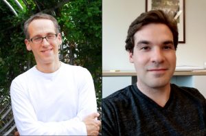
Michael Kahana (left) is principal investigator in the Defense Advanced Research Projects Agency’s RAM program and a professor in the Department of Psychology. Ethan Solomon is an M.D./Ph.D. student in the Department of Bioengineering of the School of Engineering and Applied Science and in the Perelman School of Medicine.
Now, neuroscientists at the University of Pennsylvania have discovered that the signals the brain produces during spatial navigation and episodic memory recall look similar. Low-frequency brain waves called the theta rhythm appear as people jump from one memory to the next, as many prior studies looking only at human navigation have shown. The new findings, which suggest that the brain structures responsible for helping people navigate the world may also “navigate” a mental map of prior experiences, appear in the Proceedings of the National Academy of Sciences.
The Florida Institute of Technology recently announced plans to start construction in spring 2020 on a new Health Sciences Research Center, set to further establish biomedical engineering and pre-medical coursework and research at the institute. With plans to open the new center in 2022, Florida Tech anticipates increased enrollment in the two programs, and hopes that the center will offer more opportunities in a growing professional field.
Anson Ong, Ph.D., the Associate Dean of Administration and Graduate Programs at the University of Texas at San Antonio, was recently elected to the International College of Fellows of Biomaterials Science and Engineering. With a focus on research in biomaterial implants for orthopaedic applications, Ong’s election to the college honors his advancement and contribution to the field of biomaterials research.
Nature, one of the world’s most prestigious scientific journals, recently reached out to a panel of researchers from a variety of fields, asking them what technological trends they see as having the most impact on their disciplines in the coming year.
Jennifer Phillips-Cremins, assistant professor in the Department of Bioengineering, was among these panelists. As an expert in “3D epigenetics,” or the way the genome’s highly specific folding patterns influence how and when individual genes are expressed, she highlighted a slate of new techniques that will allow researchers to take a closer look at those relationships.
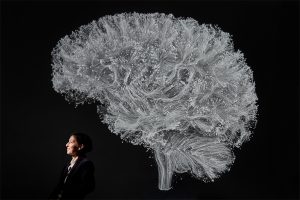
Among shots of a towering thunderstorm reaching into the stratosphere, the moon Daphnis peeking through Saturn’s rings, and an extremely close-up of a highly-endangered pangolin in Mozambique, one of Science’s favorite photos of 2019 was taken in Penn Engineering’s Raisler Lounge.
There, Danielle Bassett, J. Peter Skirkanich Professor in the Department of Bioengineering, poses underneath a giant visualization of the brain’s structural connections, projected on the wall behind her. Bassett’s research combines elements of physics, mathematics, engineering and neuroscience to provide a new look at how brain function arises from these networks of neurons.
Kelly Servick of Science profiled Bassett last year, revealing how a child whose parents discouraged her from attending college went on to become a pioneer in a highly interdisciplinary way of understanding the brain.
Read Bassett’s profile in Science here, and see the rest of the journal’s favorite photos of the year here.
Ravi Radhakrishnan has been named Chair of the Department of Bioengineering.
Radhakrishnan holds joint appointments in the Department of Bioengineering and the Department of Chemical and Biomolecular Engineering. He is a founding member and the current Director of the Penn Institute for Computational Science, as well as a member of the Penn Physical Sciences in Oncology Center, Institute for Translational Medicine and Therapeutics, and several graduate groups, including Materials Science and Engineering, Genomics and Computational Biology, and Biochemistry and Molecular Biophysics.
In addition to these roles at Penn, Radhakrishnan holds many editorial board positions in the research community, including Nature Publishing’s Scientific Reports.
Beyond being a passionate teacher and advocate for his students, Radhakrishnan’s research interests lie at the interface of chemical physics and molecular biology. His lab’s goal is to provide molecular level and mechanistic characterization of biomolecular and cellular systems and formulate quantitatively accurate microscopic models for predicting the interactions of various therapeutic agents with innate biochemical signaling mechanisms.
David F. Meaney, Solomon R. Pollack Professor of Bioengineering, has been named the Senior Associate Dean of Penn Engineering, effective January 1, 2020. This newly created leadership position will have oversight responsibilities in budget, space and infrastructure planning; facilities and research services; and will create and cultivate new interschool partnerships that will expand Penn Engineering’s footprint on campus.
Meaney is well known not only for his scholarship and innovation in neuroengineering and concussion science, but also for his leadership during his highly successful tenure as Chair of the Department of Bioengineering.
“Dave’s strong connections to the health schools will help strengthen Penn Engineering’s initiatives throughout campus,” says Vijay Kumar, Nemirovsky Family Dean of Penn Engineering. “He will have oversight of Penn Health-Tech, the Center for Engineering MechanoBiology and other efforts between engineering and the health schools, and Dave brings his unique creativity, energy and leadership experience to these collaborative efforts.”
Jason Burdick, Robert D. Bent Professor in the Department of Bioengineering, has been named a Fellow of the National Academy of Inventors (NAI), an award of high professional distinction accorded to academic inventors. Elected Fellows have demonstrated a prolific spirit of innovation in creating or facilitating outstanding inventions that have made a tangible impact on quality of life, economic development and the welfare of society.
Burdick’s research interests include developing degradable polymeric biomaterials that can be used for tissue engineering, drug delivery, and fundamental polymer studies. His lab focuses on developing polymeric materials for biomedical applications with specific emphasis on tissue regeneration and drug delivery. Burdick believes that advances in synthetic chemistry and materials processing could be the answer to organ and tissue shortages in medicine. The specific targets of his research include: scaffolding for cartilage regeneration, controlling stem cell differentiation through material signals, electrospinning and 3D printing for scaffold fabrication, and injectable hydrogels for therapies after a heart attack.
Danielle Bassett has been named the J. Peter Skirkanich Professor of Bioengineering.
Dr. Bassett is a Professor in the department of Bioengineering at the School of Engineering and Applied Science. She holds a Ph.D. in Physics from the University of Cambridge and completed her postdoctoral training at the University of California, Santa Barbara, before joining Penn in 2013.
Dr. Bassett has received numerous awards for her research, including an Alfred P Sloan Research Fellowship, a MacArthur Fellowship, an Office of Naval Research Young Investigator Award, a National Science Foundation CAREER Award and, most recently, an Erdos-Renyi Prize in Network Science to name but a few. She has authored over 190 peer-reviewed publications as well as numerous book chapters and teaching materials. She is the founding director of the Penn Network Visualization Program, a combined undergraduate art internship and K-12 outreach program bridging network science and the visual arts.
Positive results in first-in-U.S. trial of CRISPR-edited immune cells
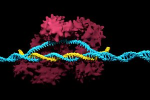
3D render of the CRISPR-Cas9 genome editing system
Genetically editing a cancer patient’s immune cells using CRISPR/Cas9 technology, then infusing those cells back into the patient appears safe and feasible based on early data from the first-ever clinical trial to test the approach in humans in the United States. Researchers from the Abramson Cancer Center have infused three participants in the trial thus far—two with multiple myeloma and one with sarcoma—and have observed the edited T cells expand and bind to their tumor target with no serious side effects related to the investigational approach. Penn is conducting the ongoing study in cooperation with the Parker Institute for Cancer Immunotherapy and Tmunity Therapeutics.
“This trial is primarily concerned with three questions: Can we edit T cells in this specific way? Are the resulting T cells functional? And are these cells safe to infuse into a patient? This early data suggests that the answer to all three questions may be yes,” says the study’s principal investigator Edward A. Stadtmauer, section chief of Hematologic Malignancies at Penn. Stadtmauer will present the findings next month at the 61st American Society of Hematology Annual Meeting and Exposition.
Tulane researchers join NIH HEAL initiative for research into opioid crisis
A Tulane University professor and researcher of biomedical engineering will join fellow researchers from over 40 other institutions in the National Institute of Health’s Help to End Addiction Long-Term (HEAL) Initiative. Of the $945 million that make up the project, Michael J. Moore, Ph.D. will receive a share of $1.2 million to advance research in modeling human pain through computer chips, with the help of fellow Tulane researchers Jeffrey Tasker, Ph.D., and James Zadina, Ph.D., each with backgrounds in neuroscience.
Because of the opioid epidemic sweeping the nation, Moore notes that there’s a rapid search going on to develop non-addictive painkiller options. However, he also sees a gap in adequate models to test those new drugs before human clinical trials are allowed to take place. Here is where he hopes to step in and bring some innovation to the field, by integrating living human cells into a computer chip for modeling pain mechanisms. Through his research, Moore wants to better understand not only how some drugs can induce pain, but also how patients can grow tolerant to some drugs over time. If successful, Moore’s work will lead to a more rapid and less expensive screening option for experimental drug advancements.
New machine learning-assisted microscope yields improved diagnostics
Researchers at Duke University recently developed a microscope that uses machine learning to adapt its lighting angles, colors, and patterns for diagnostic tests as needed. Most microscopes have lighting tailored to human vision, with an equal distribution of light that’s optimized for human eyes. But by prioritizing the computer’s vision in this new microscope, researchers enable it to see aspects of samples that humans simply can’t, allowing for a more accurate and efficient diagnostic approach.
Led by Roarke W. Horstmeyer, Ph.D., the computer-assisted microscope will diffuse light through a bowl-shaped source, allowing for a much wider range of illumination angles than traditional microscopes. With the help of convolutional neural networks — a special kind of machine learning algorithm — Horstmeyer and his team were able to tailor the microscope to accurately diagnose malaria in red blood cell samples. Where human physicians typically perform similar diagnostics with a rate of 75 percent accuracy, this new microscope can do the same work with 90 percent accuracy, making the diagnostic process for many diseases much more efficient.
Case Western Reserve University researchers create first-ever holographic map of brain
A Case Western Reserve University team of researchers recently spearheaded a project in creating an interactive holographic mapping system of the human brain. The design, which is believed to be the first of its kind, involves the use of the Microsoft HoloLens mixed reality platform. Lead researcher Cameron McIntyre, Ph.D., sees this mapping system as a better way of creating holographic navigational routes for deep brain stimulation. Recent beta tests with the map by clinicians give McIntyre hope that the holographic representation will help them better understand some of the uncertainties behind targeted brain surgeries.
More than merely providing a useful tool, McIntyre’s project also brings together decades’ worth of neurological data that has not yet been seriously studied together in one system. The three-dimensional atlas, called “HoloDBS” by his lab, provides a way of finally seeing the way all of existing neuro-anatomical data relates to each other, allowing clinicians who use the tool to better understand the brain on both an analytical and visual basis.
Implantable cancer traps reduce biopsy incidence and improve diagnostic
Biopsies are one of the most common procedures used for cancer diagnostics, involving a painful and invasive surgery. Researchers at the University of Michigan are trying to change that. Lonnie Shea, Ph.D., a professor of biomedical engineering at the university, worked with his lab to develop implants with the ability to attract any cancer cells within the body. The implant can be inserted through a scaffold placed under the patient’s skin, making it a more ideal option than biopsy for inaccessible organs like lungs.
The lab’s latest work on the project, published in Cancer Research, details its ability to capture metastatic breast cancer cells in vivo. Instead of needing to take biopsies from areas deeper within the body, the implant allows for a much simpler surgical procedure, as biopsies can be taken from the implant itself. Beyond its initial diagnostic advantages, the implant also has the ability to attract immune cells with tumor cells. By studying both types of cells, the implant can give information about the current state of cancer in a patient’s body and about how it might progress. Finally, by attracting tumor and immune cells, the implant has the ability to draw them away from the area of concern, acting in some ways as a treatment for cancer itself.
People and Places
Cesar de la Fuente-Nunez, PhD
The Philadelphia Inquirer recently published an article detailing the research of Penn’s Presidential Assistant Professor in Psychiatry, Microbiology, and Bioengineering, Cesar de la Fuente, Ph.D. In response to a growing level of worldwide deaths due to antibiotic-resistant bacteria, de la Fuente and his lab use synthetic biology, computation, and artificial intelligence to test hundreds of millions of variations in bacteria-killing proteins in the same experiment. Through his research, de la Fuente opens the door to new ways of finding and testing future antibiotics that might be the only viable options in a world with an increasing level of drug-resistant bacteria
Emily Eastburn, a Ph.D. candidate in Bioengineering at Penn and a member of the Boerckel lab of the McKay Orthopaedic Research Laboratory, recently won the Ashton fellowship. The Ashton fellowship is an award for postdoctoral students in any field of engineering that are under the age of 25, third-generation American citizens, and residents of either Pennsylvania or New Jersey. A new member of the Boerckel lab, having joined earlier this fall, Eastburn will have the opportunity to conduct research throughout her Ph.D. program in the developmental mechanobiology and regeneration that the Boerckel lab focuses on.
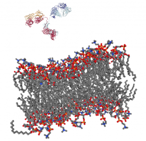
Getting a complex protein like an antibody through the membrane of a cell without damaging either is a long-standing challenge in the life sciences. Penn Engineers have found a plug-and-play solution that makes antibodies compatible with the delivery vehicles commonly used to ferry nucleic acids across that barrier.
For almost any conceivable protein, corresponding antibodies can be developed to block it from binding or changing shape, which ultimately prevents it from carrying out its normal function. As such, scientists have looked to antibodies as a way of shutting down proteins inside cells for decades, but there is still no consistent way to get them past the cell membrane in meaningful numbers.
Now, Penn Engineering researchers have figured out a way for antibodies to hitch a ride with transfection agents, positively charged bubbles of fat that biologists routinely use to transport DNA and RNA into cells. These delivery vehicles only accept cargo with a highly negative charge, a quality that nucleic acids have but antibodies lack. By designing a negatively charged amino acid chain that can be attached to any antibody without disrupting its function, they have made antibodies broadly compatible with common transfection agents.
Beyond the technique’s usefulness towards studying intracellular dynamics, the researchers conducted functional experiments with antibodies that highlight the technique’s potential for therapeutic applications. One antibody blocked a protein that decreases the efficacy of certain drugs by prematurely ejecting them from cells. Another blocked a protein involved in the transcription process, which could be an even more fundamental way of knocking out proteins with unwanted effects.


 Capillaries are one of the most important forms of vasculature in our body, as they allow our blood to transfer nutrients to other parts of our body. But for how much effect capillary functionality can have on our health, their small size makes them extremely difficult to engineer into models for a variety of diseases. Now, researchers at the University of Washington led by