Interact, Adapt, Repeat

Sophomores Linda Wu and Nova Meng spent the summer studying coevolution among plants, mutualistic bacteria, and parasitic nematodes in Corlett Wood’s biology lab.

by Katherine Unger Baillie
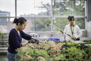
To study coevolution, the responsibilities of Nova Meng and Linda Wu included caring for plants in the Penn greenhouse. (Image: From July 2021, when masks were not required)

Coevolution is all around us. Think of the elongated blooms that perfectly accommodate a hummingbird’s slender mouth parts. But not all examples of species influencing one another’s evolutionary course accrue benefits to all parties. Tradeoffs are part of the game.

This summer, sophomores Linda Wu of Annandale, Virginia, and Nova Meng of Akron, Ohio, researched an coevolutionary scenario with benefits as well as costs for the species involved. Their work, supported by the Penn Undergraduate Research Mentoring Program (PURM) and conducted in the lab of biology professor Corlett Wood, has examined the relationship among plants in the genus Medicago, beneficial bacteria that dwell in their roots, and parasitic nematodes that try to steal the plants’ nutrients.

The Center for Undergraduate Research & Fellowships provides students in the PURM program awards of $4,500 during the 10-week summer research internship. Wu and Meng stayed busy through those weeks. Whether evaluating plants in a soybean field in Michigan or tending to hundreds—even thousands—of plants in the greenhouse at Penn, these aspiring researchers built a foundation for future scientific endeavors with hands-on practice.

“It’s been an amazing experience,” says Wu. “I’ve always been interested in genetics and evolution and have found parasitic relationships in particular really interesting. I like reading about weird parasites. This summer I’ve gotten to participate in lab meetings, read books about coevolution, and expand my knowledge about the topic.”

Mentored by Ph.D. student McCall Calvert, Wu spent the summer focused on the parasites in the Medicago model system the Wood lab uses. “I’m trying to see if those nematodes are specialists or generalists, if they’re locally adapted to their host plant or open to parasitizing on different species,” Wu says.

To do so, she’s grown pots and pots of plants in the Penn greenhouse, experimentally infecting Medicago plants as well as other species, such as carrot and daisy plants, with nematodes, to measure the degree to which the parasites flourish.
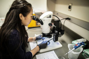
Meng, who is pursuing a bioengineering major, is examining how bacteria that dwell in plant roots affect the plants’ susceptibility to parasites.

Meng’s project looked at the bacterial side of the coevolutionary relationship. Overseen by lab manager and technician Eunnuri Yi, Meng looked at four strains of bacteria, known as rhizobia. Two strains are nitrogen-fixing, giving their associated plants a crucial nutrient to promote growth, while the other two do not seem to contribute nitrogen to the plants, and instead exist as parasites in the plants’ roots. “I’m looking at what happens when we infect the plants with nematode parasites,” Meng says, “to see if the plants that are open to mutualistic rhizobia are more susceptible to the nematode parasites.”

Read the full story in Penn Today.

Linda Wu is a sophomore pursuing an uncoordinated dual degree in business, energy, environment, and sustainability in the Wharton School and in biology with a concentration in ecology and evolution in the College of Arts and Sciences at the University of Pennsylvania.

Nova Meng is a sophomore majoring in bioengineering in the School of Engineering and Applied Science at Penn.

Penn Engineers Will Use NSF Grant to Develop ‘DReAM’ for On-demand, On-site mRNA Manufacturing

by Melissa Pappas

Daeyeon Lee, Kathleen Stebe and Michael Mitchell

COVID-19 vaccines are just the beginning for mRNA-based therapies; enabling a patient’s body to make almost any given protein could revolutionize care for other viruses, like HIV, as well as various cancers and genetic disorders. However, because mRNA molecules are very fragile, they require extremely low temperatures for storage and transportation. The logistical challenges and expense of maintaining these temperatures must be overcome before mRNA therapies can become truly widespread.

With these challenges in mind, Penn Engineering researchers are developing a new manufacturing technique that would be able to produce mRNA sequences on demand and on-site, isolating them in a way that removes the need for cryogenic temperatures. With more labs able to make and store mRNA-based therapeutics on their own, the “cold chain” between manufacturer and patient can be made shorter, faster and less expensive.

The National Science Foundation (NSF) is supporting this project, known as Distributed Ribonucleic Acid Manufacturing, or DReAM, through a four-year, $2 million grant from its Emerging Frontiers in Research and Innovation (EFRI) program.

The project will be led by Daeyeon Lee, Evan C Thompson Term Chair for Excellence in Teaching and Professor in the Department of Chemical and Biomolecular Engineering (CBE), along with Kathleen Stebe, Richer and Elizabeth Goodwin Professor in CBE and in the Department of Mechanical Engineering and Applied Mechanics. They will collaborate with Michael Mitchell, Skirkanich Assistant Professor of Innovation in the Department of Bioengineering, Drexel University’s Masoud Soroush and Michael Grady, the University of Oklahoma’s Dimitrios Papavassiliou and the University of Colorado Boulder’s Joel Kaar.

Read the full story in Penn Engineering Today.

Developing New Technologies to Solve the Mysteries of the Brain

Flavia Vitale, assistant professor of neurology, bioengineering, and physical medicine and rehabilitation, and founder of the multidisciplinary Vitale Lab. (Image: Penn Medicine News)

Neurology, bioengineering, and physical medicine and rehabilitation might not seem like three disciplines that fit together, but for Flavia Vitale, an assistant professor of all three, it makes perfect sense. As the director and principal investigator at the Vitale Lab, her research focuses on developing new technologies that help to study how the brain and neuromuscular systems function.

Years ago, while she was working at Rice University developing new materials and devices that work in the body in a safer, more effective way, former president Barack Obama launched the Brain Research Through Advancing Innovative Neurotechnologies (BRAIN) Initiative, aimed at revolutionizing the understanding of the human brain. This emphasis on how little is known about brain structure and function inspired Vitale to refocus her research on developing technology and materials that will help researchers solve the mysteries of the brain.

In 2018, she joined the faculty at the Perelman School of Medicine as an assistant professor of neurology, bioengineering, and physical medicine and rehabilitation, and founded the multidisciplinary Vitale Lab, where her team develops cutting edge materials and devices that will someday help clinicians diagnose and treat patients with complicated brain and neurological conditions. She is also one of the engineers looking forward to using new combined clinical/research facilities in neuroscience at Penn Medicine’s new Pavilion where new neurotechnoloigies will be developed and tested.

“My main goal is to create tools that can help solve mysteries of the brain, and address the needs of clinicians,” she says.

“My lab was recently awarded two grants totaling $4.5 million from the National Institute of Neurological Disorders and Stroke. In order to obtain more precise insights, noninvasively, into brain activity to improve gene therapy treatments for a range of diagnoses, from Parkinson’s disease to glioblastoma. The first grant is designated for the development of a novel surgical device for delivering gene-based therapeutics to the brain. The second is for optimization and pre-clinical validation of a novel EEG electrode technology, which uses a soft, flexible, conductive nanomaterial rather than metal and gels. We hope to confirm that these technologies work as well as, if not better than existing ones.”

Read the full story in Penn Medicine News.

Annenberg and Penn Bioengineering Research into Communication Citation Bias

Photo Credit: Debby Hudson / Unsplash

Women are frequently under-cited in academia, and the field of communication is no exception, according to research from the Annenberg School for Communication. The study, entitled “Gendered Citation Practices in the Field of Communication,” was published in Annals of the International Communication Association.

A new study from the Addiction, Health, & Adolescence (AHA!) Lab at the Annenberg School for Communication at the University of Pennsylvania found that men are over-cited and women are under-cited in the field of Communication. The researchers’ findings indicate that this problem is most persistent in papers authored by men.

“Despite known limitations in their use as proxies for research quality, we often turn to citations as a way to measure the impact of someone’s research,” says Professor David Lydon-Staley, “so it matters for individual researchers if one group is being consistently under-cited relative to another group. But it also matters for the field in the sense that if people are not citing women as much as men, then we’re building the field on the work of men and not the work of women. Our field should be representative of all of the excellent research that is being undertaken, and not just that of one group.”

The AHA! Lab is led by David Lydon-Staley, Assistant Professor of Communication and former postdoc in the Complex Systems lab of Danielle Bassett, J. Peter Skirkanich Professor in Bioengineering and in Electrical and Systems Engineering in the School of Engineering and Applied Science. Dr. Bassett and Bassett Lab members Dale Zhou and Jennifer Stiso, graduate students in the Perelman School of Medicine, also contributed to the study.

Read “Women are Under-cited and Men are Over-cited in Communication” in Annenberg School for Communication News.

Developing Endotracheal Tubes that Release Antimicrobial Peptides

by Evan Lerner

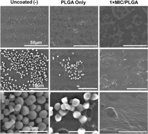
Scanning electron microscope images of endotracheal tubes at three levels of magnification. After 24 hours of Staphylococcus epidermidis exposure, tubes coated with the researchers’ AMPs (right) showed decreased biofilm production, as compared with tubes coated with just polymer (center) and uncoated tubes (left).

Endotracheal tubes are a mainstay of hospital care, as they ensure a patient’s airway is clear when they can’t breathe on their own. However, keeping a foreign object inserted in this highly sensitive part of the anatomy comes is not without risk, such as the possibility of infection, inflammation and a condition known as subglottic stenosis, in which scar tissue narrows the airway.

Broad-spectrum antibiotics are one way to mitigate these risks, but come with risks of their own, including harming beneficial bacteria and contributing to antibiotic resistance.

With this conundrum in mind, Riccardo Gottardi, Assistant Professor of Pediatrics at the Children’s Hospital of Philadelphia (CHOP) and of Bioengineering at Penn Engineering, along with Bioengineering graduate students and lab members Matthew Aronson and Paul Gehret, are developing endotracheal tubes that can provide a more targeted antimicrobial defense.

In a proof-of-concept study published in the journal The Laryngoscope, the team showed how a different type of antimicrobial agent could be incorporated into the tubes’ polymer coating, as well as preliminary results suggesting these devices would better preserve a patient’s microbiome.

Instead, the investigators explored the use of antimicrobial peptides (AMPs), which are small proteins that destabilize bacterial membranes, causing bacterial cells to fall apart and die. This mechanism of action allows them to target specific bacteria and makes them unlikely to promote antimicrobial resistance. Prior studies have shown that it is possible to coat endotracheal tubes with conventional antibiotics, so the research team investigated the possibility of incorporating AMPs into polymer-coated tubes to inhibit bacterial growth and modulate the upper-airway microbiome.

The researchers, led by Matthew Aronson, a graduate student in Penn Engineering’s Department of Bioengineering, tested their theory by creating a polymer coating that would release Lasioglossin-III, an AMP with broad-spectrum antibacterial activity. They found that Lasio released from coated endotracheal tubes, reached the expected effective concentration rapidly and continued to release at the same concentration for a week, which is the typical timeframe that an endotracheal is used before being changed. The investigators also tested their drug-eluting tube against airway microbes, including S. epidermidis, S. pneumoniae, and human microbiome samples and observed significant antibacterial activity, as well as prevention of bacterial adherence to the tube.

Read “CHOP Researchers Develop Coating for Endotracheal Tubes that Releases Antimicrobial Peptides” at CHOP News.

This post originally appeared in Penn Engineering Today.

Penn Engineers Create Faster and Cheaper COVID-19 Testing With Pencil Lead

by Melissa Pappas

César de la Fuente, PhD

Testing is key to understanding and controlling the spread of COVID-19, which has already taken more than four million lives around the world. However, current tests are limited by the tradeoff between accuracy and the time it takes to analyze a sample.

Another challenge of current COVID-19 tests is cost. Most tests are expensive to produce and require trained personnel to administer and analyze them. Testing in low-and middle-income communities has therefore been largely inaccessible, leaving individuals at greater risk of viral spread.

To address cost, time and accuracy, a new electrochemical test developed by Penn researchers uses electrodes made from graphite — the same material found in pencil lead. Developed by César de la Fuente, Presidential Assistant Professor in Bioengineering,  Microbiology and Psychiatry with a secondary appointment in Chemical and Biomolecular Engineering, these electrodes reduce the cost to $1.50 per test and require only 6.5 minutes to deliver 100-pecent-accurate results from saliva samples and up to 88 percent accuracy in nasal samples.

While his previous research highlights the invention of RAPID (Real-time Accurate Portable Impedimetric Detection prototype 1.0), a COVID-19 testing kit which uses screen-printed electrodes, this new research published in PNAS presents LEAD (Low-cost Electrochemical Advanced Diagnostic), using the same concept as RAPID but with less expensive materials. De la Fuente’s current test reduces costs from $4.67 per test (RAPID) to $1.50 per test (LEAD) just by changing the building material of the electrodes.

“Both RAPID and LEAD work on the same principle of electrochemistry,” says de la Fuente. “However, LEAD is easier to assemble, it can be used by anyone and the materials are cheaper and more accessible than those of RAPID. This is important because we are using an abundant material, graphite, the same graphite used in pencils, to build the electrode to make testing more accessible to lower-income communities.”

This figure, adapted from the paper, shows the functionalization steps of LEAD which prepares the electrodes to bind to the sample. The height of the peaks indicates whether the sample is negative or positive. Because the SARS-CoV-2 spike protein in a positive sample binds to the electrode, it inhibits the emitted signal and produces a smaller peak.

Read the full story in Penn Engineering Today.

Decoding How the Brain Accurately Depicts Ever-changing Visual Landscapes

A collaborative study finds that deeper regions of the brain encode visual information more slowly, enabling the brain to identify fast-moving objects and images more accurately and persistently.

by Erica K. Brockmeier

Busy pedestrian crossing at Hong Kong

New research from the University of Pennsylvania, the Scuola Internazionale Superiore de Studi Avanzati (SISSA), and KU Leuven details the time scales of visual information processing across different regions of the brain. Using state-of-the-art experimental and analytical techniques, the researchers found that deeper regions of the brain encode visual information slowly and persistently, which provides a mechanism for explaining how the brain accurately identifies fast-moving objects and images. The findings were published in Nature Communications.

Understanding how the brain works is a major research challenge, with many theories and models developed to explain how complex information is processed and represented. One area of particular interest is vision, a major component of neural activity. In humans, for example, there is evidence that around half of the neurons in the cortex are related to vision.

Researchers are eager to understand how the visual cortex can process and retain information about objects in motion in a way that allows people to take in dynamic scenes while still retaining information about and recognizing the objects around them.

“One of the biggest challenges of all the sensory systems is to maintain a consistent representation of our surroundings, despite the constant changes taking place around us. The same holds true for the visual system,” says Davide Zoccolan, director of SISSA’s Visual Neuroscience Laboratory. “Just look around us: objects, animals, people, all on the move. We ourselves are moving. This triggers rapid fluctuations in the signals acquired by the retina, and until now it was unclear whether the same type of variations apply to the deeper layers of the visual cortex, where information is integrated and processed. If this was the case, we would live in tremendous confusion.”

Experiments using static stimuli, such as photographs, have found that information from the sensory periphery are processed in the visual cortex according to a finely tuned hierarchy. Deeper regions of the brain then translate this information about visual scenes into more complex shapes, objects, and concepts. But how this process works in more dynamic, real-world settings is not well understood.

To shed light on this, the researchers analyzed neural activity patterns in multiple visual cortical areas in rodents while they were being shown dynamic visual stimuli. “We used three distinct datasets: one from SISSA, one from a group in KU Leuven led by Hans Op de Beeck and one from the Allen Institute for Brain Science in Seattle,” says Zoccolan. “The visual stimuli used in each were of different types. In SISSA, we created dedicated video clips showing objects moving at different speeds. The other datasets were acquired using various kinds of clips, including from films.”

Next, the researchers analyzed the signals registered in different areas of the visual cortex through a combination of sophisticated algorithms and models developed by Penn’s Eugenio Pasini and Vijay Balasubramanian. To do this, the researchers developed a theoretical framework to help connect the images in the movies to the activity of specific neurons in order to determine how neural signals evolve over different time scales.

“The art in this science was figuring out an analysis method to show that the processing of visual images is getting slower as you go deeper and deeper in the brain,” says Balasubramanian. “Different levels of the brain process information over different time scales; some things could be more stable, some quicker. It’s very hard to tell if the time scales across the brain are changing, so our contribution was to devise a method for doing this.”

Read the full story in Penn Today.

Vijay Balasubramanian is the Cathy and Marc Lasry Professor in the Department of Physics and Astronomy in the School of Arts & Sciences and a member of the Penn Bioengineering Graduate Group at the University of Pennsylvania.

New Grant Aims to Broaden Participation in Cutting-Edge Materials Research

University of Puerto Rico’s Edgardo Sánchez (left) and Penn graduate Zhiwei Liao working in the lab of Daeyeon Lee. Via the Advancing Device Innovation through Inclusive Research and Education program, researchers from Penn and the University of Puerto Rico will continue their materials science collaboration while supporting STEM career pathways for underrepresented groups. (Image credit: Felice Macera).

The National Science Foundation (NSF) has awarded grants to eight research teams to support partnerships that will increase diversity in cutting-edge materials research, education, and career development. One of those teams is Penn’s Laboratory for Research on the Structure of Matter (LRSM) and the University of Puerto Rico (UPR), whose long-running collaboration has now received an additional six years of support.

With the goal of supporting partnerships between minority-serving educational institutions and leading materials science research centers, NSF’s Partnership for Research & Education in Materials (PREM) program funds innovative research programs and provides institutional support to increase recruitment, retention, and graduation by underrepresented groups as well as providing underserved communities access to materials research and education.

‘Research at the frontier’

With this PREM award, known as the Advancing Device Innovation through Inclusive Research and Education (ADIIR) program, researchers from Penn and UPR’s Humacao and Cayey campuses will conduct research on the properties of novel carbon-based materials with unique properties, and will study the effects of surface modification in new classes of sensors, detectors, and purification devices.

Thanks to this collaboration of more than 20 years, both institutions have made significant scientific and educational progress aided by biannual symposia and regular pre-pandemic travel between both institutions before the pandemic, resulting in a rich portfolio of publications, conference presentations, patents, students trained, and outreach programs.

“Together we have been publishing good papers that have impact, and we’ve really cultivated a culture of collaboration and friendship between our institutions,” says Penn’s Arjun Yodh, former director of the LRSM. “Our goal is to carry out research at the frontier and, in the process, nurture promising students from Puerto Rico and Penn.”

Ivan Dmochowski, a chemistry professor at Penn who has been involved with PREM for several years, says that this program has helped his group connect with experts in Puerto Rico whose skills complement his group’s interests in protein engineering. Dmochowski has also hosted UPR faculty members and students in his lab and also travelled to Puerto Rico before the pandemic to participate in research symposia, seminars, and outreach events.

“I’ve had students who have benefitted from being a co-author on a paper or having a chance to mentor students, and the faculty we’ve interacted with are exceptional,” Dmochowski says. “There’s a lot of benefit for both me and my students, and I’ve enjoyed our interactions both personally and scientifically.”

Penn’s Daeyeon Lee, a chemical and biomolecular engineering professor who has been involved with PREM for several years, regularly hosts students and faculty from UPR while working on nanocarbon-based composite films for sensor applications. The success of this collaboration relies on unique materials made by researchers at UPR combined with a method for processing them into composite structures developed in Lee’s lab.

“What I really admire about people at PREM, both faculty and students, is their passion,” says Lee. “I think that’s had a really positive impact on my students and postdocs who got to interact with them because they got to see the passion that the students brought.”

Read the full story in Penn Today.

Daeyeon Lee is a professor and the Evan C Thompson Term Chair for Excellence in Teaching in the Department of Chemical and Biomolecular Engineering and a member of the Bioengineering Graduate Group in Penn’s School of Engineering and Applied Science.

Arjun Yodh is the James M. Skinner Professor of Science in the Department of Physics & Astronomy in Penn’s School of Arts & Sciences and a member of the Bioengineering Graduate Group in Penn’s School of Engineering and Applied Science.

Student Research Highlight: Colin Huber

Colin Huber, Ph.D. student

Colin Huber, a Ph.D. candidate in Bioengineering studying head impact biomechanics and concussion in sports at the Center for Injury Research and Prevention (CIRP) at the Children’s Hospital of Philadelphia (CHOP), recently published “Variations in Head Impact Rates in Male and Female High School Soccer” in Medicine & Science in Sports & Exercise with colleagues from CHOP’s Minds Matter Concussion Frontier Program and the CIRP.

Colin’s paper, the goal of which was to compare “to compare head impact exposure rates (head impacts/exposure period) in male and female high school soccer by using multiple methodological approaches,” was recently profiled in the Penn Engineering Research & Innovation Newsletter.

Read the full story in the ADRO Newsletter.

With a ‘Liquid Assembly Line,’ Penn Researchers Produce mRNA-Delivering-Nanoparticles a Hundred Times Faster than Standard Microfluidic Technologies

by Evan Lerner

Michael Mitchell, Sarah Shepherd and David Issadore pose with their new device.

The COVID vaccines currently being deployed were developed with unprecedented speed, but the mRNA technology at work in some of them is an equally impressive success story. Because any desired mRNA sequence can be synthesized in massive quantities, one of the biggest hurdles in a variety of mRNA therapies is the ability to package those sequences into the lipid nanoparticles that deliver them into cells.

Now, thanks to manufacturing technology developed by bioengineers and medical researchers at the University of Pennsylvania, a hundred-fold increase in current microfluidic production rates may soon be possible.

The researchers’ advance stems from their design of a proof-of-concept microfluidic device containing 128 mixing channels working in parallel. The channels mix a precise amount of lipid and mRNA, essentially crafting individual lipid nanoparticles on a miniaturized assembly line.

This increased speed may not be the only benefit; more precisely controlling the nanoparticles’ size could make treatments more effective. The researchers tested the lipid nanoparticles produced by their device in a mouse study, showing they could deliver therapeutic RNA sequences with four-to-five times greater activity than those made by conventional methods.

The study was led by Michael Mitchell, Skirkanich Assistant Professor of Innovation in Penn Engineering’s Department of Bioengineering, and David Issadore, Associate Professor in Penn Engineering’s Department of Bioengineering, along with Sarah Shepherd, a doctoral student in both of their labs. Rakan El-Mayta, a research engineer in Mitchell’s lab, and Sagar Yadavali, a postdoctoral researcher in Issadore’s lab, also contributed to the study.

They collaborated with several researchers at Penn’s Perelman School of Medicine: postdoctoral researcher Mohamad-Gabriel Alameh, Lili Wang, Research Associate Professor of Medicine, James M. Wilson, Rose H. Weiss Orphan Disease Center Director’s Professor in the Department of Medicine, Claude Warzecha, a senior research investigator in Wilson’s lab, and Drew Weissman, Professor of Medicine and one of the original developers of the technology behind mRNA vaccines.

It was published in the journal Nano Letters.

“We believe that this microfluidic technology has the potential to not only play a key role in the formulation of current COVID vaccines,” says Mitchell, “but also to potentially address the immense need ahead of us as mRNA technology expands into additional classes of therapeutics.”

Read the full story in Penn Engineering Today.