BE Seminar: “Deciphering the Dynamics of the Unconscious Brain Under General Anesthesia” (Emery Brown)

Emery Brown, MD, PhD

Speaker: Emery N. Brown, MD, PhD
Edward Hood Taplin Professor of Medical Engineering and of Computational Neuroscience, MIT
Warren M. Zapol Professor of Anaesthesia, Harvard Medical School
Massachusetts General Hospital

Date: Thursday, April 1, 2021
Time: 3:00-4:00 PM EDT
Zoom – check email for link or contact ksas@seas.upenn.edu

Title: “Deciphering the Dynamics of the Unconscious Brain Under General Anesthesia”

Abstract:

General anesthesia is a drug induced state that is critical for safely and humanely allowing a patient to undergo surgery or an invasive diagnostic procedure. During the last 10 years the study of the neuroscience of anesthetic drugs has been an active area of research. In this lecture we show how anesthetics create altered states of arousal by creating oscillation that impede how the various parts of the brain communicate. These oscillations, which are readily visible on the electroencephalogram (EEG), change systematically with anesthetic dose, anesthetic class and patient age. We will show how the EEG oscillations can be used to monitor the brain states of patients receiving general anesthesia, manage anesthetic delivery and learn about fundamental brain physiology.

EMERY BROWN BIO:

Emery N. Brown is the Edward Hood Taplin Professor of Medical Engineering and Professor of Computational Neuroscience at Massachusetts Institute of Technology. He is the Warren M. Zapol Professor of Anaesthesia at Harvard Medical School and Massachusetts General Hospital (MGH), and an anesthesiologist at MGH.

Dr. Brown received his BA (magna cum laude) in Applied Mathematics from Harvard College, his MA and PhD in statistics from Harvard University, and his MD (magna cum laude) from Harvard Medical School. He completed his internship in internal medicine at the Brigham and Women’s Hospital and his residency in anesthesiology at MGH. He joined the staff at MGH, the faculty at Harvard Medical School in 1992 and the faculty at MIT in 2005.

Dr. Brown is an anesthesiologist-statistician recognized for developing signal processing algorithms for neuroscience data analysis and for defining the neurophysiological mechanisms of general anesthesia.

Dr. Brown was a member of the NIH BRAIN Initiative Working Group. He is a fellow of the IEEE, the AAAS, the American Academy of Arts and Sciences and the National Academy of Inventors. Dr. Brown is a member of the National Academy of Medicine, the National Academy of Sciences and the National Academy of Engineering. He received an NIH Director’s Pioneer Award, the National Institute of Statistical Sciences Sacks Award, the American Society of Anesthesiologists Excellence in Research Award, the Dickson Prize in Science, the Swartz Prize for Theoretical and Computational Neuroscience and a Doctor of Science (honoris causa) from the University of Southern California.

Watch the Inaugural Joseph Bordogna Forum Lecture by Dr. John Brooks Slaughter

The inaugural Joseph Bordogna Forum took place on Wednesday, February 24 and featured a talk from John Brooks Slaughter, Deans’ Professor of Education and Engineering at USC’s Viterbi School of Engineering and Rossier School of Education, entitled a “Call to Action for Racial Justice and Equity in Engineering.”

Dr. Slaughter was joined by panelists Jennifer R. Lukes, Professor in Mechanical Engineering and Applied Mechanics, Oladayo Adewole, an alumnus in Robotics who recently defended his doctoral dissertation in Bioengineering, and CJ Taylor, Raymond S. Markowitz President’s Distinguished Professor in Computer and Information Science and Associate Dean, Diversity, Equity and Inclusion, who moderated the talk.

Dr. Slaughter talked about how microaggressions can often be a barrier to student success and emphasized on the importance of mentorship for underrepresented minorities: “If faculty members seek to improve the retention of underrepresented minorities, often times more has to be done than introducing science and math principles early on in their education, but instead, the unique backgrounds of these students must be understood.”

Originally posted in Penn Engineering Today.

‘RNA worked for COVID-19 vaccines. Could it be used to treat cancer and rare childhood diseases?’

William H. Peranteau, Michael J. Mitchell, Margaret Billingsley, Meghana Kashyap, and Rachel Riley (Clockwise from top left)

As COVID-19 vaccines roll out, the concept of using mRNA to fend off viruses has become a part of the public dialogue. However, scientists have been researching how mRNA can be used to in life-saving medical treatments well before the pandemic.

The “m” in “mRNA” is for “messenger.” A single-stranded counterpart to DNA, it translates the genetic code into the production of proteins, the building blocks of life. The Moderna and Pfizer COVID-19 vaccines work by introducing mRNA sequences that act as a set of instructions for the body to produce proteins that mimic parts of the virus itself. This prepares the body’s immune response to recognize the real virus and fight it off.

Because it can spur the production of proteins that the body can’t make on its own, mRNA therapies also have the potential to slow or prevent genetic diseases that develop before birth, such as cystic fibrosis and sickle-cell anemia.

However, because mRNA is a relatively unstable molecule that degrades quickly, it needs to be packaged in a way that maintains its integrity as its delivered to the cells of a developing fetus.

To solve this challenge, Michael J. Mitchell, Skirkanich Assistant Professor of Innovation in the Department of Bioengineering, is researching the use of lipid nanoparticles as packages that transport mRNA into the cell. He and William H. Peranteau, an attending surgeon in the Division of General, Thoracic and Fetal Surgery and the Adzick-McCausland Distinguished Chair in Fetal and Pediatric Surgery at Children’s Hospital of Philadelphia, recently co-authored a “proof-of-concept” paper investigating this technique.

In this study, published in Science Advances, Mitchel examined which nanoparticles were optimal in the transport of mRNA to fetal mice. Although no disease or organ was targeted in this study, the ability to administer mRNA to a mouse while still in the womb was demonstrated, and the results are promising for the next stages of targeted disease prevention in humans.

Mitchel spoke with Tom Avril at The Philadelphia Inquirer about the mouse study and its implications for treatment of rare infant diseases through the use of mRNA, ‘the messenger of life.’

Penn bioengineering professor Michael J. Mitchell, the other senior author of the mouse study, tested various combinations of lipids to see which would work best.

The appeal of the fatty substances is that they are biocompatible. In the vaccines, for example, two of the four lipids used to make the delivery spheres are identical to lipids found in the membranes of human cells — including plain old cholesterol.

When injected, the spheres, called nanoparticles, are engulfed by the person’s cells and then deposit their cargo, the RNA molecules, inside. The cells respond by making the proteins, just as they make proteins by following the instructions in the person’s own RNA. (Important reminder: The RNA in the vaccines cannot become part of your DNA.)

Among the different lipid combinations that Mitchell and his lab members tested, some were better at delivering their cargo to specific organs, such as the liver and lungs, meaning they could be a good vehicle for treating disease in those tissues.

Continue reading Tom Avril’s ‘RNA worked for COVID-19 vaccines. Could it be used to treat cancer and rare childhood diseases?’ at The Philadelphia Inquirer.

An ‘Electronic Nose’ to Sniff Out COVID-19

by Erica K. Brockmeier

Postdoc Scott Zhang at work in the Johnson lab. (Photo: Eric Sucar, University Communications)

Even as COVID-19 vaccines are being rolled out across the country, the numerous challenges posed by the pandemic won’t all be solved immediately. Because herd immunity will take some time to reach and the vaccine has not yet been approved for some groups, such as children under 16 years of age, the coming months will see a continued need for tools to rapidly track the disease using real-time community monitoring.

A team of Penn researchers is working on a new “electronic nose” that could help track the spread of COVID-19. Led by physicist Charlie Johnson, the project, which was recently awarded a $2 million grant from the NIH, aims to develop rapid and scalable handheld devices that could spot people with COVID-19 based on the disease’s unique odor profile.

Dogs and devices that can detect diseases

Long before “coronavirus” entered into the vernacular, Johnson was collaborating with Cynthia Otto, director of the Penn Vet Working Dog Center, and Monell Chemical Senses Center’s George Preti to diagnose diseases using odor. Diseases are known to alter a number of physical processes, including body odors, and the goal of the collaboration was to develop new ways to detect the volatile organic compounds (VOCs) that were unique to ovarian cancer.

The next step is to scale down the current device, and the researchers are aiming to develop a prototype for testing on patients within the next year.

Since 2012, the researchers have been developing new ways to diagnose early-stage ovarian cancer. Otto trained dogs to recognize blood plasma samples from patients with ovarian cancer using their acute sense of smell. Preti, who passed away last March, was looking for the specific VOCs that gave ovarian cancer a unique odor. Johnson developed a sensor array, an electronic version of the dog’s nose, made of carbon nanotubes interwoven with single-stranded DNA. This device binds to VOCs and can determine samples that came from patients with ovarian cancer.

Last spring, as the pandemic’s threat became increasingly apparent, Johnson and Otto shifted their efforts to see if they could train their disease-detecting devices and dogs to spot patients with COVID-19.

Continue reading at Penn Today.

N.B.: A.T. Charlie Johnson, Rebecca W. Bushnell Professor of Physics and Astronomy at the Penn School of Arts & Sciences, and Lyle Ungar, Professor in Computer and Information Science at Penn Engineering and Psychology at the School of Arts & Sciences, are both members of the Penn Bioengineering Graduate Group.

Penn Engineers’ New Bioprinting Technique Allows for Complex Microtissues

by Evan Lerner

Jason Burdick, Andrew C. Daly and Matthew Davidson

Bioprinting is currently used to generate model tissues for research and has potential applications in regenerative medicine. Existing bioprinting techniques rely on printing cells embedded in hydrogels, which results in low-cell-density constructs that are well below what is required to grow functional tissues. Maneuvering different kinds of cells into position to replicate the complex makeup of an organ, particularly at organlike cell densities, is still beyond their capabilities.

Now, researchers at the School of Engineering and Applied Science have demonstrated a new bioprinting technique that enables the bioprinting of spatially complex, high-cell-density tissues.

Using a self-healing hydrogel that allows dense clusters of cells to be picked and placed in a three-dimensional suspension, the researchers constructed a model of heart tissue that featured a mix of cells that mimic the results of a heart attack.

The study was led by Jason Burdick, Robert D. Bent Professor in the Department of Bioengineering, and Andrew C. Daly, a postdoctoral researcher in his lab. Fellow Burdick lab postdoc Matthew Davidson also contributed to the study, which has been published in the journal Nature Communications.

Even without a bioprinter, groups of cells can be made to clump into larger aggregates, known as spheroids. For Burdick and colleagues, these spheroids represented a potential building block for a better approach to bioprinting.

“Spheroids are often useful for studying biological questions that rely on the cells’ 3D microenvironments or in the construction of new tissues,” says Burdick. “However, we’d like to produce even higher levels of organization by ‘printing’ different kinds of spheroids in specific arrangements and have them fuse together into structurally complex microtissues.”

Read more at Penn Engineering Today.

Penn Dental, Penn Engineering Unite to Form Center for Innovation & Precision Dentistry

by Beth Adams

With the shared vision to transform the future of oral health care, Penn Dental Medicine and Penn’s School of Engineering and Applied Sciences have united to form the Center for Innovation & Precision Dentistry (CiPD). The new Center marked its official launch on January 22 with a virtual program celebrating the goals and plans of this unique partnership. Along with the Deans from both schools, the event gathered partners from throughout the University of Pennsylvania and invited guests, including the National Institute of Dental and Craniofacial Research Director (NIDCR) Dr. Rena D’Souza and IADR Executive Director Chris Fox.

Conceived and brought to fruition by co-directors Dr. Michel Koo of Penn Dental Medicine and Dr. Kathleen Stebe of Penn Engineering, the CiPD is bridging the two schools through cutting-edge research and technologies to accelerate the development of new solutions and devices to address unmet needs in oral health, particularly in the areas of dental caries, periodontal disease, and head and neck cancer. The CiPD will also place a high priority on programs to train the next generation of leaders in oral health care innovation.

“We have a tremendous global health challenge. Oral diseases and craniofacial disorders affect 3.5 billion people, disproportionately affecting the poor and the medically and physically compromised,” says Dr. Koo, Professor in the Department of Orthodontics and Divisions of Community Oral Health and Pediatric Dentistry, in describing their motivation to form the Center. “There is an urgent need to find better ways to diagnose, prevent, and treat these conditions, particularly in ways that are affordable and accessible for the most susceptible populations. That is our driving force for putting this Center together.”

“We have united our schools around this mission,” adds Dr. Stebe, Richer & Elizabeth Goodwin Professor in the Department of Chemical and Biomolecular Engineering. “We have formed a community of scholars to develop and harness new engineering paradigms, to generate new knowledge, and to seek new approaches that are more effective, precise, and affordable to address oral health. More importantly, we will train a new community of scholars to impact this space.”

Born through Interdisciplinary Research

A serendipitous connection born through Penn’s interdisciplinary research environment itself brought Drs. Koo and Stebe together more than five years ago, an introduction that would eventually lead to creating the CiPD.

Dr. Tagbo Niepa, now assistant professor at the University of Pittsburgh, came to Penn Engineering in 2014 as part of Penn’s Postdoctoral Fellowship for Academic Diversity, an initiative from the office of the Vice Provost for Research. His studies on the microbiome led him to reach out to Dr. Stebe and Dr. Daeyeon Lee (also at Penn Engineering), and to connect them to Dr. Koo, initiating collaboration between their labs.

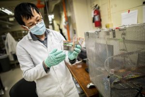
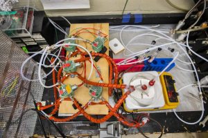
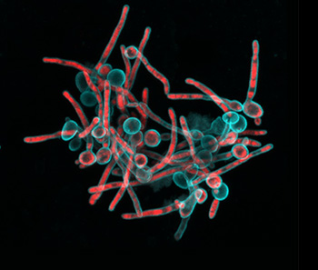
“Tagbo embodies what we are trying to do with the CiPD,” recalls Dr. Stebe. “He had initiative, he identified new tools and important context, and he did good science that may help us understand how to interrupt the disease process and identify new underlying mechanisms that can inspire new therapies.” Dr. Niepa worked on applying microfluidics and engineering to study the oral microbiome and better understand how the interactions between fungi and bacteria could impact dental caries.

“Upon meeting Michel, we became excited about the possibilities of bringing talent from the two schools together,” notes Dr. Stebe. A 2018 workshop organized by Drs. Koo and Stebe and funded by Penn’s Vice Provost of Research explored the potential for expanding cross-school research. “We invited researchers from dental medicine and engineering as well as relevant people from the arts and sciences to see if we could find a way to collaborate to advance oral and craniofacial health,” says Dr. Koo. “That was the catalyst for the Center; after the workshop, we put together a task force which would become the core members of the CiPD.”

In addition to Drs. Koo and Stebe, the CiPD Executive Committee includes Associate Directors Dr. Henry Daniell, Vice-Chair and W.D. Miller Professor, Department of Basic & Translational Sciences, Penn Dental Medicine, and Dr. Anh Le, Chair and Norman Vine Endowed Professor of Oral Rehabilitation, Department of Oral and Maxillofacial Surgery / Pharmacology, Penn Dental Medicine; as well as Dr. Andrew Tsourkas, Professor, Department of Bioengineering, Co-Director, Center for Targeted Therapeutics & Translational Nanomedicine (CT3N) and Chemical and Nanoparticle Synthesis Core, Penn Engineering; and Dr. Jason Moore, Edward Rose Professor of Informatics, Director of the Penn Institute for Biomedical Informatics. The core members of CiPD include 26 faculty from across both Penn Dental Medicine and Penn Engineering, and also from the Schools of Medicine and Arts & Sciences.

Read the full story in Penn Today.

Penn Bioengineers Develop Implantable Living Electrodes

Living Electrode Panels (image courtesy of the Cullen Lab)

Connecting the human brain to electrical devices is a long-standing goal of neuroscientists, bioengineers, and clinicians, with applications ranging from deep brain stimulation (DBS) to treat Parkinson’s disease to more futuristic endeavors such as Elon Musk’s NeuraLink initiative to record and translate brain activity. However, these approaches currently rely on using implantable metallic electrodes that inherently provoke a lasting immune response due to their non-biological nature, generally complicating the reliability and stability of these interfaces over time. To address these challenges, D. Kacy Cullen, Associate Professor in Neurosurgery and Bioengineering, and Dayo Adewole, a doctoral candidate in Bioengineering, worked with a multi-disciplinary team of collaborators to develop the first “living electrodes” as an implantable, biological bridge between the brain and external devices. In a recent article published in Science Advances, the team demonstrated the fabrication of hair-like microtissue comprised of living neuronal networks and bundled tracts of axons the signal sending fibers of the nervous system protected within soft hydrogel cylinders. They showed that these axon-based living electrodes could be fully controlled and monitored with light thus eliminating the need for electrical contact and are capable of surviving and forming synapses with the brain as demonstrated in an adult rat model. While further advancements are necessary prior to clinical use, the current findings provide the foundation for a new class of “living electrodes” as a biological intermediary between humans and devices capable of leveraging natural mechanisms to potentially provide a stable interface for clinical applications.

Cullen has a primary appointment in the Department of Neurosurgery in the Perelman School of Medicine, with a secondary appointment in the Department of Bioengineering in the School of Engineering and Applied Science, and an appointment in the Corporal Michael J. Crescenz VA Medical Center in Philadelphia.

Penn Engineering and CHOP Researchers Identify Nanoparticles that Could Be Used in Therapeutic mRNA Delivery before Birth

by Evan Lerner

William H. Peranteau, Michael J. Mitchell, Margaret Billingsley, Meghana Kashyap, and Rachel Riley (Clockwise from top left)

Researchers at Children’s Hospital of Philadelphia and the School of Engineering and Applied Science at the University of Pennsylvania have identified ionizable lipid nanoparticles that could be used to deliver mRNA as part of fetal therapy. The proof-of-concept study, published today in Science Advances, engineered and screened a number of lipid nanoparticle formulations for targeting mouse fetal organs and has laid the groundwork for testing potential therapies to treat genetic diseases before birth.

“This is an important first step in identifying nonviral mediated approaches for delivering cutting-edge therapies before birth,” said co-senior author William H. Peranteau, MD, an attending surgeon in the Division of General, Thoracic and Fetal Surgery and the Adzick-McCausland Distinguished Chair in Fetal and Pediatric Surgery at CHOP. “These lipid nanoparticles may provide a platform for in utero mRNA delivery, which would be used in therapies like fetal protein replacement and gene editing.”

Michael J. Mitchell, Skirkanich Assistant Professor of Innovation in Penn Engineering’s Department of Bioengineering, is the other co-senior author of the study. The co-first authors are Mitchell Lab members Rachel Riley, a postdoctoral fellow, and Margaret Billingsley, a graduate student, and Peranteau Lab member Meghana Kashyap, a research fellow.

Recent advances in DNA sequencing technology and prenatal diagnostics have made it possible to diagnose many genetic diseases before birth. Some of these diseases are treated by protein or enzyme replacement therapies after birth, but by then, some of the damaging effects of the disease have taken hold. Thus, applying therapies while the patient is still in the womb has the potential to be more effective for some conditions. The small fetal size allows for maximal therapeutic dosing, and the immature fetal immune system may be more tolerant of replacement therapy.

Read the full story in Penn Engineering Today.

NB: Rachel Riley is now Assistant Professor in Biomedical Engineering at Rowan University.

Studying ‘Hunters and Busybodies,’ Penn and American University Researchers Measure Different Types of Curiosity

by Melissa Pappas

Knowledge networks were created as participants browsed Wikipedia, where pages became nodes and relatedness between pages became edges. Two diverging styles emerged — “the busybody” and “the hunter.” (Illustrations by Melissa Pappas)

Curiosity has been found to play a role in our learning and emotional well-being, but due to the open-ended nature of how curiosity is actually practiced, measuring it is challenging. Psychological studies have attempted to gauge participants’ curiosity through their engagement in specific activities, such as asking questions, playing trivia games, and gossiping. However, such methods focus on quantifying a person’s curiosity rather than understanding the different ways it can be expressed.

Efforts to better understand what curiosity actually looks like for different people have underappreciated roots in the field of philosophy. Varying styles have been described with loose archetypes, like “hunter” and “busybody” — evocative, but hard to objectively measure when it comes to studying how people collect new information.

A new study led by researchers at the University of Pennsylvania’s School of Engineering and Applied Science, the Annenberg School for Communication, and the Department of Philosophy and Religion at American University, uses Wikipedia browsing as a method for describing curiosity styles. Using a branch of mathematics known as graph theory, their analysis of curiosity opens doors for using it as a tool to improve learning and life satisfaction.

The interdisciplinary study, published in Nature Human Behavior, was undertaken by Danielle Bassett, J. Peter Skirkanich Professor in Penn Engineering’s Departments of Bioengineering and Electrical and Systems Engineering, David Lydon-Staley, then a post-doctoral fellow in her lab, now an assistant professor in the Annenberg School of Communication, two members of Bassett’s Complex Systems Lab, graduate student Dale Zhou and postdoctoral fellow Ann Sizemore Blevins, and Perry Zurn, assistant professor from American University’s Department of Philosophy.

“The reason this paper exists is because of the participation of many people from different fields,” says Lydon-Staley. “Perry has been researching curiosity in novel ways that show the spectrum of curious practice and Dani has been using networks to describe form and function in many different systems. My background in human behavior allowed me to design and conduct a study linking the styles of curiosity to a measurable activity: Wikipedia searches.”

Zurn’s research on how different people express curiosity provided a framework for the study.

Read the full story in Penn Engineering Today.

Penn, Carnegie Mellon and Johns Hopkins to Develop New Turing Tests, Investigate How AI Can Become More Like Biological Intelligence

by Evan Lerner

While artificial intelligence is becoming a bigger part of nearly every industry and increasingly present in everyday life, even the most impressive AI is no match for a toddler, chimpanzee, or even a honeybee when it comes to learning, creativity, abstract thinking or connecting cause and effect in ways they haven’t been explicitly programmed to recognize.

This discrepancy gets at one of the field’s fundamental questions: what does it mean to say an artificial system is “intelligent” in the first place?

Konrad Kording, Timothy Verstynen, Joshua T. Vogelstein, and Leyla Isik (clockwise from top left)

Seventy years ago, Alan Turing famously proposed such a benchmark; a machine could be considered to have artificial intelligence if it could successfully fool a person into thinking it was a human as well. Now, many artificial systems could pass a “Turing Test” in certain limited domains, but none come close to imitating the holistic sense of intelligence we recognize in animals and people.

Understanding how AI might someday be more like this kind of biological intelligence — and developing new versions of the Turing Test with those principles in mind — is the goal of a new collaboration between researchers at the University of Pennsylvania, Carnegie Mellon University and Johns Hopkins University.

The project, called “From Biological Intelligence to Human Intelligence to Artificial General Intelligence,” is led by Konrad Kording, a Penn Integrates Knowledge Professor with appointments in the Departments of Bioengineering and Computer and Information Science in Penn Engineering and the Department of Neuroscience at Penn’s Perelman School of Medicine. Kording will collaborate with Timothy Verstynen of Carnegie Mellon University, as well Joshua T. Vogelstein and Leyla Isik, both of Johns Hopkins University, on the project.

Read the full story on Penn Engineering Today.