David F. Meaney, the Senior Associate Dean of Penn Engineering and Solomon R. Pollack Professor of Bioengineering, is known for his scholarship and innovation in neuroengineering and concussion science, his leadership as former Chair of the Department of Bioengineering, and for his marshaling of interdisciplinary research between Penn Engineering and the University’s health schools.
The Penn Engineering community has sprung into action over the course of the past few weeks in response to COVID-19. Meaney shared his perspective on those efforts and the ones that will come online as the pandemic continues to unfold.
David F. Meaney, Ph.D.
It is remarkable to think that a little more than a month ago I was saying an early goodbye to students for their spring break. In the first week of March, I was wishing everyone a happy and safe break, emphasizing safe, not knowing how prophetic that word would be. I was also looking forward to my own spring break, traveling for the first time in many years over this part of the academic calendar.
And then our campus — and world — changed.
COVID-19 is among us, in ways that we can’t exactly measure. It is among us in ways that we feel — we probably know someone that has tested positive for the virus, and others that are living with someone that is sick. And we all realize the virus will be with us for some time; the exact amount we don’t know.
Which brings up the question — what can we do to fight this pandemic? Many of us are trying to find ways to keep our connections with others vibrant and strong in the world of Zoom, Hangout, and BlueJeans. That is important. Let me also say that I can’t wait to reconnect with everyone in person, and close my laptop for a week.
But staying connected is what everyone should do. I often think about what can engineers do?
As the Senior Associate Dean, I want to let you know what I’m seeing on a quiet, but not shuttered, Penn campus. Examples of our response to the pandemic include our faculty designing personal protective equipment for health care workers, and our students, faculty and staff volunteering to assemble it. Other faculty are inventing COVID-19 test kits that can be completed at home, with the results available in less than an hour. Professors are sharing their creative mask designs with the world, for free, to make sure that we can all feel comfortable walking outside. And yet others that are collaborating to make a vaccine that will help us put COVID-19 behind us, permanently.
All of this is happening at speeds we have never seen before. Ideas move to prototypes and testing in days, not months, and to product in a week. We are not alone — our colleagues across campus are working at light speed to generate better tests, treatments, and models to fight COVID-19. This time, Nature has given us the problem. Time for us to solve it.
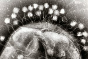
Transmission electron micrograph of multiple bacteriophages, viruses that infect bacteria, attached to a cell wall. New research describes how bacteria can optimize their “memory” of past viral infections in order to launch an effective immune response against a new invader. (Image: Graham Beards)
Before CRISPR became a household name as a tool for gene editing, researchers had been studying this unique family of DNA sequences and its role in the bacterial immune response to viruses. The region of the bacterial genome known as the CRISPR cassette contains pieces of viral genomes, a genomic “memory” of previous infections. But what was surprising to researchers is that rather than storing remnants of every single virus encountered, bacteria only keep a small portion of what they could hold within their relatively large genomes.
In recent years, CRISPR has become the go-to biotechnology platform, with the potential to transform medicine and bioengineering. In bacteria, CRISPR is a heritable and adaptive immune system that allows cells to fight viral infections: As bacteria come into contact with viruses, they acquire chunks of viral DNA called spacers that are incorporated into the bacteria’s genome. When the bacteria are attacked by a new virus, spacers are copied from the genome and linked onto molecular machines known as Cas proteins. If the attached sequence matches that of the viral invader, the Cas proteins will destroy the virus.
Bacteria have a different type of immune system than vertebrates, explains senior author Vijay Balasubramanian, but studying bacteria is an opportunity for researchers to learn more about the fundamentals of adaptive immunity. “Bacteria are simpler, so if you want to understand the logic of immune systems, the way to do that would be in bacteria,” he says. “We may be able to understand the statistical principles of effective immunity within the broader question of how to organize an immune system.”
This research was supported by the Simons Foundation (Grant 400425) and National Science Foundation Center for the Physics of Biological Function (Grant PHY-1734030).
Some medical conditions, like diabetes or limb amputation, have the potential to result in wounds that never heal, affecting patients for the rest of their lives. Though normal wound-healing processes are relatively understood by medical professionals, the complications that can lead to chronic non-healing wounds are often varied and complex, creating a gap in successful treatments. But biomedical engineering faculty from the University of Connecticut want to change that.
Ali Tamayol, Ph.D., an Associate Professor in UConn’s Biomedical Engineering Department, developed what he’s calling a “smart” bandage in collaboration with researchers from the University of Nebraska-Lincoln and Harvard Medical School. The bandage, paired with a smartphone platform, has the ability to deliver medications to the wound via wirelessly controlled mini needles. The minimally invasive device thus allows doctors to control medication dosages for wounds without the patient even having to come in for an appointment. Early tests of the device on mice showed success in wound-healing processes, and Tamayol hopes that soon, the technology will be able to do the same for humans.
A New Patch Could Fix Broken Hearts
Heart disease is by far one of the most common medical conditions in the world, and has a high risk of morbidity. While some efforts in tissue engineering have sought to resolve cardiac tissue damage, they often require the use of existing heart cells, which can introduce a variety of complications to its integration into the human body. So, a group of bioengineers at Trinity College in Dublin sought to eliminate the need for cells by creating a patch that mimics both the mechanical and electrical properties of cardiac tissue.
Using thermoelastic polymers, the engineers, led by Ussher Assistant Professor in Biomedical Engineering Michael Monaghan, Ph.D., created a patch that could withstand multiple rounds of stretching and exhibited elasticity: two of the biggest challenges in designing synthetic cardiac tissues. With the desired mechanical properties working, the team then coated the patches with an electroconductive polymer that would allow for the necessary electrical signaling of cardiac tissue without decreasing cell compatibility in the patch. So far, the patch has demonstrated success in both mechanical and electrical behaviors in ex vivo models, suggesting promise that it might be able to work in the human body, too.
3-D Printing a New Tissue Engineering Scaffold
While successful tissue engineering innovations often hold tremendous promise for advances in personalized medicine and regeneration, creating the right scaffold for cells to grow on either before or after implantation into the body can be tricky. One common approach is to use 3-D printers to extrude scaffolds into customizable shapes. But the problem is that not all scaffold materials that are best for the body will hold up their structure in the 3-D printing process.
A team of biomedical engineers at Rutgers University led by Chair of Biomedical Engineering David I. Schreiber, Ph.D., hopes to apply the use of hyaluronic acid — a common natural molecule throughout the human body — in conjunction with polyethylene glycol to create a gel-like scaffold. The hope is that the polyethylene glycol will improve the scaffold’s durability, as using hyaluronic acid alone creates a substance that is often too weak for tissue engineering use. Envisioning this gel-like scaffold as a sort of ink cartridge, the engineers hope that they can create a platform that’s customizable for a variety of different cells that require different mechanical properties to survive. Notably, this new approach can specifically control both the stiffness and the ligands of the scaffold, tailoring it to a number of tissue engineering applications.
A New Portable Chip Can Track Wide Ranges of Brain Activity
Understanding the workings of the human brain is no small feat, and neuroscience still has a long way to go. While recent technology in brain probes and imaging allows for better understanding of the organ than ever before, that technology often requires immense amounts of wires and stationary attachments, limiting the scope of brain activity that can be studied. The answer to this problem? Figure out a way to implant a portable probe into the brain to monitor its everyday signaling pathways.
That’s exactly what researchers from the University of Arizona, George Washington University, and Northwestern University set out to do. Together, they created a small, wireless, and battery-free device that can monitor brain activity by using light. The light-sensing works by first tinting some neurons with a dye that can change its brightness according to neuronal activity levels. Instead of using a battery, the device relies on energy from oscillating magnetic fields that it can pick up with a miniature antenna. Led in part by the University of Arizona’s Gutruf Lab, the new device holds promise for better understanding how complex brain conditions like Alzheimer’s and Parkinson’s might work, as well as what the mechanisms of some mental health conditions look like, too.
People & Places
Each year, the National Academy of Engineering (NAE) elects new members in what is considered one of the highest professional honors in engineering. This year, NAE elected 87 new members and 18 international members, including a former Penn faculty member and alumna Susan S. Margulies, Ph.D. Now a professor of Biomedical Engineering at Georgia Tech and Emory University, Margulies was recognized by the NAE for her contributions to “elaborating the traumatic injury thresholds of brain and lung in terms of structure-function mechanisms.” Congratulations, Dr. Margulies!
Nimmi Ramanujam, Ph.D., a Distinguished Professor of Bioengineering at Duke University, was recently announced as having one of the highest-scoring proposals for the MacArthur Foundation’s 100&Change competition for her proposal “Women-Inspired Strategies for Health (WISH): A Revolution Against Cervical Cancer.” Dr. Ramanujam’s proposal, which will enter the next round of competition for the grant, focuses on closing the cervical cancer inequity gap by creating a new model of women-centered healthcare.
A blood test may be able to detect the most common form of pancreatic cancer while it is still in its early stages while also helping doctors accurately stage a patient’s disease and guide them to the appropriate treatment. A multidisciplinary study found the test—known as a liquid biopsy—was more accurate at detecting disease in a blinded study than any other known biomarker alone, and was also more accurate at staging disease than imaging is capable of alone. The team, which includes researchers from the Perelman School of Medicine, the Abramson Cancer Center, and the School of Engineering and Applied Science, published their findings in Clinical Cancer Research, a journal of the American Association for Cancer Research.
Pancreatic ductal adenocarcinoma (PDAC), the most common form of pancreatic cancer, is the third leading cause of cancer deaths. The overall five-year survival rate is just 9%, and most patients live less than one year following their diagnosis. One of the biggest challenges is catching the disease before it has progressed or spread. If the disease is caught early, patients may be candidates for surgery to remove the cancer, which can be curative. For locally advanced patients—meaning patients whose cancer has not spread beyond the pancreas but who are not candidates for surgery based on the size or location of the tumor—treatment involves three months of systemic therapy like chemo or radiation, then reassessing to see if surgery is an option. For patients whose disease has spread, there are currently no curative treatment options.
“Right now, the majority of patients who are diagnosed already have metastatic disease, so there is a critical need for a test that can not only detect the disease earlier but also accurately tell us who might be at a point where we can direct them to a potentially curative treatment,” says the study’s co-senior author Erica L. Carpenter,director of the Liquid Biopsy Laboratory and a research assistant professor of medicine. The study’s other co-senior author is David Issadore, an associate professor of bioengineering and electrical and systems engineering.
A message from Penn Bioengineering Professor and Chair Ravi Radhakrishnan:
In response to the unprecedented challenges presented by the global outbreak of the novel coronavirus SARS-CoV-2, Penn Bioengineering’s faculty, students, and staff are finding innovative ways of pivoting their research and academic projects to contribute to the fight against COVID-19. Though these projects are all works in progress, I think it is vitally important to keep those in our broader communities informed of the critical contributions our people are making. Whether adapting current research to focus on COVID-19, investing time, technology, and equipment to help health care infrastructure, or creating new outreach and educational programs for students, I am incredibly proud of the way Penn Bioengineering is making a difference. I invite you to read more about our ongoing projects below.
RESEARCH
Novel Chest X-Ray Contrast
David Cormode, Associate Professor of Radiology and Bioengineering
The Cormode and Noel labs are working to develop dark-field X-ray imaging, which may prove very helpful for COVID patients. It involves fabricating diffusers that incorporate gold nanoparticles to modify the X-ray beam. This method gives excellent images of lung structure. Chest X-ray is being used on the front lines for COVID patients, and this could potentially be an easy to implement modification of existing X-ray systems. The additional data give insight into the health state of the microstructures (alveoli) in the lung. This new contrast mechanics could be an early insight into the disease status of COVID-19 patients. For more on this research, see Cormode and Noel’s chapter in the forthcoming volume Spectral, Photon Counting Computed Tomography: Technology and Applications, edited by Katsuyuki Taguchi, Ira Blevis, and Krzysztof Iniewski (Routledge 2020).
Immunotherapy
Michael J. Mitchell, Skirkanich Assistant Professor of Innovation in Bioengineering
Mike Mitchell is working with Saar Gill (Penn Medicine) on engineering drug delivery technologies for COVID-19 mRNA vaccination. He is also developing inhalable drug delivery technologies to block COVID-19 internalization into the lungs. These new technologies are adaptations of prior research published Volume 20 of Nano Letters (“Ionizable Lipid Nanoparticle-Mediated mRNA Delivery for Human CAR T Cell Engineering” January 2020) and discussed in Volume 18 of Nature Reviews Drug Discovery (“Delivery Technologies for Cancer Immunotherapy” January 2019).
Respiratory Distress Therapy Modeling
Ravi Radhakrishnan, Professor, and Chair of Bioengineering and Professor of Chemical and Biomolecular Engineering
Computational Models for Targeting Acute Respiratory Distress Syndrome (ARDS). The severe forms of COVID-19 infections resulting in death proceeds by the propagation of the acute respiratory distress syndrome or ARDS. In ARDS, the lungs fill up with fluid preventing oxygenation and effective delivery of therapeutics through the inhalation route. To overcome this major limitation, delivery of antiinflammatory drugs through the vasculature (IV injection) is a better approach; however, the high injected dose required can lead to toxicity. A group of undergraduate and postdoctoral researchers in the Radhakrishnan Lab (Emma Glass, Christina Eng, Samaneh Farokhirad, and Sreeja Kandy) are developing a computational model that can design drug-filled nanoparticles and target them to the inflamed lung regions. The model combines different length-scales, (namely, pharmacodynamic factors at the organ scale, hydrodynamic and transport factors in the tissue scale, and nanoparticle-cell interaction at the subcellular scale), into one integrated framework. This targeted approach can significantly decrease the required dose for combating ARDS. This project is done in collaboration with Clinical Scientist Dr. Jacob Brenner, who is an attending ER Physician in Penn Medicine. This research is adapted from prior findings published in Volume 13, Issue 4 of Nanomedicine: Nanotechnology, Biology and Medicine: “Mechanisms that determine nanocarrier targeting to healthy versus inflamed lung regions” (May 2017).
Diagnostics
Sydney Shaffer, Assistant Professor of Bioengineering and Pathology and Laboratory Medicine
Arjun Raj, David Issadore, and Sydney Shaffer are working on developing an integrated, rapid point-of-care diagnostic for SARS-CoV-2 using single molecule RNA FISH. The platform currently in development uses sequence specific fluorescent probes that bind to the viral RNA when it is present. The fluorescent probes are detected using a iPhone compatible point-of-care reader device that determines whether the specimen is infected or uninfected. As the entire assay takes less than 10 minutes and can be performed with minimal equipment, we envision that this platform could ultimately be used for screening for active COVID19 at doctors’ offices and testing sites. Support for this project will come from a recently-announced IRM Collaborative Research Grant from the Institute of Regenerative Medicine with matching funding provided by the Departments of Bioengineering and Pathology and Laboratory Medicine in the Perelman School of Medicine (PSOM) (PI’s: Sydney Shaffer, Sara Cherry, Ophir Shalem, Arjun Raj). This research is adapted from findings published in the journal Lab on a Chip: “Multiplexed detection of viral infections using rapid in situ RNA analysis on a chip” (Issue 15, 2015). See also United States Provisional Patent Application Serial No. 14/900,494 (2014): “Methods for rapid ribonucleic acid fluorescence in situ hybridization” (Inventors: Raj A., Shaffer S.M., Issadore D.).
HEALTH CARE INFRASTRUCTURE
Penn Health-Tech Coronavirus COVID-19 Collaborations
Brian Litt, Professor of Bioengineering, Neurology, and Neurosurgery
In his role as one of the faculty directors for Penn Health-Tech, Professor Brian Litt is working closely with me to facilitate all the rapid response team initiatives, and in helping to garner support the center and remove obstacles. These projects include ramping up ventilator capacity and fabrication of ventilator parts, the creation of point-of-care ultrasounds and diagnostic testing, evaluating processes of PPE decontamination, and more. Visit the Penn Health-Tech coronavirus website to learn more, get involved with an existing team, or submit a new idea.
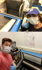
BE Educational Labs staff members Dana Abulez (BE ’19, Master’s BE ’20) and Matthew Zwimpfer (MSE ’18, Master’s MSE ’19) take shifts to laser-cut face shields.
The George H. Stephenson Foundation Educational Laboratory & Bio-MakerSpace staff have donated their PPE to Penn Medicine. Two staff members (Dana Abulez, BE ’19, Master’s BE ’20 and Matthew Zwimpfer, MSE ’18, Master’s MSE ’19) took shifts to laser-cut face shields in collaboration with Penn Health-Tech. Dana and Matthew are also working with Dr. Matthew Maltese on his low-cost ventilator project (details below).
Low-Cost Ventilator
Matthew Maltese, Adjunct Professor of Medical Devices and BE Graduate Group Member
Dr. Maltese is rapidly developing a low-cost ventilator that could be deployed in Penn Medicine for the expected surge, and any surge in subsequent waves. This design is currently under consideration by the FDA for Emergency Use Authorization (EUA). This example is one of several designs considered by Penn Medicine in dealing with the patient surge.
Face Shields
David F. Meaney, Solomon R. Pollack Professor of Bioengineering and Senior Associate Dean
Led by David Meaney, Kevin Turner, Peter Bruno and Mark Yim, the face shield team at Penn Health-Tech is working on developing thousands of rapidly producible shields to protect and prolong the usage of Personal Protective Equipment (PPE). Learn more about Penn Health-Tech’s initiatives and apply to get involved here.
Update 4/29/20: The Penn Engineering community has sprung into action over the course of the past few weeks in response to COVID-19. Dr. Meaney shared his perspective on those efforts and the ones that will come online as the pandemic continues to unfold. Read the full post on the Penn Engineering blog.
OUTREACH & EDUCATION
Student Community Building
Yale Cohen, Professor of Otorhinolaryngology, Department of Psychology, BE Graduate Group Member, and BE Graduate Chair
Yale Cohen, and Penn Bioengineering’s Graduate Chair, is working with Penn faculty and peer institutions across the country to identify intellectually engaging and/or community-building activities for Bioengineering students. While those ideas are in progress, he has also worked with BE Department Chair Ravi Radhakrishnan and Undergraduate Chair Andrew Tsourkas to set up a dedicated Penn Bioengineering slack channel open to all Penn Bioengineering Undergrads, Master’s and Doctoral Students, and Postdocs as well as faculty and staff. It has already become an enjoyable place for the Penn BE community to connect and share ideas, articles, and funny memes.
Undergraduate Course: Biotechnology, Immunology, Vaccines and COVID-19 (ENGR 35)
Daniel A. Hammer, Alfred G. and Meta A. Ennis Professor of Bioengineering and Chemical and Biomolecular Engineering
This Summer Session II, Professor Dan Hammer and CBE Senior Lecturer Miriam R. Wattenbarger will teach a brand-new course introducing Penn undergraduates to a basic understanding of biological systems, immunology, viruses, and vaccines. This course will start with the fundamentals of biotechnology, and no prior knowledge of biotechnology is necessary. Some chemistry is needed to understand how biological systems work. The course will cover basic concepts in biotechnology, including DNA, RNA, the Central Dogma, proteins, recombinant DNA technology, polymerase chain reaction, DNA sequencing, the functioning of the immune system, acquired vs. innate immunity, viruses (including HIV, influenza, adenovirus, and coronavirus), gene therapy, CRISPR-Cas9 editing, drug discovery, types of pharmaceuticals (including small molecule inhibitors and monoclonal antibodies), vaccines, clinical trials. Some quantitative principles will be used to quantifying the strength of binding, calculate the dynamics of enzymes, writing and solving simple epidemiological models, methods for making and purifying drugs and vaccines. The course will end with specific case study of coronavirus pandemic, types of drugs proposed and their mechanism of action, and vaccine development.
Update 4/29/20: Read the Penn Engineering blog post on this course published April 27, 2020.
Neuromatch Conference
Konrad Kording, Penn Integrates Knowledge University Professor of Bioengineering, Neuroscience, and Computer and Information Science
Dr. Kording facilitated Neuromatch 2020, a large virtual neurosciences conferences consisting of over 3,000 registrants. All of the conference talk videos are archived on the conference website and Dr. Kording has blogged about what he learned in the course of running a large conference entirely online. Based on the success of Neuromatch 1.0, the team are now working on planning Neuromatch 2.0, which will take place in May 2020. Dr. Kording is also working on facilitating the transition of neuroscience communication into the online space, including a weekly social (#neurodrinking) with both US and EU versions.
Neuromatch Academy
Konrad Kording, Penn Integrates Knowledge University Professor of Bioengineering, Neuroscience, and Computer and Information Science
Dr. Kording is working to launch the Neuromatch Academy, an open, online, 3-week intensive tutorial-based computational neuroscience training event (July 13-31, 2020). Participants from undergraduate to professors as well as industry are welcome. The Neuromatch Academy will introduce traditional and emerging computational neuroscience tools, their complementarity, and what they can tell us about the brain. A main focus is not just on using the techniques, but on understanding how they relate to biological questions. The school will be Python-based making use of Google Colab. The Academy will also include professional development / meta-science, model interpretation, and networking sessions. The goal is to give participants the computational background needed to do research in neuroscience. Interested participants can learn more and apply here.
Journal of Biomedical Engineering Call for Review Articles
Beth Winkelstein, Vice Provost for Education and Eduardo D. Glandt President’s Distinguished Professor of Bioengineering
The American Society of Medical Engineers’ (ASME) Journal of Biomechanical Engineering (JBME), of which Dr. Winkelstein is an Editor, has put out a call for review articles by trainees for a special issue of the journal. The call was made in March 2020 when many labs were ramping down, and trainees began refocusing on review articles and remote work. This call continues the JBME’s long history of supporting junior faculty and trainees and promoting their intellectual contributions during challenging times.
Update 4/29/20: CFP for the special 2021 issue here.
Are you a Penn Bioengineering community member involved in a coronavirus-related project? Let us know! Please reach out to ksas@seas.upenn.edu.
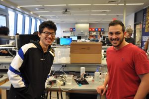
Andrew Chan (left, M.S.E. in Robotics ‘19) and Omar Abdoun (right, BE M.D./Ph.D. student) present “Cryogripper”
Almost every engineering school in the country offers a course in mechatronics — the overlap of mechanical, electrical, and computer engineering in electromechanical system design — but how many offer a course in biomechatronics? Taught by LeAnn Dourte, Ph.D., a Practice Associate Professor in Bioengineering, Penn Engineering’s Biomechatronics course (BE 570) gives students the chance to think about how the principles of mechatronic design can be used in biological settings involving orthopaedics, cardiovascular systems, and respiration, to name a few.
Throughout the course, students engage in different projects related to circuitry, signal processing, mechanics, motors, and analog controls, eventually applying all of these to biological examples before working on a final culminating project in design teams of two. In a simulation meant to mimic the sort of thinking and design processes that go behind innovations in robotic surgery, students create an electromechanical device that acts as a robotic hand. The catch? The “hand” has to have enough dexterity to pick up a water bead with a slipperiness similar to that of human tissue.
In addition to successfully performing this mechanical task using skills that the students learned throughout the semester, design teams also have to incorporate biological interfaces into the final project, such as using EMG signals to move part of the robotic hand, to give one example. Furthermore, each team needs to have a unique element to their design, whether in the use of a second biological interface, the application of Bluetooth to the system, or even a physical extension of the robotic hand to include the electromechanical equivalents of a shoulder, elbow, or wrist joint.
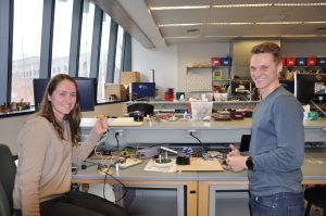
Carolyn Godone and Mike Furr (both M.S.E. in Bioengineering ‘19) model their design
Students Carolyn Godone and Mike Furr (both M.S.E. in Bioengineering ‘19) created a design inspired by the mechanical iris of a camera lens, using gears to push 3-D printed slices together in a symmetrical pattern to close around an object for pickup. They controlled their unique gripper with a thermal sensing camera that could employ a heat map of the device’s user to rotate, raise, and lower the gripper. Another pair of students, Omar Abdoun (BE M.D./Ph.D. student) and Andrew Chan (M.S.E. in Robotics ‘19), made what they called a “cryogripper”: a tissue moistened with water that freezes on demand when it contacts its target hydrogel. The ice allows the target to be lifted without falling, and the tissue can later be thawed with pumps of warm water to release hydrogel.
After weeks of working on their projects in the George H. Stephenson Foundation Educational Laboratory and Bio-MakerSpace, the class presented their final robotic hands during an open demonstration day (or Demo Day) in the lab. To see all the devices live and in action, watch the Facebook video below!
Spencer Glantz (left) examines a scheme for light-activated protein cleavage with Dr. Brian Chow (middle) and 2014 iGEM team member Daniel Cabrera (right).
Spencer Glantz, a graduate of the Penn Bioengineering doctoral program and former member of the Brian Chow Lab, was mentioned in a recent WHYY piece highlighting the efforts of Penn labs to develop rapid, at-home testing for COVID-19. Glantz is currently a co-leader of the molecular biology team for 4Catalyzer, a medical device incubator founded by National Medal of Technology and Innovation recipient, and sponsor of the annual Rothberg Catalyzer Makerthon competition, Jonathan Rothberg. 4Catalyzer is developing the testing technology while Penn researchers are working to evaluate its effectiveness.
Once hailed as medical miracles, antibiotics are losing their effectiveness due to the rapid increase of bacterial immunity.
Researchers are scrambling to keep up with evolution, and they are currently exploring how machine learning can be applied to microbiology to develop more effective treatments.
In the past, researchers have studied bacteria behavior and used their findings to work against the natural patterns of bacterial life. In the 1980s, computer-assisted screening methods helped researchers in their efforts but few developments surfaced from their work. It seemed that there were no new antibiotics to be found using traditional methods, and pharmaceutical companies stepped away from funding antibiotic development in favor of more profitable drugs used to treat chronic conditions. But a new field of research shows a way forward, thanks to the massive advances in computing that have occurred over the intervening decades.
Among the pioneering researchers in this field is César de La Fuente, Presidential Assistant Professor in Psychiatry, Microbiology and Bioengineering. De La Fuente is accelerating the discovery of new antibiotics with his Drug Repurposing Hub, a library of more than 6,000 compounds that is using machine learning algorithms to seek out possible solutions for human disease. With his compound library, de La Fuente is able to examine drugs already approved by the FDA and hunt for new, more effective applications.
In addition to this work, de La Fuente and his colleagues are interested in using machine learning to innovate drug design itself. His lab uses a machine learning platform to generate new molecules in silico and perform experiments on them. Once the results of the experiments come in, they are fed back into the computer so the machine learning platform can continuously learn and improve its findings from the data.
In a recent interview with Katherine Harmon Courage in Quanta Magazine, de La Fuente said:
“The hypothesis is that nature has run out of inspiration in terms of providing us with new antibiotics. That’s why we think that machines … could diversify natural molecules to convert them to synthetic versions that would be much more effective.”
Originally posted on the Penn Engineering blog. Read more about de La Fuente’s work and other researchers exploring the computational design of new antibiotics in Quanta Magazine or The Atlantic.
Though the coronavirus situation is changing daily, even hourly, by now the need for physical separation from those not in your household is clear. That doesn’t mean it’s easy, says Penn psychologist Melissa Hunt.
“We’re social animals,” says Hunt, associate director of clinical training in Penn’s Psychology Department. “We have an entire neuroendocrine system that responds to touch and social proximity with people we care about, that contributes to our sense of well-being and connection in the world. Losing out on that is really hard.”
It’s also not something we’ve really been asked to do before, says Lyle Ungar, a Penn computer scientist who is part of the World Well-Being Project, an initiative that uses social media language to measure psychological well-being and physical health. “This is an experiment on a scale that we’ve never seen in the United States,” he says.
Ungar and Hunt offer some suggestions to stay positive and healthy in the face of this new social isolation.
1. Maintain a connection with the people you love, even if it can’t be a physical one.
“Social distance does not mean no social contact,” Ungar says. Psychologically, face-to-face conversations are best, but right now they’re not likely possible. Instead, Ungar suggests video calls. “They’re second best in terms of emotional bonding,” he says. “Phone calls aren’t as good as video chats, and texting is even worse. But of course, being totally isolated is the worst.”
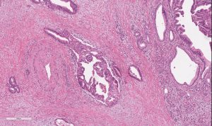
A microscopic view of pancreatic adenocarcinoma. (Image: Emma Furth)
Swept up in a pancreatic cancer diagnosis is inevitably a sense of fear and sadness.
But at Penn, researchers are bringing new hope to this disease. And with patients like Nick Pifani, it’s clear that they’re moving in the right direction.
Pifani, from Delran, New Jersey, first noticed some lingering stomach upset in February 2017. He called his family doctor, concerned—especially given that he was an otherwise healthy marathon runner who was only 42. He was sent to a gastrointestinal specialist. A few weeks later, some crippling stomach pain sent him back to the emergency room and he received an MRI that showed a mass on his pancreas—Stage Three, inoperable, he was told.
He was treated with chemotherapy, along with radiation and, eventually, and after receiving advice from doctors at Penn, his tumor was removed. Thereafter, he realized he had a PALB2 mutation—a cousin of the BRCA gene mutation. At that moment, his long-term needs changed and he found himself seeking specialized care at Penn, where he met Kim Reiss Binder, assistant professor of medicine at the Hospital of the University of Pennsylvania (HUP).
“I’m a planner; I want to understand what [my] potential options are,” Pifani says. “[Reiss Binder] asked why I was there to see her and I explained and quickly I could tell she was—outside of her being remarkably intelligent—a great listener and a compassionate doctor.”
“I have a feeling she worries about me more than I do,” he laughs.
Pifani has now been in remission for two years and four months; he sees Reiss-Binder every three months for checkups. His survival story is inspiring and a sign of momentum, even if a world without pancreatic cancer is still frustratingly out of reach.
Pancreatic cancer at Penn
Pancreatic cancer is the third-leading cause of cancer-related death in the United States, outmatched only by lung cancer (No. 1) and colorectal cancer (No. 2). A person diagnosed with pancreatic cancer is still unlikely to survive past five years—only 9% of survivors do, giving it the highest mortality rate among every major cancer.
In short, pancreatic cancer seldom paves the way for optimistic narratives. Some of the hope that has surfaced, though, is thanks to some talent, dedication to the cause, and hard work at Penn.
A key point of progress in the battle against the disease was made in 2002, when former Assistant Professor of Medicine David Tuveson established a standard model for examining human development of this disease in mice. This model has allowed for a reliable way to study the disease and has influenced progress made here at Penn and elsewhere since.
“There’s been a burst of activity in translational research, from bench to bedside,” explains Ben Stanger, the Hanna Wise Professor in cancer research and director of the Penn Pancreatic Cancer Research Center (PCRC) at the Abramson Cancer Center.
“And there’s a lot of momentum with community building, a dramatic increase in patient volumes, and a dramatic increase in what we know about the cancer,” he says of the status of pancreatic cancer today.
Reiss Binder, meanwhile, explains that one mark of progress at Penn and beyond has been learning about people like Pifani, who have the PALB2 gene, and why they respond differently to treatments than those without it. Platinum-based chemotherapies, for example, are especially effective for people with the PALB2 gene who are battling pancreatic cancer. An ongoing trial at Penn has tested and found some success with using PARP inhibitors—taken orally as an enzyme that fixes single-stranded breaks of DNA—as a maintenance therapy in that same PALB2 demographic after they’ve had chemotherapy. These are less toxic than chemotherapy for patients with the same mutations.
It’s all been slow progress toward better treatments, but there has been progress.
“This is the tip of the iceberg for a disease that we historically have treated with perpetual chemotherapy,” Reiss Binder says. “We owe it to patients to find better options to suppress the cancer but not ruin their quality of life.”
Catching cancer earlier
The consensus on why pancreatic cancer is so deadly? It just can’t be spotted fast enough.
Pancreatic cancer often presents well after it has developed and metastasized, and does so in a way that is not easy to recognize as cancer. Common symptoms include, for example, stomach upset and back pain. And by the time a harder-to-ignore symptom of the cancer surfaces, a sort of yellowing of the skin (a result of a bile duct blockage), it’s likely too late to stop the cancer in its tracks.
One approach to improved detection being tested at Penn, by Research Assistant Professor of Medicine Erica Carpenter, is a liquid biopsy—drawn from a standard blood test. Current means to test for pancreatic cancer—imaging through an endoscopic tube—are invasive and expensive, meaning a common liquid test could transform how many cases are detected early.
Carpenter explains that circulating tumor cells (CTCs) can shed from a tumor that’s adjacent to the wall of a blood vessel; what’s shed then shows up in a blood test. The cells, if detected, can explain more about the nature of the tumor, giving doctors an opportunity to examine characteristics of cancerous cells and decide how to effectively treat a tumor if it can’t be surgically removed. It also allows interpretations of disease burden and the effectiveness of medications—through genome sequencing—that imaging does not.
Ultimately, this gives doctors the potential to track the growth of a tumor before it’s fully developed, all through one tube of blood—detected through an innovative use of technology.
David Issadore, Ph.D.
David Issadore, associate professor of bioengineering and electrical and systems engineering in the School of Engineering and Applied Science, has worked since 2017 to develop a chip that detects cancer in the blood, using machine learning to sort through literally hundreds of billions of vesicles and cells, looking for these CTCs. The chip retrieves data and the machine learning developed interprets that data, attempting to make a diagnosis that not only finds pancreatic cancer but also provides information about its progression—and, importantly, whether a patient might benefit from surgery.


 Some medical conditions, like diabetes or limb amputation, have the potential to result in wounds that never heal, affecting patients for the rest of their lives. Though normal wound-healing processes are relatively understood by medical professionals, the complications that can lead to chronic non-healing wounds are often varied and complex, creating a gap in successful treatments. But biomedical engineering faculty from the University of Connecticut want to change that.
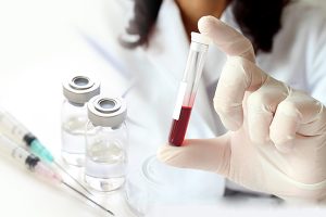 A blood test may be able to detect the most common form of pancreatic cancer while it is still in its early stages while also helping doctors accurately stage a patient’s disease and guide them to the appropriate treatment. A multidisciplinary study found the test—known as a liquid biopsy—was more accurate at detecting disease in a blinded study than any other known biomarker alone, and was also more accurate at staging disease than imaging is capable of alone. The team, which includes researchers from the
 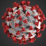 In response to the unprecedented challenges presented by the global outbreak of the novel coronavirus SARS-CoV-2,