Dr. LeAnn Dourte Appointed Practice Associate Professor

LeAnn Dourte, Ph.D.

The Department of Bioengineering at the University of Pennsylvania would like to congratulate LeAnn Dourte, Ph.D. on her recent promotion to Practice Associate Professor. Formerly a Senior Lecturer, this promotion reflects Dr. Dourte’s innovative approaches to pedagogy since arriving at Penn in 2011. The title of Practice Associate Professor reflects exceptional accomplishment in teaching, leadership, and educational programs. As an official member of the faculty, this formalizes Dourte’s role as a leader in pedagogy and educational scholarship, furthering empowering her to think creatively and progressively about higher education.

As a key member of the BE teaching faculty, Dourte has regularly taught core undergraduate subjects such as Intro to Biomechanics (BE 200) and Biomaterials (BE 220) as well as popular graduate electives Biomechanics and Biotransport (BE 510) and Biomechatronics (BE 570). In particular, she spearheaded the department’s initiative to improve classroom and learning experiences through experimentation with the Structured, Active, In-Class Learning (or SAIL) model of education which emphasizes teamwork and dynamic problem-solving. Dourte worked with the Center for Teaching and Learning to understand this model and assess how it would best be applicable for BE students, and then presented her findings at national conferences and to the BE faculty, helping to introduce them to these techniques and advise them on best practices. Other BE faculty such as Chris Fang-Yen, Ph.D., Jennifer Philips-Cremins, Ph.D., and Danielle Bassett, Ph.D., have experimented with and incorporated these ideas into their courses. Thanks to Dourte’s efforts, BE has been integral in demonstrating the success of SAIL classes to the School of Engineering and then spreading this philosophy to other schools at Penn. In addition to her pedagogical interests, Dourte is also highly involved in the Department’s student wellness initiative, serving on the Department’s Climate Committee and as the Wellness Ambassador.

Now that she has achieved this latest milestone, Dourte has set her sights on goals for the future. She intends to work with Associate Dean for Undergraduate Education Dr. Russ Composto to strengthen initiatives to assist SEAS’s First-Generation, Low-Income (FGLI) students. She will be working with Dr. Bassett, who specializes in network science, to learn more about how students learn, and what tools can be developed to assess students in addition to traditional exams and homework; to tell more easily when students are missing key concepts; and to intervene sooner in moments of crisis. And finally, Dourte will also be one of three representatives from Penn at an upcoming national education summit this May to discuss the future of Bioengineering curricula.

“Our best educators are teachers for the rest of the faculty, as well as the students,” says Department Chair Dr. David Meaney. “We are enormously proud of the prestige and expertise that LeAnn shares with all of us. I was fortunate to teach biomechanics with LeAnn for many years, and saw her outstanding ability as an educator in person.”

Once again, we would like to extend hearty congratulations to Dr. Dourte on this well-deserved recognition of her leadership and both in and out of the classroom.

Week in BioE (February 28, 2019)

by Sophie Burkholder

Louisiana Tech Sends First All-Female Team to RockOn

A team of faculty and students from Louisiana Tech University will participate in RockOn, a NASA-sponsored workshop on rocketry and engineering. Mechanical Engineering Lecturer Krystal Corbett, Ph.D., and Assistant Professor of bioengineering Mary Caldorera-Moore, Ph.D., will work together to lead the university’s first team of three all-female students at the event. At the program, they will have the chance to work on projects involving components of spacecraft systems, increasing students’ experience in hands-on activities and real-world engineering.

Refining Autism Treatments Using Big Data

Though treatments like therapy and medication exist for patients with autism, one of the biggest challenges that those caring for these patients face is in measuring their effects over time. Many of the markers of progress are qualitative, and based on a given professional’s opinion on a case-by-case basis. But now, a team of researchers from Rensselaer Polytechnic Institute (RPI) hopes to change that with the use of big data.

Juergen Hahn, Ph. D., and his lab recently published a paper in Frontiers in Cellular Neuroscience discussing their findings in connecting metabolic changes with behavioral improvements in autistic patients. Their analysis looks for multiple chemical and medical markers simultaneously in data from three distinct clinical trials involving metabolic treatment for patients. Being able to quantitatively describe the effects of current autism treatments would revolutionize clinical trials in the field, and lead to overall better patient care.

Penn Engineers Can Detect Ultra Rare Proteins in Blood Using a Cellphone Camera

One of the frontiers of medical diagnostics is the race for more sensitive blood tests. The ability to detect extremely rare proteins could make a life-saving difference for many conditions, such as the early detection of certain cancers or the diagnosis of traumatic brain injury, where the relevant biomarkers only appear in vanishingly small quantities. Commercial approaches to ultrasensitive protein detection are starting to become available, but they are based on expensive optics and fluid handlers, which make them relatively bulky and expensive and constrain their use to laboratory settings.

Knowing that having this sort of diagnostic system available as a point-of-care device would be critical for many conditions — especially traumatic brain injury — a team of engineers led by Assistant Professor in the Department of Bioengineering, David Issadore, Ph.D., at the University of Pennsylvania have developed a test that uses off-the-shelf components and can detect single proteins with results in a matter of minutes, compared to the traditional workflow, which can take days.

Read the full story on Penn Engineering’s Medium blog.

Treating Cerebral Palsy with Battery-Powered Exoskeletons

Cerebral palsy is one of the most common movement disorders in the United States. The disorder affects a patient’s control over even basic movements like walking, so treatments for cerebral palsy often involve the use of assistive devices in an effort to give patients better command over their muscles. Zach Lerner, Ph.D., is an Assistant Professor of Mechanical Engineering and faculty in Northern Arizona University’s Center for Bioengineering Innovation whose research looks to improve these kinds of assistive devices through the use of battery-powered exoskeletons.

Lerner and his lab recently received three grants, one each from the National Institute of Health (NIH), the National Science Foundation (NSF), and the Arabidopsis Biological Resource Center, to continue their research in developing these exoskeletons. Their goal is to create devices with powered assistance at joints like the ankle or knee to help improve patient gait patterns in rehabilitating the neuromuscular systems associated with walking. The team hopes that their work under these new grants will help further advance treatment for children with cerebral palsy, and improve overall patient care.

People & Places

David Aguilar, a 19-year-old bioengineering student at Universitat Internacional de Catalunya made headlines recently for a robotic prosthetic arm that he built for himself using Lego pieces. Due to a rare genetic condition, Aguilar was born without a right forearm, a disability that inspired him to play with the idea of creating his own prosthetic arm from age nine. His design includes a working elbow joint and grabber that functions like a hand. In the future, Aguilar hopes to continue improving his own prosthetic designs, and to help create similar versions of affordable devices for other patients who need them.

This week, we would like to congratulate two recipients of the National Science Foundation’s Career Awards, given to junior faculty that exemplify the role of teacher-scholars in their research. The first recipient we’d like to acknowledge is the University of Arkansas’ Kyle Quinn, Ph.D., who received the award for his work in developing new image analysis methods and models using the fluorescence of two metabolic cofactors. Dr. Quinn completed his Ph.D. here at Penn in Dr. Beth Winkelstein’s lab, and received the Solomon R. Pollack Award for Excellence in Graduate Bioengineering Dissertation Research for his work.

The second recipient of the award we wish to congratulate is Reuben Kraft, Ph.D., who is an Assistant Professor in Mechanical and Biomedical Engineering at Penn State. Dr. Kraft’s research centers around developing computational models of the brain through linking neuroimaging and biomechanical assessments. Dr. Kraft also collaborates with Kacy Cullen, Ph.D., who is a secondary faculty member in Penn’s bioengineering department and a member of the BE Graduate Group faculty.

Finally, we’d like to congratulate Dawn Elliott, Ph.D., on being awarded the Orthopaedic Research Society’s Adele L. Boskey, PhD Award, awarded annually to a member of the Society with a commitment to both mentorship and innovative research. Dr. Elliott’s spent 12 years here at Penn as a member of the orthopaedic surgery and bioengineering faculty before joining the University of Delaware in 2011 to become the founding director of the bioengineering department there. Her research focuses primarily on the biomechanics of fibrous tissue in tendons and the spine.

Bioengineering Welcomes New Faculty Member Sydney Shaffer

Sydney Shaffer, MD, PhD

The Department of Bioengineering at the University of Pennsylvania is proud to announce the appointment of Sydney Shaffer, Ph.D., as an Assistant Professor of Bioengineering. She shares a joint appointment with Pathology and Laboratory Medicine. Originally from Atlanta, GA, Dr. Shaffer received her B.S. in Chemical Engineering from Georgia Institute of Technology, coming to Penn to complete her M.D./Ph.D. work in Bioengineering and the Perelman School of Medicine. After graduating in 2018, she conducted her postdoctoral work at Penn in Cancer Biology with Dr. Junwei Shi.

Dr. Shaffer’s research is is focused on understanding how differences present in single-cells can generate phenotypes such as drug resistance in cancer, oncogenesis, differentiation, and invasion. Our approach leverages cutting-edge technologies including high-throughput imaging, single-molecule RNA FISH, fluorescent protein tagging, CRISPR/Cas9 screening, and flow cytometry to investigate rare single-cell phenomena. Further information can be found at www.sydshafferlab.com.

In addition to her exciting research, Dr. Shaffer will be an enthusiastic new member of the Bioengineering Department community. In the short term, she will be taking over the popular class BE 400 (Preceptorships in Bioengineering) which gives undergraduates the rare chance to shadow renowned physicians over a period of ten weeks. She will also serve as a faculty advisor as well as a mentor to the lucky students in her classes and lab.

Dr. Shaffer says that, “With my research interests and training at the interface of engineering and medicine, I am thrilled to be part of the highly interdisciplinary community of Penn Bioengineering.”

“Sydney has a unique combination of creativity and impact in her work,” says Solomon R. Pollack Professor and Chair Dr. David Meaney. “Her work to untangle the secrets of how single cancer cells can develop resistance to a cancer drug  therefore leading to a return of the cancer  is nothing short of stunning. We are incredibly fortunate to have her on our faculty. ”

Week in BioE (February 21, 2019)

by Sophie Burkholder

Detecting Infectious Diseases with Paper-Based Devices

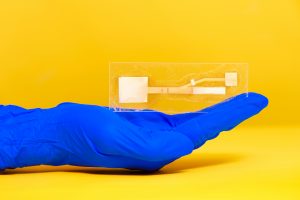
Dr. Linnes’ paper device. Image used courtesy of Erin Easterling, Purdue College of Engineering.

Despite great advancements in diagnostics technology over the past few decades, patient accessibility to these technologies remains one of the biggest challenges of the field today. Particularly in low-resource areas, even simple processes can end up taking weeks or months to return results from tests that are normally completed in days. But what if these tests could be simplified to smaller, at-home tests based on properties of microfluidics – something like a pregnancy test but for infectious diseases like HIV?

Jacqueline Linnes, Ph.D., and her team of researchers at Purdue University are working towards finding a way to do just that by creating paper-based devices that use microfluidics to help carry out the necessary diagnostic tests. Specifically, her lab designed such a paper-based system that can detect HIV nucleic acids within 90 minutes of receiving a drop of patient blood. The success of this design shows promise for producing devices for diseases whose diagnostics process involve similar pathways of pathogen detection, opening the door to more applications of at-home tests based in the properties of paper microfluidics.

Here at Penn, undergraduate bioengineering students enrolled in the two-semester laboratory course Bioengineering Modeling, Analysis, and Design (BE 309 & BE 310) have the chance to create their own models of paper microfluidics delivery systems based on given time constraints in a multi-step process. Though the students’ challenge only involves water as a substrate, Linnes’ research demonstrates the later implications of studying fluid flow through a medium as cheap and accessible as paper.

Watch the video below demonstrating Dr. Linnes’ device:

Funding for Cancer Research in Tumor Mimicry and Imaging

Two of the deadliest forms of cancer today are breast cancer and pancreatic cancer, with the latter having a five-year survival rate of only about 8%. Because cancer treatments are often adjusted according to a unique patient-to-patient basis, learning how to improve predictions of tumor behavior could help determine proper therapies sooner.

Chien-Chi Lin, Ph.D., an associate professor of biomedical engineering at Indiana University – Purdue University Indianapolis, recently received a grant from the National Institute of Health to advance his research in pancreatic cancer treatment. His project under the grant involves the development of bio-inspired, responsive, and viscoelastic (BRAVE) cell-laden hydrogels to help understand cell interactions in pancreatic ductal adenocarcinoma, which is the most common form of malignancy in the pancreas. These hydrogels mimic tumor tissue, as well as model tumor development over time, helping to eventually find better ways of treating pancreatic cancer.

In other news surrounding cancer-related research, a team of researchers led by Kenneth Tichauer, Ph.D., at the Illinois Institute of Technology won the university’s Nayar Prize for their development of the Agent-Dependent Early Photon Tomography (ADEPT) Cancer Imager, a machine designed to find early tumor development in the lymph nodes of breast cancer patients. Through the use of a special dyeing process that now dyes the entire lymph node, providing a sharper image that allows for a quicker discovery of smaller tumors.

Penn’s Women in Computer Science (WiCS) Hosts FemmeHacks

Penn President Amy Gutmann and Penn Engineering Dean Vijay Kumar stopped by FemmeHacks at the Pennovation Center Feb. 9. The annual event is a beginner-friendly collegiate hackathon for women-identifying people with an interest in computer programming, and featured a day of all-levels workshops Feb. 8. The event is sponsored by Penn’s Women in Computer Science student organization.

Though the event is not specifically tailored towards applications in bioengineering, skills relating to coding and software development are increasingly important for those interested in pursuing a career in medical device design. In fact, in the evaluation of new medical devices, the FDA often focuses more on software over hardware, as the former is associated with more security liabilities, due to its relative novelty.

Read the full story and see pictures on FemmeHacks on Penn Today.

People & Places

In December, the department of biomedical engineering at the University of Alabama at Birmingham received the Ruth L. Kirschstein National Research Service Awards Institutional Research Training Grant, which will support predoctoral students enrolled in the university’s biomedical engineering graduate programs. The department plans to use the grant for research in cardiovascular tissue engineering.

Case Western Reserve University and Cleveland Clinic announced the launch of an alliance last year with the goal of creating better synergy across the two renowned institutions, hoping to provide more opportunities for students with interest in medicine at all levels, from high school to postdoctoral education. Though researchers from both institutions frequently partner on projects, this new alliance will create a more structured platform for future collaborations.

We would like to commend Steven George, M.D./Ph.D., on his new position as the chair of the Department of Biomedical Engineering at the University of California at Davis. His research involves the development of “organ-on-a-chip” technologies using stem cells and microfluidics to mimic human organ functions of vascularized cardiac, tumor, and pancreatic tissues.

Finally, we want to congratulate Paul Yock, M.D., on his being chosen to receive the National Academy of Engineering’s 2019 Fritz J. and Dolores H. Russ Prize. The prize honors two of Dr. Yock’s inventions from his research in interventional cardiology, one of which is Rapid Exchange, which is a kind of stenting and balloon angioplasty system. Dr. Yock is the Martha Meier Weiland Professor in the School of Medicine and Professor of Bioengineering.


Week in BioE (February 13, 2019)

by Sophie Burkholder

Bioengineers Tackle Heart Disease

Heart disease is currently the leading cause of death in the United States, resulting in about 630,000 deaths every year according to the Center for Disease Control. One of the most common side effects of heart disease is damage to blood vessels and cardiac tissue, which can ultimately lead to conditions like high blood pressure, arrhythmia, and even cardiac arrest. In serious cases of irreversible heart damage, often the only option for patients is a full heart transplant, and efforts to engineer vascularized cardiac tissue grafts have proved challenging in research so far.

But researchers Ying Zheng, Ph.D., and Charles Murry, M.D., Ph.D., both of whom have joint appointments in Bioengineering at the University of Washington, have found success in using human microvascular grafts to create working blood vessels in vitro to treat infarcted rat hearts. The new heart muscle, developed from human embryonic stem cell-derived endothelial cells in petri dishes, was grown with a focus on not only being able to easily integrate it in vivo, but also in creating a patch of vasculature that closely mirrored that of the heart. In concentrating more on the mechanical aspects of the blood vessel network, Zheng and Murry were able to better restore normal blood flow to the damaged rat hearts after integration of the grafts. The study appears in a recent edition of Nature Communications.

Another team of bioengineers, led by Michael Sacks, Ph.D. at the University of Texas at Austin, recently invented a software-based method for repairing mitral valves in the heart. Their work, published in the International Journal for Numerical Methods in Biomedical Engineering, uses computational modeling techniques to create a noninvasive way of simulating repairs to the mitral valve, which will allow for a better prediction of surgical procedures and postoperative side effects on a more patient-specific basis. This ability to know which treatment plan may be best-suited for a given patient is important especially for valve repair, as heart valves are notoriously difficult to model or image due to the complexity of their functions. But through the use of advanced technology in 3D echocardiography, Sacks and his team say that their new model is accurate enough to rely on in clinical settings.

Virtual Reality Assists in the Evaluation of Surgery

Any form of surgery is always a high risk procedure, as it is subject to a wide variety of sources of human error and irregularity, even with the best surgeons. Certainly, there should be a system in place to not only continually assess the knowledge of surgeons throughout their careers, but also to evaluate their practices and techniques during operation. Such an evaluation, however, would put patients at risk during the assessment of the surgeon.

But now a team of researchers from Rensselaer Polytechnic Institute has developed a way of simulating colorectal surgical procedures using virtual reality technology. Suvranu De, Sc.D. — the J. Erik Jonsson ‘22 Distinguished Professor of Engineering and Head of the Department of Mechanical, Aerospace and Nuclear Engineering with joint appointments in Biomedical Engineering and Information Technology and Web Science —leads the project which incorporates both visual and tactile feedback for users to employ as a tool for both training and evaluating colorectal surgeons. While virtual reality simulators have been used for similar applications related to procedures like the colonoscopy, they have yet to be fully developed for open surgical procedures, because of the difficulties in creating a fully engaged and immersive environment. Nonetheless, De and his team hope that their work will lead to the creation of the first “Virtual Intelligent Preceptor,” which will allow for more advanced technological innovations in aspects of surgical education that have so far been difficult to standardize. Support for the project comes from the National Institute of Biomedical Imaging and Bioengineering (NBIB).

Penn BE’s Dr. Bassett on Understanding Knowledge Networks in the Brain

Dr. Danielle Bassett, Ph.D., Eduardo D. Glandt Faculty Fellow and Associate Professor of Bioengineering

As a network neuroscientist, Danielle Bassett, Ph.D., Eduardo D. Glandt Faculty Fellow and Associate Professor in the Department Bioengineering, brings together insights from a variety of fields to understand how the brain’s connections form and change: mathematics, physics, electrical engineering and developmental biology, to name a few. Bassett’s recent work on the learning process also draws from linguistics, educational theory and other domains even further afield.

The intersection and interaction of knowledge from multiple sources doesn’t just describe Bassett’s methodology; it’s at the heart of her research itself. At the Society for Industrial and Applied MathematicsAnnual Meeting last year, Bassett provided an address on how the structure of knowledge networks can influence what our brains can do when it comes to learning new things.

Read the full story on Penn Engineering’s Medium blog.

People & Places:

Tammy Dorsey, a graduate student at Wichita State University, created a non-invasive in utero tool to help read the oxygen levels of unborn babies as part of her senior design project. Dorsey says the inspiration for the project came from complications during the birth of her middle child, who despite having a normal heart rate throughout the entire pregnancy, was born blue. The device Dorsey created uses measurements of the baby’s pH to read fetal oxygen levels. She hopes that the design will help doctors better detect when a fetus is in distress during pregnancy and childbirth.

The field of bioengineering is constantly growing, and new programs are always in development. Boise State University has announced the launch of a new doctoral program in bioengineering that will begin in the fall of 2019. Developed through the collaboration of the university’s College of Health Sciences, College of Engineering, Graduate College, and College of Arts and Sciences, this new opportunity to do research in the field of bioengineering will have three study tracks available in biomechanics, mechanobiology, and human performance.

The new biomedical engineering department at the University of Massachusetts Amherst has announced the department’s first faculty appointments. The founding department head will be Professor Tammy L. Haut Donahue, Ph.D., whose research focus is on the biomechanics of the musculoskeletal system. Another professor joining the department’s new faculty is Seth W. Donahue, Ph.D., who has also done research in the field of biomechanics, and specifically how it pertains to tissue regeneration.

Since we last posted, there have also been several significant academic appointments in the field of Bioengineering. This week, we would like to congratulate Bruce Tromberg, Ph.D., on his appointment as the director of the National Institute of Biomedical Imaging and Bioengineering (NIBIB). Dr. Tromberg is currently a Professor with appointments in Biomedical Engineering and Surgery at the University of California at Irvine, where he leads research in bioimaging and biophotonics. He has also served on the External Advisory Board of NIH P41 Center for Magnetic Resonance and Optical Imaging here at Penn since 2009, and has also given several lectures here on his work in bioimaging.

Secondly, we congratulate the University of Toronto’s Professor Warren Chan, Ph.D., who was recently named as a Tier 1 Canada Research Chair in Nanobioengineering. Professor Chan, who is also the director of the Institute of Biomaterials and Biomedical Engineering at the University of Toronto, conducts research in the field of nanotechnology for applications in the treatment and diagnosis of cancer and viral diseases.

And finally, we also want to congratulate Frank Pintar, Ph.D., on his appointment as the Founding Chair of the Marquette University and Medical College of Wisconsin. Dr. Pintar’s research in bioengineering involves the study of the biomechanics involved with brain and spinal cord injury, with a focus on motor vehicle crash trauma.

Penn BE Undergraduates’ Plate Reader Design Published

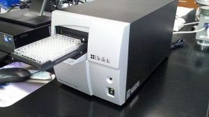
Microplate reader, Wikimedia Commons

In a paper recently published in Biochemistry, a group of University of Pennsylvania Bioengineering students describe the results of their work designing a new, open-source, low-cost microplate reader. Plate readers are instruments designed to measure light absorption and fluorescence emission from molecules useful for clinical biomarker analyses and assays in a diverse array of fields including synthetic biology, optogenetics, and photosensory biology. This new design costs less than $3500, a significantly lower price than other commercially available alternatives. As described in the paper’s abstract, this design is the latest in a growing trend of open-source  hardware to enhance access to equipment for biology labs. The project originated as part of the annual International Genetically Engineering Machine Competition (iGEM), an annual worldwide competition focusing on “push[ing] the boundaries of synthetic biology by tackling everyday issues facing the world” (iGEM website).

The group consists of current junior Andrew Clark (BSE ’20) and recent graduates Karol Szymula (BSE ’18), who works in the lab of Dr. Danielle Bassett, and Michael Patterson (BSE ’18), a Master’s student in Bioengineering and Engineer of Instructional Laboratories. Assistant Professor of Bioengineering Dr. Brian Chow served as their faculty mentor alongside Director of Instructional Labs Sevile Mannickarottu and Michael Magaraci, a Ph.D. candidate in Bioengineering, all of whom serve as co-authors on the published article. The research and design of the project was conducted in the Stephenson Foundation Bioengineering Educational Laboratory here at the University of Pennsylvania’s Department of Bioengineering.

Bioengineering Graduate Group Symposium – January 2019

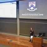
On January 8, 2019 the Department of Bioengineering at Penn held its annual Graduate Group Research Symposium to great success.

Thank you to everyone who attended and participated, our student volunteers, our faculty who participated as judges for the student talks and poster competition, and especially to our keynote speaker, Dr. Sujata Bhatia, Professor of Chemical and Biomolecular Engineering at the University of Delaware.

Congratulations to the award winners:

Student Talks:

  • First prize – Meagan Ita
  • Second prize – Nicolette Driscoll
  • Third prize – Minna Chen

Poster Presentations:

  • First prize – Mariia Alibekova
  • Second prize – Jonathan Galarraga, Sonia Kartha, and John Viola
  • Third prize – Andrei Georgescu

Annual Bioengineering Graduate Group Research Symposium: January 8th

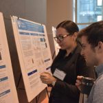
The annual Bioengineering Graduate Symposium provides an opportunity for current bioengineering graduate students to showcase their research to faculty and peers through poster sessions and short talks.

Date: January 8th
Location: Wu & Chen Auditorium and Levine Hall Lobby
Time: Check-in opens 12:45, event starts 1 pm.

RSVP: https://goo.gl/forms/r4le7koqYnMj3W4m1

Dr. Sujata Bhatia

Keynote Speaker: Dr. Sujata Bhatia,
Professor of Chemical and Bimolecular Engineering, faculty director, McNair Scholars Program,
University of Delaware

  “Bioengineering to Alleviate the Global Burden of Disease”

Biochemical and biomedical engineers face an unprecedented opportunity to improve and save the lives of millions worldwide.  Both high-income and low-income countries are experiencing an explosion in the incidence of chronic diseases, including coronary artery disease, stroke, diabetes, and cancer.  At the same time, low-income countries continue to be plagued by infectious diseases, including HIV, tuberculosis, diarrheal disease, and pneumonia; low-income countries are often said to experience a dual burden of chronic and infectious diseases.  Innovative biomedical materials will only reach the clinic if these technologies solve pressing clinical problems.  This talk will describe opportunities for bioengineers to alleviate the global burden of disease.  The talk will additionally highlight specific examples of unmet clinical needs.  Bioengineers in industry, academia, and government can all make a difference, not only by designing novel biomedical products, but also by training the next generation of bioengineers and shaping the future direction of research and development.

Michael Mitchell Receives BMES Rising Star Award

Michael Mitchell, PhD, Skirkanich Assistant Professor of Innovation in the Department of Bioengineering at Penn, has been honored with a Rising Star Award in Cellular and Molecular Bioengineering from the Biomedical Engineering Society (BMES). According to the BMES website, “The BMES Cellular and Molecular Bioengineering Special Interest Group brings together researchers with diverse scientific and clinical interests with a common goal of understanding and engineering molecules, cells, their interactions and microenvironments in the pursuit of controlling biological processes and improving the practice of medicine.” Dr. Mitchell received the award and delivered a lecture at the 2019 Cellular and Molecular Bioengineering Conference in San Diego, California in January, 2019.

One of six early-stage investigators from across the nation to receive the honor, Dr. Mitchell was recognized for his work on engineering delivery technologies for cancer gene therapy and immunotherapy, which is helping to lay the foundation for a new class of therapeutic strategies against hematologic cancers such as multiple myeloma and leukemia. In 2018, Dr. Mitchell was awarded the NIH Director’s New Innovator Award for this research, and received the Burroughs Wellcome Fund Career Award at the Scientific Interface) in 2016. He joined the Penn faculty in January 2018 after completing an NIH NCI postdoctoral fellowship with Dr. Robert Langer at the Koch Institute for Integrative Cancer Research at MIT.

BE Seminar Series: November 29th

The BE Seminar Series continues this week. We hope to see you there!

Speaker: Alison Pouch, Ph.D.
Research Associate, Penn Image Computing & Science Lab, Department of Radiology
Gorman Cardiovascular Research Group, Department of Surgery, University of Pennsylvania

Date: November 29, 2018
Time: 12:00 pm
Location: Room 337, Towne Building

“Shaping Innovation in Heart Valve Surgery with Image-Based Modeling”

A number of challenging questions routinely arise before heart valve surgery. Should a regurgitant valve be repaired or replaced? If repaired, which techniques and maneuvers should be employed? When is the optimal time to perform surgery? This is just the beginning of a series of questions whose answers rely on an individualized assessment of heart valve morphology and function. With advanced imaging technology like 3D echocardiography already in the operating room, we have a window into patient-specific disease characteristics that cannot otherwise be appreciated immediately before surgery. Unfortunately, imaging resources are often not tapped to their full potential, which contributes to delays in surgical innovation. This presentation introduces an image-based modeling approach to creating enhanced visualizations and quantification of heart valves from real-time 3D echocardiography. The goal of this approach is to fundamentally change the way that surgeons interact with and utilize images in the operating room. Pre-operative image-based heart valve models can be used to characterize mechanisms of disease prior to cardiopulmonary bypass and to identify connections between pre-operative image features and clinical outcomes. Image-based modeling provides a means for surgeons to innovate and master new surgical techniques like bicuspid aortic valve repair and to devise new standardized approaches for surgical treatment of ischemic mitral regurgitation. Image analysis and surgical innovation are linked within and beyond the cardiovascular domain, and this work aims to optimize the potential of imaging and shape modeling for pre-surgical planning.