Late last semester, Penn Bioengineering Department chair David Meaney and senior lecturer LeAnn Dourte held a second roundtable with BE undergrads Eric Helfgott, Joseph Maggiore, Kayla Prezelski, and Margaret Schroeder. They picked up on topics from the last roundtable, extending the topics to balancing an engineering workload and other commitments.
One of the key processes in embryonic development and growth through childhood and adolescence is that of how tissue folds into the specific shapes required for them to function in the body. For instance, mesenchymal stem cells, which form a variety of tissues including bones, muscles, and fat, are required to “know” what shapes to take on as they form organ systems and other structures. Therefore, a big concern in tissue engineering is determining how to control these processes of tissue folding.
Just in time for his arrival at Penn Bioengineering, Dr. Alex Hughes, a new assistant professor in the department, is the lead author on a new paper in Developmental Cell that explores this concern. The study was coauthored with Dr. Zev Gartner of the University of California, Berkeley, where Dr. Hughes just finished a postdoctoral fellowship. In the paper, the authors used three-dimensional cell-patterning techniques, embryonic tissue explants, and finite element modeling to determine that the folding process involves the interaction of a protein called myosin II with the extracellular matrix, itself the molecular material that provides a structural framework for developing tissues. With the knowledge gained in the initial experiments, the authors were then able to reproduce the tissue folding process in the lab.
“Bioengineers are currently thinking about building tissues,” Dr. Hughes says, “not just at the level of organoids, but at the level of organs in the body. One of my interests at Penn is to harness developmental principles that link these length scales, allowing us to design medically relevant scaffolds and machines.”
Equipped with the knowledge gleaned from this research, future studies could contribute further to the ability to generate tissues and even organ systems in laboratories. Ultimately, this knowledge could revolutionize transplant medicine, as well as variety of other fields.
Dr. Konrad Kording, a University of Pennsylvania PIK Professor in Bioengineering and Neuroscience, has been named an associate fellow by the Canadian Institute for Advanced Research (CIFAR), an advanced study institute headquartered in Toronto and partially funded by the government of Canada. Dr. Kording’s fellowship is in the institute’s Learning in Machines & Brains area, which has been one of CIFAR’s 14 interdisciplinary study fields since 2004. He joins 32 other fellows currently supported by the institute for their work in this area.
“The CIFAR program in Learning in Machines & Brains brings together many of the world’s leading deep learning scientists,” Dr. Kording says. “I look forward to collaborate with them to figure out how the brain learns.”
CIFAR was founded in 1982. Over the last 35 years, the institute has supported the work of scientists in 133 countries, including 18 Nobel Prize laureates.
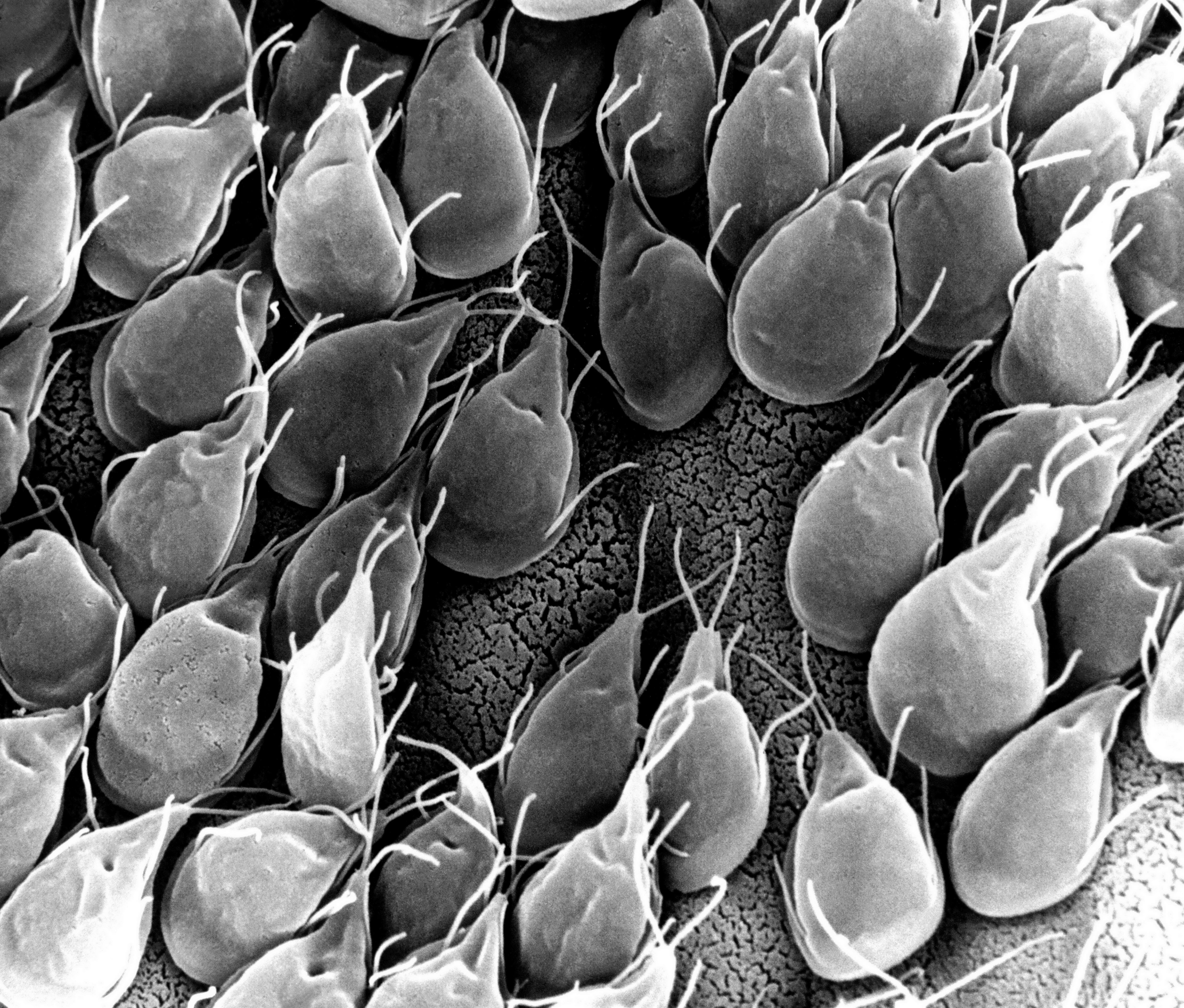
Small intestinal mucosa infested with Giardia lamblia parasites
Diseases of the small intestine, including Crohn’s disease and microbial infections, impose a huge burden on health. However, finding treatments for these diseases is challenged by the lack of optimal models for studying disease. Animal models are only so close to human disease states, and laboratory models using cell lines do not completely mimic the environment inside the gut.
However, these limitations might be overcome soon thanks to the research of scientists at Tufts University. In an article recently published in PLOS ONE, a team led by David L. Kaplan, Ph.D., of the Tufts Department of Biomedical Engineering, describes how they used donor stem cells and a compartmentalized biomimetic scaffold to model and generate small intestine cells that could differentiate into the broad variety of cell types common to that organ.
The study team tested the response of its cell model to E. coli, a common pathogen. At the genetic level, the model matched the reaction of the human small intestine when exposed to this bacterium. The success of the model could translate into its use in the near future to better understand the digestive system’s response to infection, as well as to test individualized treatments for inflammatory bowel diseases such as Crohn’s.
Saving Battle-wounded Eyes
The increase in combat survival rates has led to a higher incidence of veterans with permanent vision loss due to catastrophic damage to the eye. Globe injuries will recover of some vision, if caught in time. However, combat care for eye injuries often occurs hundreds or thousands of miles away from emergency rooms with attending ophthalmologists. With this unavoidable delay in treatment, people with globe injuries suffer blindness and often enucleation.
However, battle medics might soon have something in their arsenals to prevent such blinding injuries immediately in the combat theater. As reported recently in Science Translational Medicine, engineers at the University of Southern California (USC) and ophthalmologists from USC’s Roski Eye Institute have collaborated in creating a new material for temporary sealing of globe injuries. The study authors, led by John J. Whalen, III, Ph.D., used a gel called poly(N-isopropylacrylamide) (PNIPAM), already under investigation for treating retinal injuries. PNIPAM is a thermoresponsive sealant, meaning it is a liquid at cooler temperature but an adhesive gel at warmer temperature. These interesting properties mean PNIPAM can be applied as a liquid and then solidifies quickly on the eye. The authors manipulated PNIPAM chemically to make it more stable at body temperature. As envisioned, the gel, when used with globe injuries, could be applied by medics and then removed with cold water just before the eye is treated.
The study team has tested the gel in rabbits, where it showed statistically significant improvement in wound sealing and no negative effects on the eyes or overall health of the rabbits. The authors believe the material will be ready for human testing in 2019.
Predicting Seizures in Epilepsy
Epilepsy is a central nervous system disorder characterized by seizure activity that can range in severity from mild to debilitating. Many patients with epilepsy experience adequate control of seizures with medications; however, about a third of epileptic patients have intractable cases requiring surgery or other invasive procedures.
In what could be a breakthrough in the treatment of refractive epilepsy, scientists from Australia in collaboration with IBM Research-Australia have used big data from epilepsy patients to develop a computer model that can predict when seizures will occur. So far, the technology predicts 69% of seizures in patients. While it’s still short of a range of accuracy making it feasible for use in patients outside of experimental settings, the acquisition of ever-increasing amounts of data will render the model more accurately.
The Art of Genetic Engineering
Among the techniques used in genetic engineering is protein folding, which is one of the naturally occurring processes that DNA undergoes as it takes on three dimensions. Among the major developments in genetic engineering was the discovery of the ability to fold DNA strands artificially, in a process called DNA origami.
Now, as suggested by the name “origami,” some people have begun using the process in quasi-artistic fashion. In an article recently published in Nature, bioengineers at CalTech led by Lulu Qian, Ph.D., assistant professor of bioengineering, showed they were able to produce a variety of shapes and designs using DNA origami, including a nanoscale replica of Leonardo da Vinci’s Mona Lisa.
DNA now also has another unique artistic application — tattoos, although people’s opinions of whether tattooing constitutes art might vary. Edith Mathiowitz, Ph.D., of Brown University’s Center for Biomedical Engineering, is among the patenters of Everence, a technology that takes DNA provided by a customer and incorporates it into tattoo ink. Potential tattooees can now have the DNA of loved ones incorporated into their bodies permanently, if they should so wish.
People and Places
The University of Washington has launched its new Institute for Nano-engineered Systems, cutting the ribbon on the building on December 4. The center will house facilities dedicated to scalable nanomanufacturing and integrated photonics, among others. Meanwhile, at the University of Chicago, Rama Ranganathan, M.D., Ph.D., a professor in the Department of Biochemistry and Molecular Biology and the Institute for Molecular Engineering, will lead that college’s new Center for Physics of Evolving Systems. Congratulations!
This interview with Dr. LeAnn Dourte is a collaboration between Double Shelix and the University of Pennsylvania Department of Bioengineering! Thanks to Kayla and Sally for conducingt this interview! If there’s someone else at UPenn BioE (or elsewhere!) that you think they should feature, let them know!
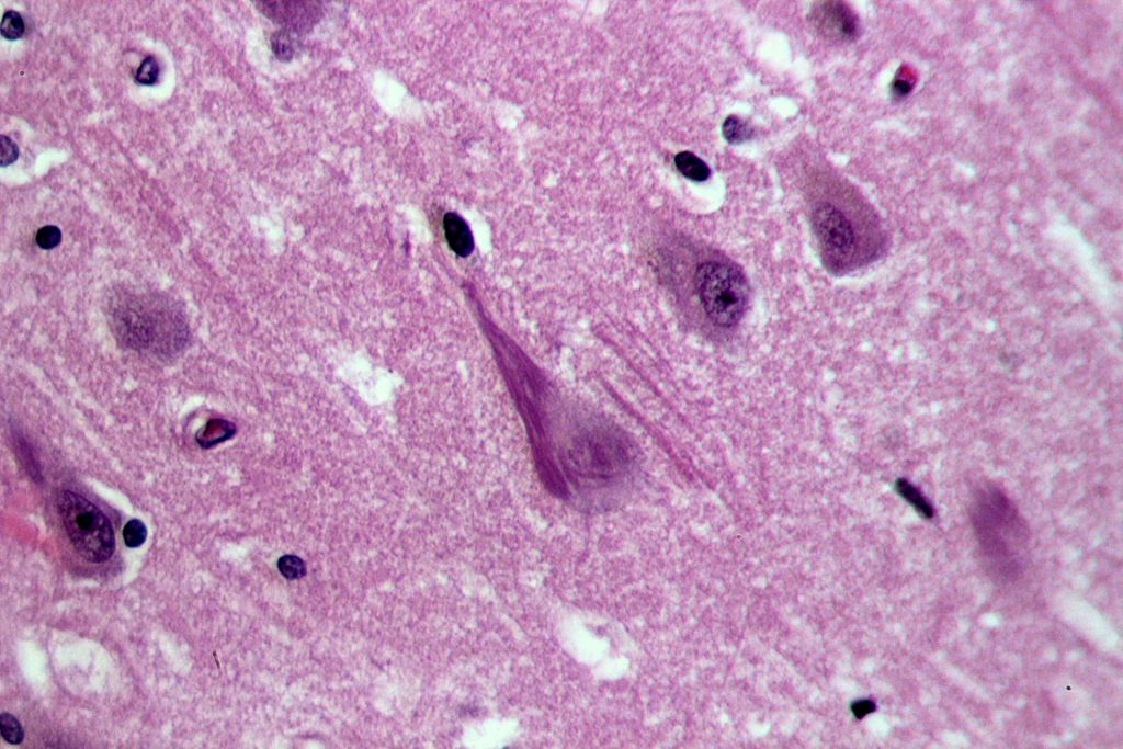
Neurofibrillary tangles in the hippocampus of a patient with Alzheimer’s disease.
The dramatic increase in life expectancy over the past couple of generations has one unfavorable consequence: an increase in the incidence of age-related dementias that include Alzheimer’s disease. Drugs like donepezil, which inhibits hydrolization of acetylcholine and thus increases its presence at the neural synapses, is one treatment that can slow the progression of these diseases, but there is currently no cure.
An alternative technology that directly stimulates the brain with an implantable chip holds promise to reverse the effects of Alzheimer’s. At the annual meeting of the Society for Neuroscience, held last month in Washington, D.C., Dong Song, Ph.D., Research Associate Professor of Biomedical Engineering at USC’s Viterbi School of Engineering, gave a lecture on his lab’s device, which uses an array of implantable electrodes to improve human memory.
Dr. Song tested his device in epilepsy patients, who often receive implants designed to control their seizures in intractable cases. Twenty such patients volunteered to receive Dr. Song’s implant, and data from these patients showed that short-term memory increased by 15% and working memory by 25%. While additional testing is needed on more patients, it might not be long before implants like Dr. Song’s become the standard of care in treatment dementias.
Genetic Variation in the Human Microbiome
The human body is host to a veritable universe of microbes that play important roles in the organ systems and other bodily processes. E. coli, for example, is present in the large intestine and it participates in the breaking down of food for energy. Like all other forms of life, these microbes evolve. creating variations in genetic information and, ultimately, new bacteria species. Within any given species of bacteria, the number of differences in the genome sequences can vary broadly; with E. coli, some areas of the genome can vary radically between strains and cannot be explained by DNA copying errors.
To determine why the genome of E. coli subject to such variation, scientists at the University of Illinois, Urbana-Champaign (UIUC), led by Sergei Maslov, Ph.D., professor of bioengineering and physics at UIUC, investigated the issue by developing computational models using Multi Locus Sequence Typing (MLST). In their findings, published in Genetics, they concluded that the variation can be ascribed to the process of recombination, by which different sequences from different sources are combined into the same chromosome. When such events are frequent, they result in a sort of genetic stability in which variation in genetic information increases without speciation.
The study provides an important contribution to basic science in helping to better explain how different strains of bacteria develop, including virulent and drug-resistant strains. In addition, it sheds further light on the mechanisms underlying evolution.
A Step Closer to Water-efficient Agriculture
Drought and famine are closely related phenomena. Some plants are more resistant to drought than others, but few of these plants are fit for human consumption. Determining how plants resist drought could provide a key to engineering crops to become drought-resistant.
Investigating this topic, scientists at the Oak Ridge National Laboratory of the U.S. Department of Energy sought to understand better the process of crassulacean acid metabolism (CAM), by which drought-resistant plants keep their stomata, or pores, closed during sunlight hours to retain water and open them at night. The team reports in Nature Communications that they compared the genomes of three drought-resistant plants — orchid, pineapple, and Kalanchoë fedtschenkoi, a species of plant native to Madagascar. Among the authors’ discoveries was a variation in a gene encoding phosphoenolpyruvate carboxylase, an enzyme that plays a role in CAM.
With this increased knowledge of the evolutionary development of drought resistance, we come a step closer to being able to expedite the evolution of plants that are typically not resistant to drought to developing the CAM mechanism and developing this resistance.
Computer Model Can Mimic Heart Attack
Heart disease remains the leading cause of death in developed countries. A major obstacle in reducing the deaths due to cardiac arrest is the inability to determine the precise mechanics unfolding in the heart when it stops suddenly. Abnormal heart rhythms (arrythmias) are a major cause of death, but the reasons how arrhythmias occur at the cellular level is poorly understood.
In a recent study published in PLOS Computational Biology, Raimond L. Winslow, Ph.D. who is Raj and Neera Singh Professor in the Department of Biomedical Engineering at Johns Hopkins University, and his colleagues developed a computer model of calcium dynamics in cardiac cells. The model predicted a new mechanism for arrythmia that would occur when cardiac cells expelled calcium, creating an electrical charge outside the cell that could evoke an arrhythmia.
The authors believe that their research will facilitate the development of drugs to prevent cardiac arrhythmias and treatments for sudden cardiac arrest. In addition, the work shows that it could be easier to predict the statistical relationship between arrhythmias and cardiac arrest on the basis of far less data.
People and Places
Stevens Institute of Technology in Hoboken, N.J., has announced plans to divide its Department of Biomedical Engineering, Chemistry and Biological Sciences (BCB) into two new departments: the Department of Biomedical Engineering and the Department of Chemistry and Chemical Biology. Hongjun Wang, Ph.D., associate professor in the BCB department, will be the new chair of BME. Congratulations Hongjun!
Pretreatment Determination of Cancer Therapy Efficacy
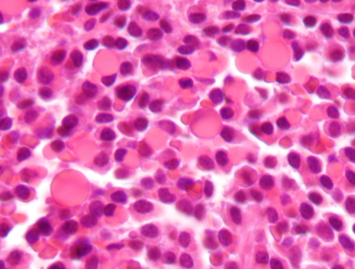
Micrography showing malignant plasma cells with Russell bodies (eosinophilic uniformly staining membrane bound bodies containing immunoglobulin).
Multiple myeloma is a type of blood cell cancer affecting the blood plasma cells. Although significant advances have improved the treatment of multiple myeloma, the 5-year survival rate remains only 50%. Among the obstacles to increasing survival is that some patients do not respond to drugs for the disease. Similarly, there are only limited ways to predict whether a patient will respond to any given drug.
However, that limitation might be a thing of the past. In a study published recently in Nature Communications, MIT engineers and scientists showed that the mass accumulation rate (MAR) of cancer cells predicted the likelihood of cancerous cells responding to specific drugs. The lead author of the study, Scott R. Manalis of MIT’s Department of Biological Engineering, coauthored the paper with scientists from the Koch Institute for Integrative Cancer Research at MIT, the Dana-Farber Cancer Institute in Boston, and Harvard Medical School.
Dr. Manalis and his colleagues found in previous studies that the MAR, which is the rate at which single cells increase in mass, was predictive of drug sensitivity. The authors used a cleverly designed device called a suspended microchannel resonator to measure the cells’ MAR — itself an impressive feat of engineering given the microscopic size of the myeloma cells. The device was used to analyze multiple myeloma cells obtained from nine patients with the disease. The authors concluded that the MAR could predict the cells’ sensitivity to standard treatment, as well as combination therapies and investigational drugs. If this technology proves effective in larger cohorts, it could significantly increase survival rates for patients with this disease.
Glaucoma Treatment Implant Could Replace Eye Drops
Glaucoma is a common eye disease in which there are abnormal increases in intraocular pressure (IOP), which is a causal predictor for damage to the optic nerve and a precursor to permanent vision loss. Luckily, there are many available treatments for this disease, many of which involve the use of eye drops. However, given the correlation between advanced age and glaucoma, ophthalmologists find that many patients are unable to administer eye drops on their own. Unless they have someone who can administer the drops for them, the patients will lose their vision.
Scientists at the University of California, San Francisco (UCSF), have made a significant advance in solving this problem. The UCSF team, led by Tejal A. Desai, Ph.D., Professor and Chair of the Department of Bioengineering and Therapeutic Sciences, developed a long-term implant for glaucoma patients using polycaprolactone, a type of biodegradable polyester, to eliminate the need for eye drops to treat glaucoma. The authors report in the Journal of Controlled Release that the device could effectively administer a glaucoma drug in rabbits over a six month period. The authors will continue testing, first in larger animals and ultimately, if all goes well, in humans.
Using Deep Learning to Develop Better Microscopes
Artificial neural networks are one type of technology used by scientists to develop machine learning — the process by which computers are designed to learn on their own without being programmed beforehand. In deep learning, a subtype of machine learning, computers process raw data to determine the characteristics they need to know, rather than being “taught.”
The applications of deep learning are potentially limitless. In one application, researchers from the Bioengineering Department at the University of California, Los Angeles (UCLA), are using deep learning to develop more accurate microscopes. Aydogan Ozcan, Ph.D., Chancellor’s Professor and HHMI Professor at UCLA, is lead author of a paper published in Optica describing how he and his colleagues created a deep neural network trained to increase resolution based on visual information. Using images obtained with a regular microscope as their initial data, their network produced significantly higher-quality images that resembled images obtained with higher-magnification lenses. Their findings show that deep learning could improve the quality of low resolution microscopy images, which could significantly enhance point of care applications.
Keeping Bioengineering Ethical
If you’re a frequent reader of this blog, you know we’ve begun producing podcasts. However, a recent podcast produced by Russ Altman, MD, PhD, Professor of Bioengineering, Genetics, Medicine and Biomedical Data Science at Stanford, caught our interest. In the podcast, Dr. Altman interviews Dr. Megan J. Palmer, a Senior Research Scholar at the Center for International Security and Cooperation at Stanford, and they discuss the security challenges faced by scientists involved in biotechnology. Enjoy!
People and Places
Two institutions announced new centers recently for areas related to bioengineering. First, the Texas Medical Center in Houston has opened its Center for Device Innovation — a collaboration between TMC and Johnson & Johnson to facilitate the development of new devices from idea to marketing. In addition, Saint Vincent College near Pittsburgh dedicated a new engineering and biomedical sciences building, the $6 million James F. Will Engineering and Biomedical Science Hall, which will house the college’s biomedical science program.
Last week, we reported on researchers at Purdue University studying how the brain processes visual data. A recent report from biomedical engineers at Washington University-St. Louis studies another intriguing aspect of brain function: how we detect and interpret sound. The popular perception is that that neurons in the brain’s auditory cortex first identify that a sound is present (introductory reaction) and then determine the sound content (secondary reaction). Dennis Barbour, MD, Ph.D., associate professor of biomedical engineering at WashU and lead author on this study, tested whether the accuracy of the information encoded during the first process was less accurate than that recorded during the second process. While animals were exposed to auditory stimuli, the activity of neurons in their auditory cortices was measured and recorded using event-related potentials and functional MRI.
Dr. Barbour refuted the popular assumption of less accuracy earlier in sound processing. The group’s data showed that neurons were equally accurate in communicating sound information regardless of whether it was an introductory or secondary reaction. Therefore, it is likely that these two reactions serve a different purpose than initially suspected. Whether this model of neuron reaction to stimuli pertains to the other sensory organs remains to be seen.
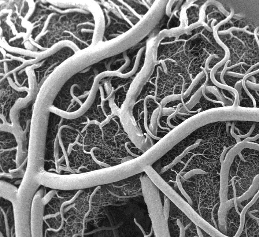
Stem Cells Regenerate Damaged Arteries
Peripheral artery disease (PAD) is one complication of diabetes, characterized by a narrowing of blood vessels in the peripheral circulation. PAD can lead to poor oxygenation of tissue in the limbs, and in the most severe cases, it can lead to limb amputation. Therefore, there is a great unmet clinical need to reverse the poor circulation caused in PAD. In a recent issue of Theranostics, mesenchymal stem cells (MSCs) were used to regrow blood vessels damaged by PAD. Led by Wawrzyniec Lawrence Dobrucki, Ph.D., professor of bioengineering and of medicine and head of the Experimental Molecular Imaging Laboratory at the Beckman Institute for Advanced Science and Technology at the University of Illinois, Urbana-Champaign, this report showed the development of new blood vessels (angiogenesis) could be accelerated by injecting MSCs into mice following limb ischemia. The authors found that angiogenesis was 80% greater than the angiogenesis in untreated animals. These changes in the blood vessel network were also matched with functional improvement, as blood perfusion increased by 42% and muscle strength by 70% in animals treated with MSCs.
The study provides additional evidence for the multiple medical applications of stem cells. Dr. Dobrucki believes the technology tested in this study could eventually be applied not only to regenerate damaged vascular tissue but also to diagnose diseases like PAD.
Saliva Test for Lupus
Blood testing provides a simple and effective way to diagnose many diseases. But what can healthcare professionals do if obtaining a blood sample isn’t possible? Children and patients who fear needles pose the biggest problems here, but collecting blood can also be difficult for patients in remote areas. To both reduce the discomfort and increase patient accessibility to diagnostic tests, there is a great interest in replacing blood-based diagnostic tests with tests using other fluids like saliva and urine. Using a grant from the National Institutes of Health, Chandra Mohan, Ph.D., Hugh Roy and Lillie Cranz Cullen Endowed Professor of biomedical engineering at the University of Houston, intends to address this issue by developing a saliva-based test for lupus, an autoimmune disorder that affects approximately 1.5 million Americans. Based on the discovery that anti-double stranded DNA antibodies appear in the blood and saliva of lupus patients, Dr. Mohan will develop and then test the new diagnostic method to evaluate the potential of replacing blood-based detection with saliva samples.
Engineering Better Plastics
Along with concerns about climate change, environmental concerns regarding pollution have been an emphasis of scientists and activists for decades. Garbage poses a particular problem because most of the plastic in garbage is not biodegradable.
In response to this environmental concern, a team of engineering students at the University of Iowa have used genetic engineering to develop sensors for biodegradable plastics. Bacteria already produce a biodegradable plastic – 3-hydroxypropionate (3HP) – that could be a replacement for the non-degradable plastics that are used in the market today. However, manufacturing 3HP is more expensive, and new production methods would be more efficient if there were a sensor available to determine 3HP amounts during the manufacturing process. The Iowa team engineered bacteria that emit light based on the 3HP present in the microenvironment. By monitoring the emitted light during the manufacture of 3HP, we could control and optimize the production of 3HP and eventually make it an affordable alternative to non-degradable plastics. The team presented its research last week at the Giant Jamboree sponsored by the International Genetically Engineered Machine Foundation in Boston.
People and Places
The University of California, Santa Barbara, opened its new bioengineering building recently. The building will house at least a dozen faculty and their research groups and both the Center for Bioengineering and the Institute for Collaborative Biotechnologies. At the University of Southern California, officials announced the creation of a new center: the USC Michelson Center for Convergent Bioscience, designed to take advantage of collaborative research teams to tackle major health problems, including cancer, infection and drug development. The center will be run by chemistry faculty member Valery Fokin and by Peter Kuhn, Ph.D., Professor of Aerospace and Mechanical Engineering & Biomedical Engineering at USC. Finally, last week, Tulane University’s Department of Biomedical Engineering celebrated its 40th anniversary. Happy anniversary, Tulane!
Last week, for our latest podcast, Penn Bioengineering Department Communications Coordinator Andrew Mathis sat down with a roundtable of five undergraduate students — Lamis Elsawah, Eric Helfgott, Joseph Maggiore, Kayla Prezelski, and Margaret Schroeder — to talk about how they chose Penn, what majoring in BE has been like so far, and other things.
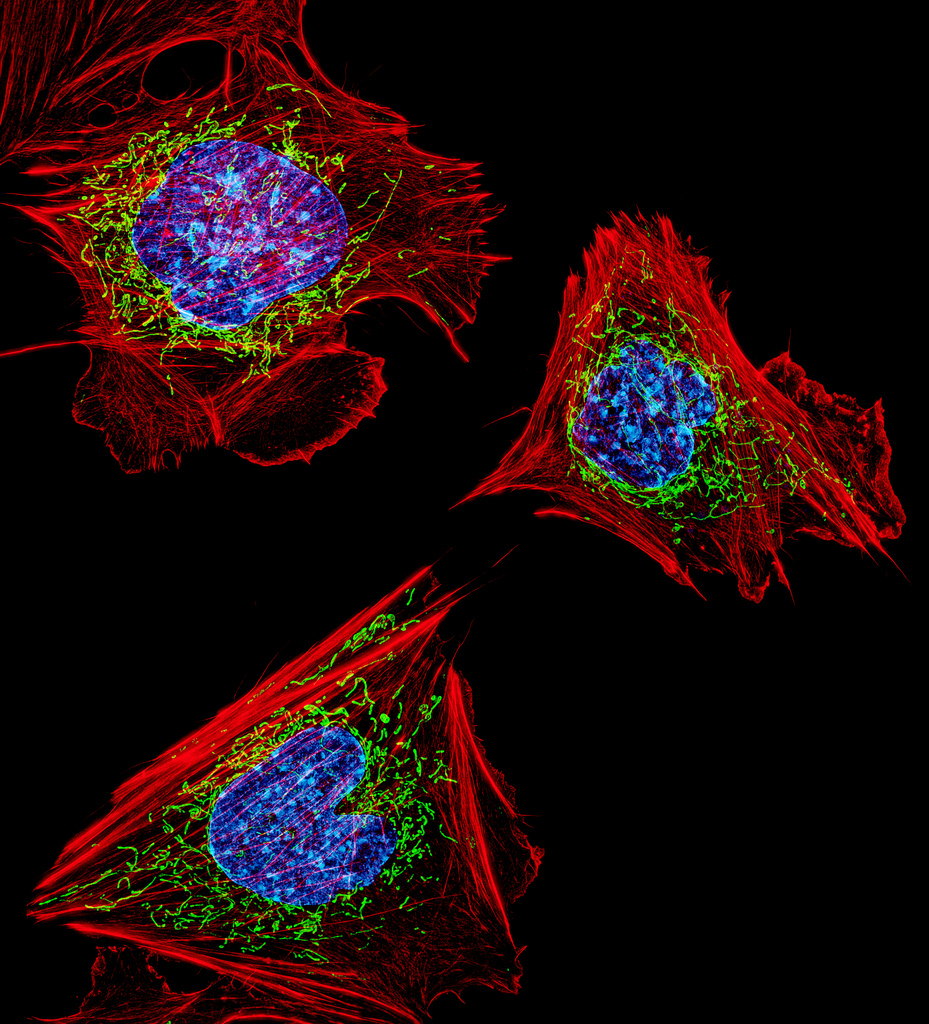
We’ve known for many years that exercise is good for you, but it was less clear how muscle strength and stamina were assembled at the molecular level. Based the principle that the health of mitochondria – a key organelle within the muscle cell – regulates muscle health, recent work identifies some of the key signaling pathways in vivo that can switch a cell between degrading damaged mitochondria or creating new mitochondria. Zhen Yan, M.D., Ph.D., of the University of Virginia, used a fluorescent reporter gene (MitoTimer) to “report back” the information for individual mitochondria in muscle cells prior to and following exercise. The results reported in a recent issue of Nature Communications show very clearly that mitochondria can switch a muscle cell’s fate. Dr. Yan’s research team identified a new signaling pathway within skeletal muscle that is essential to mitophagy. Knowledge of this pathway could help to develop a variety of therapies for diseases of the muscles or damage to the muscles due to injury.
Understanding How the Brain Processes Visual Data
As a model for how the brain “computes” the information surrounding all of us, researchers have studied how visual information is processed by the brain. One method for investigating this question is the use of artificial neural networks to recognize visual information that they have previously “seen.” A recent article in Cerebral Cortex details how a team at Purdue University, led by Zhongming Liu, Ph.D., Assistant Professor of Electrical and Computer Engineering and Biomedical Engineering, used an artificial neural network to predict and decode information obtained with functional magnetic resonance imaging (fMRI). By collecting fMRI brain activation data when people watch movies, the artificial neural network could generate feature maps that strongly resembled the objects depicted by the initial stimuli. Available now in open access format, the team at Purdue intends to repeat these experiments with more complex networks and more detailed imaging modalities
Preventing Prosthesis-related Infection
Prostheses have improved by leaps and bounds over the years, with the development of osseointegrated prostheses — which are fused directly to the existing bones — a major step in this evolution. However, these prostheses can lead to severe infections that would require the removal of the prosthesis. These problems have been seen more commonly over the last decade or so in the military, where wounded soldiers have received prostheses but suffered subsequent infections.
In a major step forward to address this issue, Mark Ehrensberger, PhD, assistant professor of biomedical engineering at SUNY Buffalo, is the principal investigator on a two-year $1.1 million grant from the Office of Naval Research in the U.S. Department of Defense, awarded for the purpose of investigating implant-related infections. Initial research by Dr. Ehrensberger, who shares the grant award with scientists from the departments of orthopaedics and microbiology and immunology, showed that delivering electrical stimulation to the site of the prosthesis could be effective. One method the team will investigate is using titanium from within the implants themselves to conduct the current to the site.
Success with this grant could mean that patients receiving prostheses show better recovery rates and much lower rates of rejection. It could also reduce the antibiotics used by such patients, which would be a welcome outcome given the increasing rates of antibiotic resistance in health care.
Bioengineering Treatments for Depression
Depression is a largely invisible illness, but it brings with it a massive burden on both the patient and society, with health care costs exceeding $200 billion per year in the U.S. alone. Different drugs are used to treat depression, but all have significant side effects. Psychotherapy also has some effectiveness, but not all patients are helped with therapy.
One promising alternative to treat depression uses transcranial magnetic stimulation, but the devices used in this treatment are often cumbersome. In response to calls to develop more accessible forms of therapy for depression, a startup company in Sweden called Flow Neuroscience has developed a wearable device that uses transcranial direct-current stimulation targeted at the left frontal lobe. The device is noninvasive and is smaller than a sun visor, and the company claims it will be relatively inexpensive (estimated at $750). Flow Neuroscience is in the process of applying for regulatory approval in the European Union.
People and Places
United Kingdom Chancellor of the Exchequer Philip Hammond has announced that the British government will provide £7 million (approximately $9.2 million) in funding to create the UK Centre for Engineering Biology, Metrology and Standards. The government is collaborating with the the Francis Crick Institute in London, with the goal of supporting startup companies in Great Britain dedicated to using engineering and the biological sciences to develop new products.
Closer to home, the Universities of Shady Grove — a partnership of nine Maryland public universities where each university provides its most heavily demanded program — have begun construction on a $162 million biomedical sciences building. The building is slated for completion in 2019 and is expected to nearly double the enrollment at Shady Grove.
Here at Penn, Adam Pardes, a current Ph.D. candidate in our own Department of Engineering, is one of the cofounders of NeuroFlow, a company developing a mobile platform to track and record biometric information obtained from wearables. NeuroFlow recently received $1.25 million in investments to continue developing its technology and ultimately bring it to market. Congratulations, Adam!
Finally, California State University, Long Beach, is our newest national BME program this fall. Burkhard Englert, Ph.D., professor and chair of the Department of Computer Engineering and Computer Science at CSULB, heads the new program as interim chair until a permanent chair is hired.

 Late last semester, Penn Bioengineering Department chair David Meaney and senior lecturer LeAnn Dourte held a second roundtable with BE undergrads Eric Helfgott, Joseph Maggiore, Kayla Prezelski, and Margaret Schroeder. They picked up on topics from the last roundtable, extending the topics to balancing an engineering workload and other commitments.