As we reported earlier, Dan Huh, Wilf Family Term Chair & Assistant Professor in the Department of Bioengineering, has been awarded a $1 million grant from the Cancer Research Institute (CRI), along with its first CRI Technology Impact Award.
Recently, the Penn Engineering Blog featured a story on Dr. Huh’s grant and the research it will support for the next three years. You can read the story at the SEAS blog.
Here’s the promised interview with new faculty member Mike Mitchell, who starts as assistant professor of bioengineering at Penn in the Spring 2017 semester. Mike and editor Andrew E. Mathis discuss Mike’s background and education, where cancer research is now and where it’s heading, and just how big the radius is on the cheesesteak zone of impact around Philadelphia.
While brainstorming and writing a proposal for a device to detect pediatric tuberculosis has been extremely valuable, we recognize the challenge of developing our devices as undergraduate/graduate students. This acknowledgement led us to try to identify a healthcare problem in Ghana and to come up with a solution that undergraduates could potentially pursue. The process began after we arrived in Ghana, with each student independently identifying a problem and brainstorming a solution. Next, we played an entrepreneurial game, in which each student gave a pitch for an idea, and everyone gave hypothetical money to his or her favorite idea. The ideas with the most hypothetical monetary investments would move on to the next round. After two rounds of pitches, we narrowed our list down to two ideas: Big Data and the Multi-Cot. Splitting up our group between the two ideas, we then prepared a presentation to give to Kumasi Center for Collaborative Research in Tropical Medicine (KCCR) researchers. Yesterday and today, we present the summaries of our ideas.
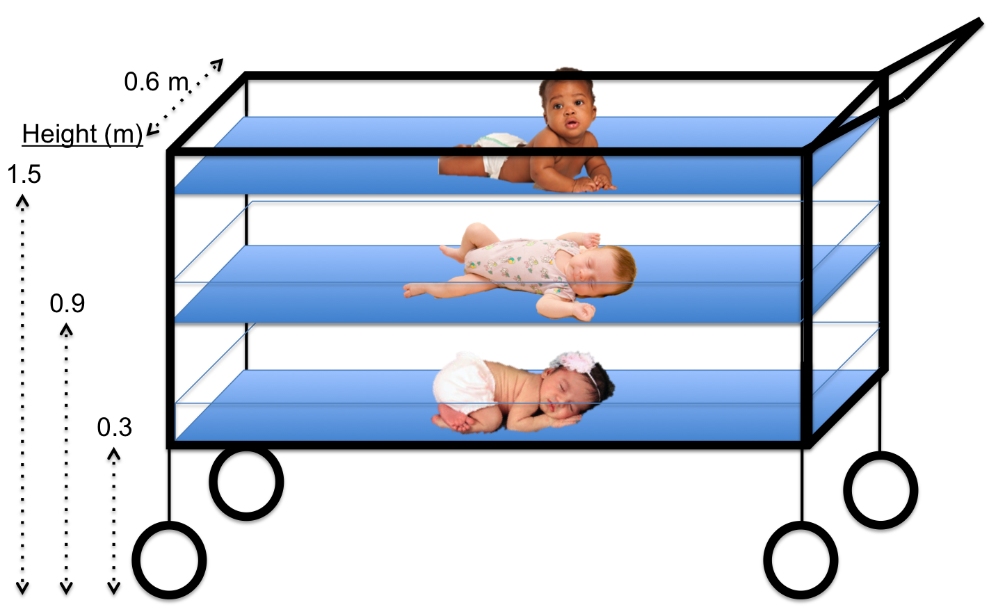
The Multi-Cot aims to tackle the issue of limited space in large regional hospitals within Ghana and other similar situations of overcrowding.
Kate Panzer (gave first-round pitch) ’18, Katharine Cocherl ’20, Kaila Helm ’20, Hope McMahon ’18
Throughout our time in Ghana, we had the opportunity to visit many hospitals and smaller health clinics. While visiting Komfo Anokye Teaching Hospital (KATH) in Kumasi, Ghana, we noticed that there was a poster on a pediatrician’s wall for the “One Baby One Cot” initiative. We soon learned that there is very limited space per patient at the large regional hospitals — certainly not enough space for each individual baby to occupy his or her own cot. For example, in some hospitals, there can be up to eight babies in one cot! This can be problematic when trying to prevent the spread of infection but also difficult for mothers who have little to no space to watch over their newborns when they stay at the hospital to breastfeed.
There are several implications of having multiple babies in a single cot that we would like to address. First, the risk of hospital-acquired infections greatly increases because of the close contact of the babies. This close contact also makes it difficult for nurses and caretakers to monitor each baby. In addition, many babies may need to be transported to other hospitals because of a lack of bed space, moving the patients and their caretakers farther from home.
The horizontal sliding mechanism of the Multi-Cot allows each newborn to be safely removed from the structure, regardless of the cot level.
After learning about this problem, we began thinking of ways to decrease the complications associated with having multiple newborns in one cot. During the brainstorming session, the key element that led to our solution was actually how we view the problem. We started to see the issue as a lack of horizontal space – meaning the inability to add more cots horizontally without physically expanding the newborn ward. If expanding the horizontal space is not possible, then why not try to make better use of the vertical space that is already available? This concept of vertical space led us to the idea of the Multi-Cot, which involves three smaller newborn cots stacked vertically, with space between each cot to provide proper airflow. With clear plastic sides and an open top, each baby would be easily seen from every direction. Finally, to ensure safety when removing newborns from the lower levels, we added a sliding mechanism to our design to allow the lower cots to slide horizontally and eliminate any vertical obstructions when picking up the baby.
As we anticipate developing the Multi-Cot, we must consider multiple factors. Our main consideration is safety, which includes the Multi-Cot’s stability, the visibility of every child, and the ability to be sanitized. Other factors to be considered include the cost, as well as the ease of physical construction and dismantling; however, we would address these details later in the design process.
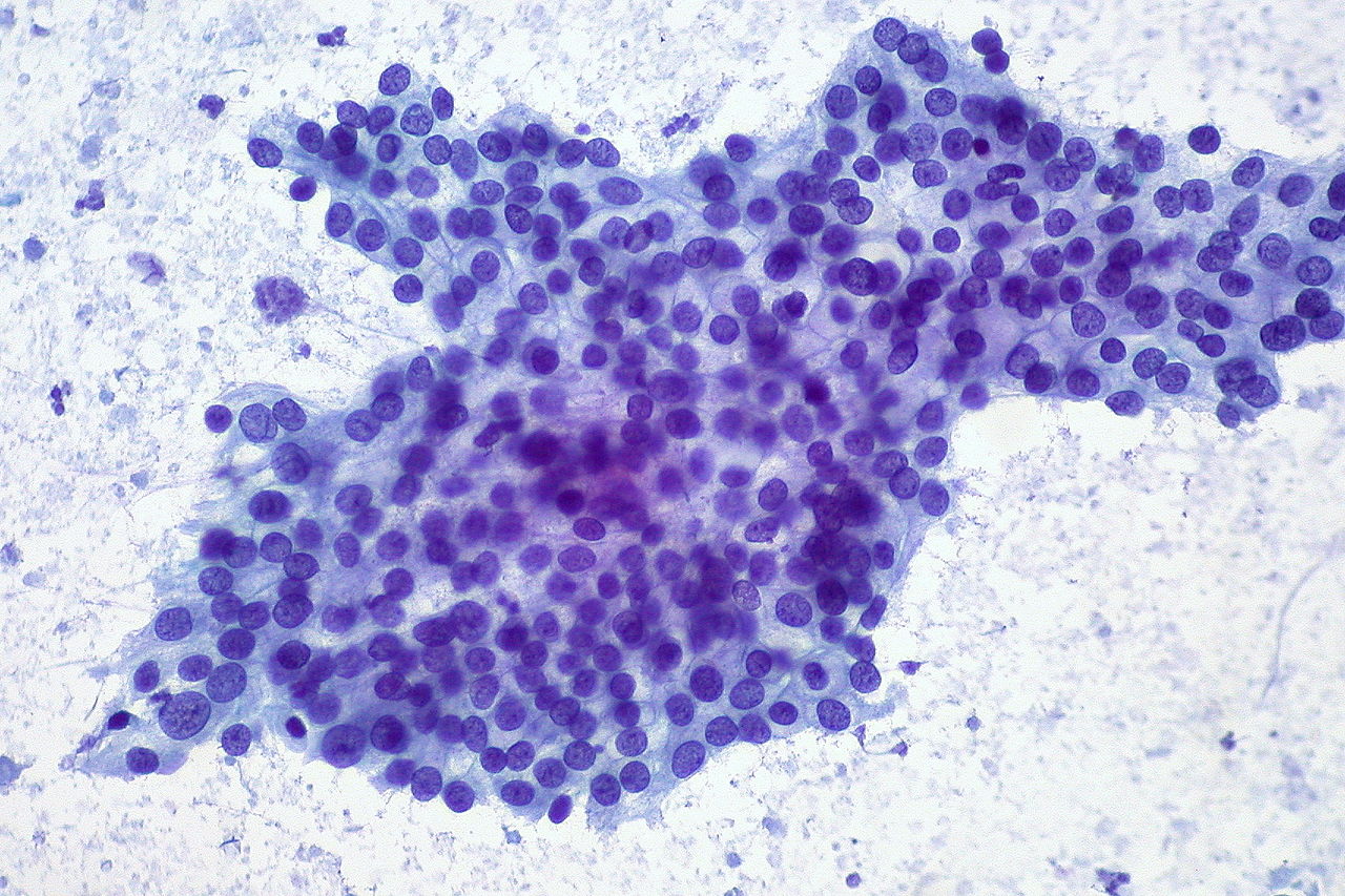
Adenocarcinoma of the pancreas, stained and viewed under a microscope.
How smartphones reveal the world of physical activity
We all know lack of exercise adversely affects our health. Policy experts often cite that exercise is such an infrequent part of people’s lives that it now constitutes a public health crisis. However, we never had a global view on how physical activity differs among all of us.
In an article recently published in Nature, scientists used smartphones to collect data on how much variation occurs in the daily activity of people from more than 100 countries. Bioengineering professor Scott Delp and his team from Stanford used smartphones to collect 68 million days of physical activity from 717,527 people living in 111 countries.
Some of the conclusions made intuitive sense, which is always good when dealing with large data sets. For example, inactivity was strongly predictive of obesity, and this correlation was stronger in women than in men. In addition, people walked more in countries where the terrain was easier to navigate. When people were more physically active in a country, the differences in obesity rates between men and women decreased. All of this means that if it is easier to walk, people will walk, and the gender disparity in activity will shrink. Geographic data also produced interesting findings, with East Asian countries (China and Japan) walking the most, and countries near the equator walking less. Perhaps the most significant finding is the importance of the activity gap within a country’s population, defined as the difference between highly active and inactive individuals within a country. The larger the gap, the higher likelihood for high obesity rates. Unfortunately, the US ranks among the highest in activity gap among its population and, in turn, among the highest in national obesity rates.
While we all ponder little tricks to make us walk more (at Penn we schedule classes across campus to make the professors get up at least once a day), other work is making the task of climbing stairs easier. Reporting in PLOS One, Lena Ting and her team from Georgia Tech developed energy-conserving stairs, using springs that store and release energy when the user ascends. Their design means that someone can save 40% energy going up their flight of stairs when compared to the traditional design. Innovations like these could be a real help to people recovering from surgeries or with age-related joint deterioration.
Networking a human
As we start to unleash the power of smartphones on health and wellness, many predict the next disruption is networking inexpensive monitoring technologies together for a single person via their smartphone. One main benefit of creating a ‘networked human’ is to monitor an individual continuously for the earliest signs of health trouble, rather than waiting for the individual to experience a significant health episode (e.g., heart attack) and unleash the powerful (but expensive) army of technologies and people for saving their life. A recent symposium was held at Northwestern University discuss the future of wearable electronics in this future. Inevitably, this will evolve the Internet of Things (IoT) into the ‘Internet of Me’ for health technologies. John Rogers of Northwestern gave a presentation showing the wearable wireless electronics he developed to monitor bodily functions in babies. The adhesive devices, which resemble temporary tattoos, are far more comfortable than many monitoring devices. Other presentations at the symposium showcased technologies for monitoring concussion, cellphone apps to facilitate psychotherapy, and more intuitive touch screens for electronics.
A Blood Test for Early Pancreatic Cancer
Pancreatic cancer is one of the deadliest cancers because it is usually only detected after it has become too advanced to treat efficaciously. However, a collaboration between Penn and Mayo Clinic scientists may have made a key advance in mitigating this threat.
A team led by Kenneth S. Zaret, Ph.D., of Penn’s Institute for Regenerative Medicine reports in Science Translational Medicine that they were able to identify thrombospondin-2, a protein, as a biomarker of pancreatic cancer. Most impressively, plasma measurements of the protein detected cancer in patients in stage I of the disease, when it can still be treated surgically. The predictive power of the biomarker test increased significantly when combined with measurements of a previously identified marker, cancer antigen 19-9, to detect pancreatic cancer at a much earlier stage.
People in the News
Our colleagues at Carnegie Mellon named a new chair of their Department of Biomedical Engineering: Bin He, Ph.D.. His appointment begins February 1, 2018. Closer to home, the Rutgers Biomedical Engineering named David I. Shreiber as its new chair. Dr. Shreiber earned his Ph.D. in Bioengineering from Penn in 1998. Congratulations to Drs. He and Shreiber!
Speaking of Penn alums, we’d like to congratulate Dr. Spencer Szczesny, who was hired as a new assistant professor at Penn State to start in the fall 2017 semester. We’re very proud of Spencer and wish him the best of luck.
Last not but not least, if you’ve flown in or out of Washington’s Dulles Airport recently, you might have seen the exhibit Life: Magnified, selections of which are available online. One of the images featured, showing skin cancer cells connected by actin, a normally occurring protein that also facilitates cancer metastasis, was created by Dr. Catherine Galbraith, who earned her BS (1986) and MS (1985) in Penn Bioengineering. Congratulations to Cathy for such wonderful visibility!
While brainstorming and writing a proposal for a device to detect pediatric tuberculosis has been extremely valuable, we recognize the challenge of developing our devices as undergraduate/graduate students. This acknowledgement led us to try to identify a healthcare problem in Ghana and to come up with a solution that undergraduates could potentially pursue. The process began after we arrived in Ghana, with each student independently identifying a problem and brainstorming a solution. Next, we played an entrepreneurial game, in which each student gave a pitch for an idea, and everyone gave hypothetical money to his or her favorite idea. The ideas with the most hypothetical monetary investments would move on to the next round. After two rounds of pitches, we narrowed our list down to two ideas: Big Data and the Multi-Cot. Splitting up our group between the two ideas, we then prepared a presentation to give to Kumasi Center for Collaborative Research in Tropical Medicine (KCCR) researchers. Today and Friday we present the summaries of our ideas.
Big Data: Deciphering Acoustic Trends in Tuberculosis, Pneumonia and Healthy Coughs
David Pontoriero (gave first-round pitch) ’18, Kathleen Givan ’20, Jason Grosz ’19, Danielle Tsougarakis ’20, Ethan Zhao ’19
Our goal was to think of a project that a team of undergraduates at Penn could complete in one year to produce something of value to KCCR in the scope of Ghanaian healthcare. We turned our attention toward big data science and the difficulties in tuberculosis diagnosis. One of the difficulties identified was the lack of diagnostic tools in more remote arms of the healthcare system. This lack leads to unnecessary and numerous referrals to larger care centers, inconveniencing the patient and placing a burden on the efficiency of the healthcare system.
Specifically, the only standard-of-care diagnostic ubiquitous throughout all clinics was patient-reported symptoms — the most notable of which is prolonged coughing. Moreover, this symptom can often be confused with asthma or pneumonia. However, asthma involves bronchial constriction, and TB and pneumonia have different sputum distribution profiles. We theorized that this difference would correlate with differentiated sound profiles for patient coughs or baseline breathing and, subsequently, measurable biomarkers. The idea proposed was that, if blind data could be collected from KCCR with sound recordings of patients coughing and breathing, along with their demographics and final diagnoses, then analyses could be run to produce an algorithm capable of differentiating between each cough or breath. This algorithm could then be extended to a phone app that could be used to more empirically diagnose patients in any setting and increase overall healthcare efficiency.
Uncertainty is part of life, but the underlying neuroscience of how we make decisions under conditions of uncertainty is only beginning to be understood. In a paper published Monday by Nature Human Behaviour, new Penn Bioengineering faculty member and Penn Integrates Knowledge Professor Konrad Kording, Ph.D., and his coauthor, Iris Vilares, Ph.D., of University College London, offer additional evidence that dopamine lies at the heart of how the brain operates when there is a lack of certainty.
Drs. Kording and Vilares devised a simple computerized test that examined the extent to which test takers relied on previous knowledge vs. what they saw at the present moment. They then administered the test to a cohort of patients with Parkinson’s disease, a condition associated with depleted dopamine levels. The patients were tested both while taking dopaminergic medication and while off it. They found that dopaminergic medication caused the patients to pay greater attention to sensory (i.e., visual) information — an effect that diminished as the patients learned. Ultimately, the study provided evidence that dopamine levels were related to the tendency to rely on new information, also called likelihood uncertainty.
“Scientists believe that understanding uncertainty is key to understanding how the brain computes,” Dr. Kording says. “There are many theories in this space. We provide fairly clean evidence for one of them, which is that dopamine encodes likelihood uncertainty. This information could change the way people think about the manner in which the brain deals with uncertainty.”
Throughout the Spring 2017 semester, our professor, Dr. David Issadore, taught us (a class of eight undergraduates students and one graduate student) about microfluidics and point-of-care diagnostics. The next phase of the course was to come up with a new diagnostic for pediatric tuberculosis. At the end of the semester, our final assignments included submitting an NIH Research Project Grant (R01) proposal and giving a 20-minute presentation for our devices. These assignments greatly prepared us for our trip to Ghana, as we were able to ask questions and get feedback on our proposed devices by speaking to healthcare professionals at Ghanaian hospitals, clinics, and research facilities. The semester course was mainly focused on the technical design of our devices, which enabled us to hone in on the practical and real-world implementation of the devices while in Ghana. This week, the BE Blog will publish our summaries.
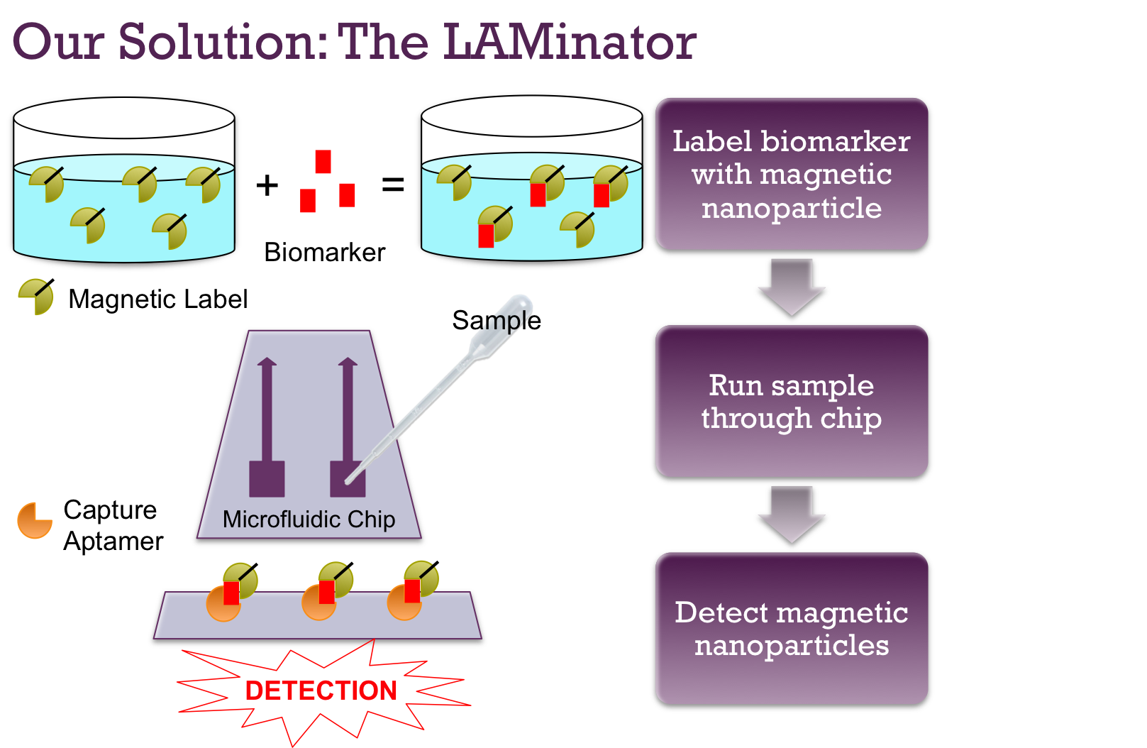
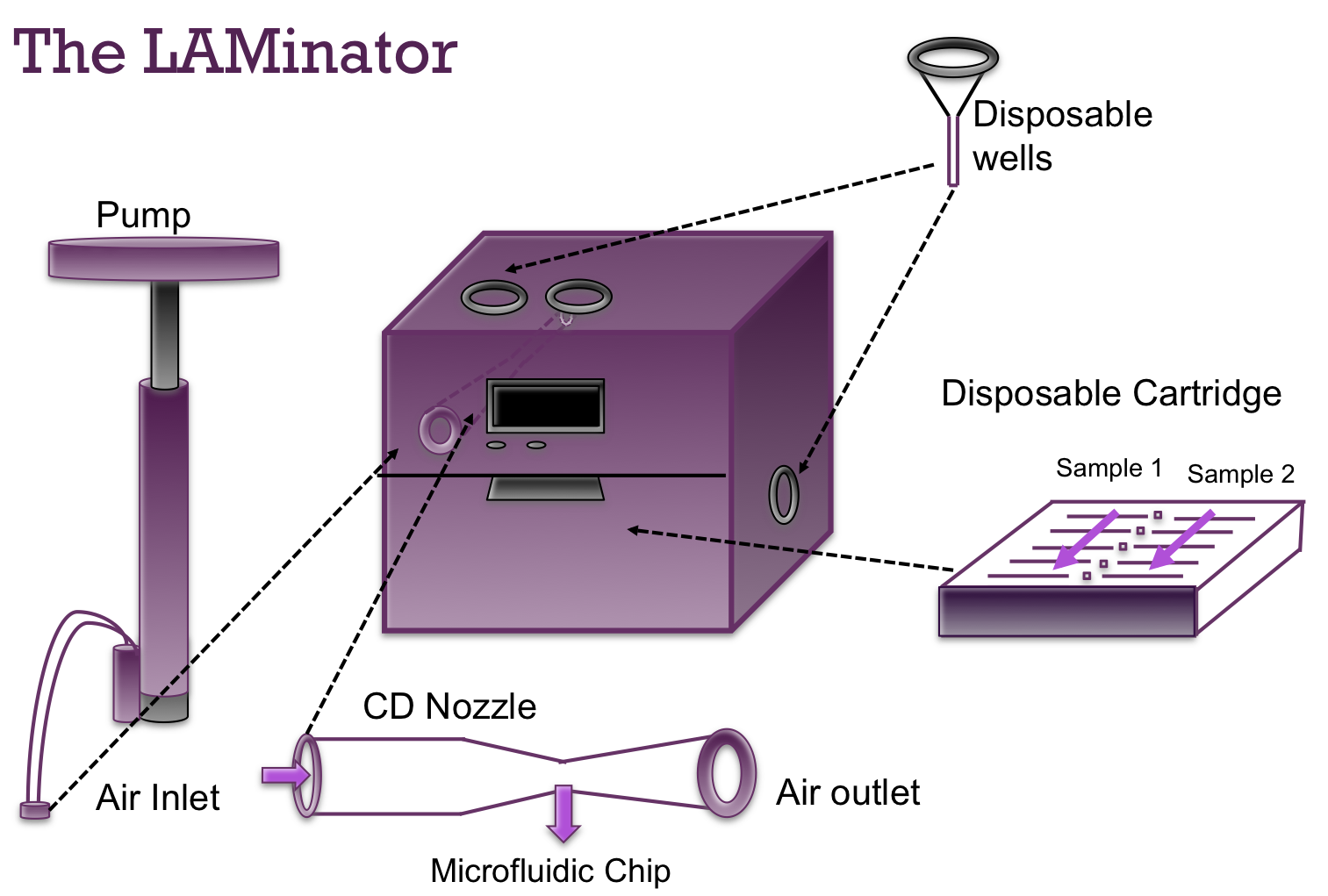
The LAMinator: Urine Diagnostic for Pediatric Tuberculosis
Danielle Tsougarakis ’20, Ethan Zhao ’19, Jason Grosz ’19, Kate Panzer ’18
Current devices that detect Mycobacterium tuberculosis include chest X-ray, smear microscopy, and GeneXpert. Although the combination of these techniques can lead to a proper diagnosis for adults, there are three main limitations of their use: (1) necessary infrastructure; (2) required sputum samples; and (3) time. First, many clinics in rural Ghana do not currently have the infrastructure or electricity sources to support these machines. Second, both smear microscopy and the GeneXpert rely on analyzing sputum samples (bacteria-containing phlegm), but children have difficulty providing sufficient samples. Finally, since sputum samples are best taken in the morning, these techniques often require patients to go home and return the next day to provide a sample.
Since all biological molecules are inherently non-magnetic, these magnetic nanoparticles can be attached to ManLAM using aptamers to allow for detection by the spin-valve sensor.
To address these limitations in our own design, we proposed a diagnostic device that does not require electricity, relies on a urine sample instead of a sputum sample, and is anticipated to take one hour to obtain a diagnosis. By incorporating these three characteristics, we propose a device that can be used to more easily diagnose children during their first initial visit at any healthcare facility in Ghana.
This overview of our device shows how the biomarker will be magnetically labeled, pushed through microfluidic channels, captured on the surface, and detected by the spin-valve sensor.
After doing a literature search of publications on pediatric tuberculosis, we learned that M. tuberculosis sheds a glycolipid called lipoarabinomannan (ManLAM) that is excreted in the urine. Therefore, ManLAM is the biomarker we hope to detect. Next, after learning that biology is inherently nonmagnetic, we figured that we could detect ManLAM specifically and sensitively if we could label it magnetically. Our proposed design does this labeling by adding magnetic nanoparticles (MNPs) to the ManLAM. This magnetic labeling involves aptamers, which are synthetic oligonucleotides that can be created to bind to a specific target. By combining the MNPs with aptamers that bind only to ManLAM, we can ultimately give the urine biomarker a magnetic property.
The LAMinator has a reusable box component to house the electronics as well as a disposable cartridge to hold the microfluidic chip and disposable wells to avoid sample contamination.
Therefore, the first step of our device is treating the urine sample with the custom aptamer-bound MNPs. The electronic components of our diagnostic device consist of specialized sensors, called spin-valve sensors, that can detect the presence of magnetic particles. Small fluid channels containing the urine sample traverse the surface of these sensors. If ManLAM is present in the urine as it passes by the spin-valve sensors, the surface-bound aptamers bind to the magnetically labeled ManLAM and capture them on the surface. The presence of these magnetic particles activates the spin-valve sensors and produces a change in voltage that can be detected by computer-like microprocessors. If ManLAM is not in the sample, then nothing will bind to the capture aptamers and no TB will be detected.
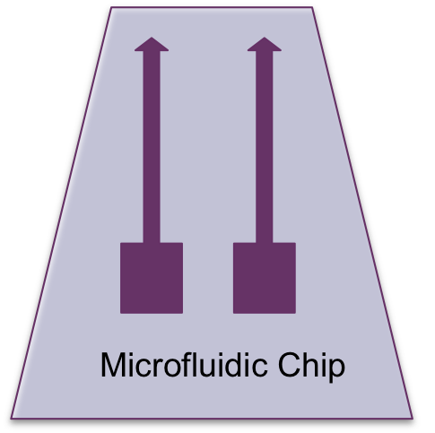
The microfluidic chip design has two channels to allow for two urine samples to be analyzed at the same time.
We would like to thank Penn Engineering and everyone who has helped to make this program possible. As you can see from our blog posts, our time in the classroom and the month in Ghana have been an unforgettable academic and cultural experience. The APOC program has been an amazing opportunity to get out of our comfort zones and to see the potential of engineering solutions in the world around us.
Throughout the Spring 2017 semester, our professor, Dr. David Issadore, taught us (a class of eight undergraduates students and one graduate student) about microfluidics and point-of-care diagnostics. The next phase of the course was to come up with a new diagnostic for pediatric tuberculosis. At the end of the semester, our final assignments included submitting an NIH Research Project Grant (R01) proposal and giving a 20-minute presentation for our devices. These assignments greatly prepared us for our trip to Ghana, as we were able to ask questions and get feedback on our proposed devices by speaking to healthcare professionals at Ghanaian hospitals, clinics, and research facilities. The semester course was mainly focused on the technical design of our devices, which enabled us to hone in on the practical and real-world implementation of the devices while in Ghana. This week, the BE Blog will publish our summaries.
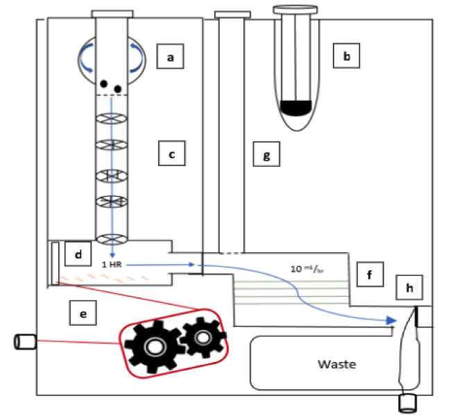
In order to address the numerous diagnostic problems specific to pediatric tuberculosis in low-resource settings, we have designed a device that uses a fecal sample rather than the current method of sputum samples. Because many children cannot produce sputum samples with the required quality and quantity of sputum, we decided to use stool samples. This noninvasive substitute will ideally allow us to collect all the bacteria swallowed by the patient. The bacterium that causes the disease, Mycobacterium tuberculosis (MTB), is very hardy and has been found to appear in fecal matter. However, this method may be difficult because there are many other substances in fecal matter that need to be removed. By filtering out these impurities, the presence of the bacteria can be detected.
The device we designed is essentially a disposable cartridge that separates virulent TB bacteria from all other fecal material. This collection can be performed with no power and minimal technician input and can be obtained in any desired volume. The total operation time is predicted to be 90 minutes.
The figure shows an overview of the steps (a-h) for use of the fecal diagnostic for pediatric TB (click to enlarge).
The first aim of our project is to identify a target protein on the surface of the bacteria so that the bacteria can be isolated from the solution. Next, the MTB will be enriched from fecal samples with a single-use filtration device so that a final sample can be provided in a similar form as a sputum sample. This final sample can then be used for smear microscopy, in which technicians look for the presence of the bacteria under a microscope, or for use with the GeneXpert. The GeneXpert is an automated diagnostic test that can identify MTB DNA and resistance to the most potent TB drug, rifampicin. These devices have been distributed to labs and hospitals across Ghana, but they are not yet widely used for general diagnostics.
Because the number of GeneXperts available and the infrastructure supporting them are increasing, we are hopeful that, in the near future, our diagnostic will be able to be used in conjunction with this technology. Upon integration with the GeneXpert system, our device would be able to increase sample specificity for the underserved demographic of pediatric TB patients. In addition, as technology becomes available in smaller, more local clinics, we foresee lower travel burdens for families and lower operational costs for healthcare facilities.
We are beyond grateful for the opportunity to engage with Ghana’s medical system. Before traveling to Ghana, we created a proposal for our fecal diagnostic for pediatric TB. After learning more about the current medical system and infrastructure in place, we were able to revise our ideas in a meaningful way. It is our hope that one day a project of this magnitude can come to fruition.
Dennis E. Discher, Ph.D., Robert D. Bent Professor in the Department of Chemical and Biomolecular Engineering and a secondary faculty member in the Department of Bioengineering, was the lead author on a recent study that showed that engineered macrophages (a type of immune cell) could be injected into mice, circulate through their bodies, and invade solid tumors in the mice, engulfing human cancers cells in the tumors.
According to Cory Alvey, a graduate student in pharmacology who works in Professor Discher’s lab and the first author on the paper, said, “Combined with cancer-specific targeting antibodies, these engineered macrophages swarm into solid tumors and rapidly drive regression of human tumors without any measurable toxicity.”
Abdominal surgery can lead to complications when the intestines are accidentally damaged. One key point in the surgery is during wound closure, when the surgeon must place the final sutures without knowing where the underlying intestine is located. A new material designed by bioengineers can be inserted into the abdomen and protect the intestines from perforations by the surgical needle. Key features of this material include its flexibility to fit into the small incision made during laprascopy and its ability to dissolve naturally within hours upon insertion into the abdominal cavity. Before it dissolves, the material is tough enough to protect the intestines from puncture, allowing the surgeon to close the incision with much less risk of perforation. So the material is ready when it is needed and disappears soon thereafter.
New materials appear frequently to perform the functions of naturally occurring biological tissues. For decades, several researchers attempted to re-create cartilage outside of the body. Although these artificial cartilage tissues may contain all of the right ‘ingredients’ — i.e., molecules and cells — the tissue is commonly not strong enough to withstand the forces normally experienced by the target tissue. Recently, researchers invented a process to load the cartilage tissue surrogate while it was fabricated, a departure from the traditional process in which the tissue substitute is mechanically loaded after it is built. Both techniques are designed to make the artificial tissue stronger. However, this subtle new design step to mechanically load during fabrication makes the cartilage substitute six times stronger than any existing manufacturing technique, raising the possibility that we can build tissue outside the body for use inside the body.
An emu egg
Finally, the field is constantly discovering new ways to use historical observations in science. One example was an observation from the analysis of 19,000-year-old DNA from an emu egg, made possible because the DNA was protected from degradation by the calcified material present in the eggshell. This scientific observation to understand the origin of the species inspired bioengineer Bill Murphy at the University of Wisconsin-Madison to create a new method to protect proteins from degradation by incorporating these proteins into mineralized materials. Reminiscent of the mineralized matrix found in the emu eggshell that protected DNA for 19,000 years, the charged mineralized matrix stabilizes the protein structure and significantly improves the stability of the protein. By designing the mineralized material to degrade slowly, this work shows that one can stabilize and release therapeutic proteins over much longer periods than previously possible.
Technological Advances in Cancer Diagnosis and Treatment
Despite tremendous advances in diagnosis and treatment, cancer remains a major public health threat. Surgery is often a key part of cancer treatment, but tumor removal is complicated by the difficulty in producing surgical margins that are free of cancer cells. If cancer cells remain in the margins, it is common for the cancer to return. However, a team of researchers at the University of Washington developed a light-sheet microscope capable of imaging these surgical margins quickly – about 30 minutes. The technology could go a long way toward reducing or eliminating the 20% to 40% of cases of breast cancer in which relapse occurs.
While breast cancer remains the most common cancer among women, proliferation of HPV has resulted in a steadily increasing rate of cervical cancer over past decades. Early screening here is a key to successful treatment, but gynecological examinations are uncomfortable for many women. Failure to schedule a follow-up colposcopy is common following an abnormal Pap smear, resulting in persistently higher rates. A pocket colposcope developed by Duke Biomedical Engineering Professor Nimmi Ramanujam could close this gap in treatment. Although the colposcope must still be rigorously tested, a small group of 15 volunteers who tested the device reported that it was 80% accurate.
New BioE dept at Lehigh
Lehigh University in Bethlehem, Pa., has announced the creation of a Department of Bioengineering. Anand Jagota, a professor formerly in the Department of Chemical Engineering, founded the department and will act as its first chair. In addition to Professor Jagota, 16 professors form the core department faculty.