A Novel Method for Monitoring the ‘Engine’ of Pregnancy

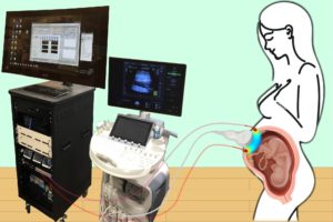
Combining optical measurements with ultrasound, an interdisciplinary team from the School of Arts & Sciences, Perelman School of Medicine, and CHOP developed a device to better measure blood flow and oxygenation in the placenta. (Image: Lin Wang)

A study published in Nature Biomedical Engineering details a novel method for imaging the placenta in pregnant patients as well as the results of a pilot clinical study. By combining optical measurements with ultrasound, the findings show how oxygen levels can be monitored noninvasively and provides a new way to generate a better understanding of this complex, crucial organ. This research was the result of a collaboration of the groups of the University of Pennsylvania’s Arjun Yodh and Nadav Schwartz with colleagues from the Children’s Hospital of Philadelphia (CHOP) and was led by postdoc Lin Wang.

Schwartz describes the placenta as the “engine” of pregnancy, an organ that plays a crucial role in delivering nutrients and oxygen to the fetus. Placental dysfunction can lead to complications such as fetal growth restriction, preeclampsia, and stillbirth. To increase knowledge about this crucial organ, the National Institute of Child Health and Human Development launched the Human Placenta Project in 2014. One focus of the program is to develop tools to assess human placental structure and function in real time, including optical devices.

For three years, the researchers optimized the design of their instrument and tested it in preclinical settings. The process involved integrating optical fibers with ultrasound probes, exploring various ultrasound transducers, and improving the multimodal technology so that measurements were stable, accurate, and reproducible while collecting data at the bedside. The resulting instrumentation now enables researchers to study the anatomy of the placenta while also collecting detailed functional information about placenta blood flow and oxygenation, capabilities that existing commercially devices do not have, the researchers say.

Because the placenta is located far below the body’s surface, one of the key technical challenges addressed by Wang, a postdoc in Yodh’s lab, was reducing background noise in the opto-electronic system. Light is scattered and absorbed when it travels through thick tissues, Yodh says, and the key for success was to reduce background interference so that the small amount of light that penetrates deep into the placenta and then returns is still large enough for a high-quality measurement.

“We’re sending a light signal that goes through the same deep tissues as the ultrasound. The extremely small amount of light that returns to the surface probe is then used to accurately assess tissue properties, which is only possible with very stable lasers, optics, and detectors,” says Yodh. “Lin had to overcome many barriers to improve the signal-to-noise ratio to the point where we trusted our data.”

Read the full story in Penn Today.

The authors are Lin Wang, Jeffrey M. Cochran, Kenneth Abramson, Lian He, Venki Kavuri, Samuel Parry, Arjun G. Yodh, and Nadav Schwartz from Penn; Tiffany Ko, Wesley B. Baker, and Rebecca L. Linn from the Children’s Hospital of Philadelphia, and David R. Busch, previously a research associate at Penn and now at the University of Texas Southwestern Medical School.

Arjun Yodh is the James M. Skinner Professor of Science in the Department of Physics and Astronomy in the School of Arts & Sciences at the University of Pennsylvania. He is a member of the Penn Bioengineering Graduate Group.

Nadav Schwartz is an Associate Professor in the Department of Obstetrics and Gynecology in Penn’s Perelman School of Medicine.

Lin Wang is a postdoc in the Department of Physics and Astronomy in Penn’s School of Arts & Sciences.

This research was supported by National Institutes of Health grants F31HD085731, R01NS113945, R01NS060653, P41EB015893, P41EB015893, T32HL007915, and U01HD087180.

Week in BioE (May 31, 2019)

by Sophie Burkholder

Vector Flow Imaging Helps Visualize Blood Flow in Pediatric Hearts

A group of biomedical engineers at the University of Arkansas used a new ultrasound-based imaging technique called vector flow imaging to help improve the diagnosis of congenital heart disease in pediatric patients. The study, led by associate professor of biomedical engineering Morten Jensen, Ph.D., collaborated with cardiologists at the local Children’s Hospital in Little Rock to produce images of the heart in infants to help potentially diagnose congenital heart defects. Though the use of vector flow imaging has yet to be developed for adult patients, this type of imaging could possibly provide more detail about the direction of blood flow through the heart than traditional techniques like echocardiography do. In the future, the use of both techniques could provide information about both the causes and larger effects of heart defects in patients.

Using Stem Cells to Improve Fertility in Leukemia Survivors

One of the more common side effects of leukemia treatment in female patients is infertility, but researchers at the University of Michigan want to change that. Led by associate professor of biomedical engineering Ariella Shikanov, Ph.D., researchers in her lab found ways of increasing ovarian follicle productivity in mice, which directly relates to the development of mature eggs. The project involves the use of adipose-derived stem cells, that can be found in human fat tissue, to surround the follicles in an ovary-like, three-dimensional scaffold.  Because the radiation treatments for leukemia and some other cancers are harmful to follicles, increasing their survival rate with this stem cell method could reduce the rate of infertility in patients undergoing these treatments. Furthermore, this new approach is innovative in its use of a three-dimensional scaffold as opposed to a two-dimensional one, as it stimulates follicle growth in all directions and thus helps to increase the follicle survival rate.

Penn Engineers Look at How Stretching & Alignment of Collagen Fibers Help Cancer Cells Spread

Cancer has such a massive impact on people’s lives that it might be easy to forget that the disease originates at the cellular level. To spread and cause significant damage, individual cancer cells must navigate the fibrous extracellular environment that cells live in, an environment that Penn Engineer Vivek Shenoy has been investigating for years.

Shenoy is the Eduardo D. Glandt President’s Distinguished Professor with appointments in Materials Science and Engineering, Mechanical Engineering and Applied Mechanics, and Bioengineering. He is also the Director of the Center for Engineering MechanoBiology (CEMB), one of the NSF’s twelve Science and Technology Centers.

Shenoy’s most recent study on cancer’s mechanical environment was led by a postdoctoral researcher in his lab, Ehsan Ban. Paul Janmey, professor in Physiology and Bioengineering, and colleagues at Stanford University also contributed to the study. Shenoy also received the Heilmeier Award this March and delivered the Heilmeier Award Lecture in April.

Read the rest of this story on Penn Engineering’s Medium Blog.

Controlled Electrical Stimulation Can Prevent Joint Replacement Infections

Joint replacements are one of the most common kinds of surgery today, but they still require intense post-operative therapy and have a risk of infection from the replacement implant. These infections are usually due to the inflammatory response that the body has to any foreign object, and can become serious and life-threatening if left untreated. Researchers at the University of Buffalo Jacobs School of Medicine and Biomedical Sciences hope to offer a solution to preventing infections through the use of controlled electrical stimulation. Led by Mark Ehrensberger, Ph.D., Kenneth A. Krackow, M.D., and Anthony A. Campagnari, Ph.D., the treatment system uses the electrical signal to create an antibacterial environment at the interface of the body and the implant. While the signal does not prevent infections completely, these antibacterial properties will prevent infections from worsening to a more serious level. Patented as the Biofilm Disruption Device TM, the final product uses two electrode skin patches and a minimally invasive probe that delivers the electrical signal directly to the joint-body interface. The researchers behind the design hope that it can help create a more standard way of effectively treating joint replacement infections.

People and Places

TBx: Gabriel Koo, Ethan Zhao, Daphne Cheung, and Shelly Teng

For their senior design project, four bioengineering seniors Gabriel Koo, Ethan Zhao, Daphne Cheung, and Shelly Teng created a low-cost tuberculosis diagnostic that they called TBx. Using their knowledge of the photoacoustic effect of certain dyes, the platform the group created can detect the presence of lipoarabinomannan in patient urine. The four seniors presented TBx at the Rice360 Design Competition in Houston, Texas this spring, which annually features student-designed low-cost global health technologies.

Week in BioE: March 29, 2019

by Sophie Burkholder

New Studies in Mechanobiology Could Open Doors for Cellular Disease Treatment

When we think of treatments at the cellular level, we most often think of biochemical applications. But what if we began to consider more biomechanical-oriented approaches in the regulation of cellular life and death? Under a grant from the National Science Foundation (NSF),Worcester Polytechnic Institute’s (WPI) Head of the Department of Biomedical Engineering Kristen Billiar, Ph.D., performs research that looks at the way mechanical stimuli can affect and trigger programmed cell death.

Billiar, who received his M.S.E. and Ph.D. from Penn, began his research by first noticing the way that cells typically respond to the mechanical stimuli in their everyday environment, such as pressure or stretching, with behaviors like migration, proliferation, or contraction. He and his research team hope to find a way to eventually predict and control cellular responses to their environment, which they hope could open doors to more forms of treatment for disorders like heart disease or cancer, where cellular behavior is directly linked to the cause of the disease.

Self-Learning Algorithm Could Help Improve Robotic Leg Functionality

Obviously, one of the biggest challenges in the field of prosthetics is the extreme difficulty in creating a device that perfectly mimics whatever the device replaces for its user. Particularly with more complex designs that involve user-controlled motion for joints in the limbs or hands, the electrical circuits implemented are by no means a perfect replacement of the neural connections in the human body from brain to muscle. But recently at the University of Southern California Viterbi School of Engineering, a team of researchers led by Francisco J. Valero-Cuevas, Ph. D.,  developed an algorithm with the ability to learn new walking tasks and adapt to others without any additional programming.

The algorithm will hopefully help to speed the progress of robotic interactions with the world, and thus allow for more adaptive technology in prosthetics, that responds to and learns with their users. The algorithm Valero-Cuevas and his team created takes inspiration from the cognition involved with babies and toddlers as they slowly learn how to walk, first through random free play and then from pulling on relevant prior experience. In a prosthetic leg, the algorithm could help the device adjust to its user’s habits and gait preferences, more closely mimicking the behavior of an actual human leg.

Neurofeedback Can Improve Behavioral Performance in High-Stress Situations

We’re all familiar with the concept of being “in the zone,” or the feeling of extraordinary focus that we can sometimes have in situations of high-stress. But how can we understand this shift in mindset on a neuroengineering level? Using the principal of the Yerkes-Dodson law, which says that there is a state of brain arousal that is optimal for behavioral performance, a team of biomedical engineering researchers at Columbia University hope to find ways of applying neurofeedback to improving this performance in demanding high-stress tasks.

Led by Paul Sajda, Ph.D., who received his doctoral degree from Penn, the researchers used a brain-computer interface to collect electroencephalography (EEG) signals from users immersed in virtual reality aerial navigation tasks of varying difficulty levels. In doing so, they were able to make connections between stressful situations and brain activity as transmitted through the EEG data, adding to the understanding of how the Yerkes-Dodson law actually operates in the human body and eventually demonstrating that the use of neurofeedback reduced the neural state of arousal in patients. The hope is that neurofeedback may be used in the future to help treat emotional conditions like post-traumatic stress disorder (PTSD).

Ultrasound Stimulation Could Lead to New Treatments for Inflammatory Arthritis

Arthritis, an autoimmune disease that causes painful inflammation in the joints, is one of the more common diseases among older patients, with more than 3 million diagnosed cases in the United States every year. Though extreme measures like joint replacement surgery are one solution, most patients simply treat the pain with nonsteroidal anti-inflammatory drugs or the adoption of gentle exercise routines like yoga. Recently however, researchers at the University of Minnesota led by Daniel Zachs, M.S.E., in the Sensory Optimization and Neural Implant Coding Lab used ultrasound stimulation treatment as a way to reduce arthritic pain in mice. In collaboration with Medtronic, Zachs and his team found that this noninvasive ultrasound stimulation greatly decreased joint swelling in mice who received the treatment as opposed to those that did not. They hope that in the future, similar methods of noninvasive treatment will be able to be used for arthritic patients, who otherwise have to rely on surgical remedies for serious pain.

People and Places

Leadership and Inspiration: EDAB’s Blueprint for Engineering Student Life

To undergraduates at a large university, the administration can seem like a mysterious, all-powerful entity, creating policy that affects their lives but doesn’t always take into account the reality of their day-to-day experience. The Engineering Deans’ Advisory Board (EDAB) was designed to bridge that gap and give students a platform to communicate with key decision makers.

The 13-member board meets once per week for 60 to 90 minutes. The executive board, comprised of four members, also meets weekly to plan out action items and brainstorm. Throughout his interactions with the group, board president Jonathan Chen, (ENG ‘19, W ‘19), has found a real kinship with his fellow board members, who he says work hard and enjoy one another’s company in equal measure.

Bioengineering major Daphne Cheung (ENG’19) joined the board as a first-year student because she saw an opportunity to develop professional skills outside of the classroom. “For me, it was about trying to build a different kind of aptitude in areas such as project management, and learning how to work with different kinds of people, including students and faculty, and of course, the deans,” she says.

Read the full story on Penn Engineering’s Medium Blog. Media contact Evan Lerner.

Purdue University College of Engineering and Indiana University School of Medicine Team Up in New Engineering-Medicine Partnership

The Purdue University College of Engineering and the Indiana University School of Medicine recently announced a new Engineering-Medicine partnership, that seeks to formalize ongoing and future collaborations in research between the two schools. One highlight of the partnership is the establishment of a new M.D./M.S. degree program in biomedical engineering that will allow medical students at Indiana University to receive M.S.-level training in engineering technologies as they apply to clinical practice. The goal of this new level of collaboration is to further involve Purdue’s engineering program in the medical field, and to exhibit the benefits that developing an engineering mindset can have for medical students. The leadership of this new partnership includes

Week in BioE (June 19, 2018)

Dolphin Echolocation Could Improve Ultrasound

dolphin echolocationDolphins are among the most intelligent creatures on earth, showing behaviors such as teaching, learning, cooperation, delayed gratification, and other markers of high intelligence. Dolphins communicate vocally with one another, although we aren’t sure exactly what they communicate. While this communication isn’t “language” as humans define it, it uses echolocation — finding objects and orienteering on the basis of reflected sound — which humans don’t use in their communications.

Now, we have new information about dolphin echolocation thanks to an article recently published in the Journal of the Acoustical Society of America by mathematicians and biomedical engineers in Sweden.  On the basis of earlier research finding that dolphin echolocation signals consist of two tones, rather than one, the new study finds that these two tones are emitted at slightly different times and that the sound waves have a Gaussian shape, similar to a bell curve. Using a mathematical algorithm, the authors successfully simulated echolocation signals in the lab.

The findings explain how dolphins use echolocation effectively but could also contribute to more accurate sound-based diagnostic techniques — particularly ultrasound, which relies heavily on methods similar to echolocation to provide images of moving tissues within the body, e.g., prenatal imaging and heart contraction.

Modeling Diseased Blood Vessels for Drug and Device Testing

Drugs and devices require extensive testing before they are approved by regulatory agencies and used to treat human patients. Tissue engineering has helped bridge the gap between a promising idea and its use in a patient by creating technologies that mimic the complex structure of human tissue. Most of these technologies focus on the engineering of healthy tissues and much less on constructing models of diseased tissue. These models of diseased tissue may be useful for designing treatments for diseases and understanding how diseases are caused.

In this light, Marsha W. Rolle, PhD, Associate Professor of Biomedical Engineering at Worcester Polytechnic Institute (WPI), is working to create engineered blood vessels that are already diseased as a way to test possible treatments. With three years of funding from the National Institutes of Health’s National Heart Lung and Blood Institute amounting to nearly $500,000, Dr. Rolle and her research team create these damaged vessels by engineering smooth muscle cells to form tubes 2 mm in diameter. These synthetic vessels are then modified to resemble features of diseases. For example, growth factors attached to microspheres can encourage the growth of tissue in small parts of the vessel wall, eventually becoming areas of narrowing in the vessel. Similarly, other factors could lead to changes in the vessel that resemble aneurysms. In both cases, the function of the microengineered vessel could be measured as the change happens, providing insight into either vascular stenosis or aneurysms, neither of which is possible in humans.

Dr. Rolle’s first step will be to test the damaged engineered vessels with existing medications. If successful, this new technique could be used for testing of new drugs and devices prior to testing in animals.

New Heart Implant Can Deliver Drug

Speaking of damage to the circulatory system, a new article in Nature Biomedical Engineering details how engineers at MIT, Harvard, and Trinity College, Dublin, created a heart implant that can deliver targeted therapy to damaged heart tissue. The authors, led in part by Conor J. Walsh, PhD, and David J. Mooney, PhD, of Harvard, created a device called Therepi, approximately 4 mm in size, which is deployed with a hypodermic. Once placed, a reservoir of medicine within the Therepi treats the damaged heart muscle. In addition, it can be refilled without needing to remove the implant. The Nature Biomedical Engineering study is limited to testing in rats, but the authors see testing in humans in the near future.

Erdős-Rényi Prize for Penn Professor

Danielle S. Bassett, PhD, Eduardo D. Glandt Faculty Fellow and Associate Professor of Bioengineering at the University of Pennsylvania, has been named the 2018 recipient of the Erdős-Rényi Prize in Network Science by the Network Science Society (NetSci). NetSci has recognized Dr. Bassett for “fundamental contributions to our understanding of the network architecture of the human brain, its evolution over learning and development, and its alteration in neurological disease.” Dr. Bassett will receive the award and deliver a lecture on June 14 at the International Conference on Network Science in Paris. She is the seventh scientist and fourth American to receive the prize.

The Erdős-Rényi Prize is awarded annually to a scientist younger than 40 years old for his/her achievements in the field of network science. It is named for the Hungarian mathematicians Paul Erdős, whose surname provided a measurement for research collaboration by academic mathematicians, and Alfréd Rényi, whose work focused on probability and graph theory. In network science, an Erdős-Rényi model is a model for generating random graphs. Dr. Bassett’s research applies the principles of network science in neuroscience, with the intention of understanding the brain better by modeling the networks and circuits of our most complex organ.

People and Places

Two new centers dedicated to health sciences are opening. Western New England University opened its new Center for Global Health Engineering in April, with Michael J. Rust, PhD, Associate Professor of Biomedical Engineering, as the codirector under director Christian Salmon, PhD. Elsewhere, Northwestern University launched a new center — the Center for Advanced Regenerative Engineering — with Guillermo Ameer, PhD, Daniel Hale Williams Professor of Biomedical Engineering and Surgery at Northwestern, as founding director.

Finally, Joseph J. Pancrazio, Ph.D., Professor of Bioengineering at the University of Texas at Dallas and Associate Provost,  has been named Vice President for Research. Before moving to UT Dallas in 2015, Dr. Pancrazio was the founding chair of Bioengineering at George Mason University in Virginia.