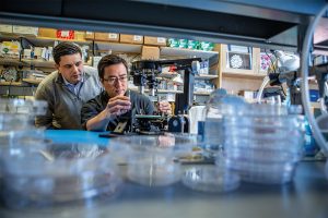
Dan Huh, Associate Professor in the Department of Bioengineering, has been steadily growing a collection of organs-on-chips. These devices incorporate human cells into precisely engineered microfluidic channels that mimic an organ’s natural environment, providing a way to conduct experiments that would not otherwise be feasible.
Huh’s previous research has involved using a placenta-on-a-chip to study which drugs are able to reach a developing fetus; investigating microgravity’s effect on the immune system by sending one of his chips to the International Space Station; and testing treatments for dry eye disease using an eye-on-a-chip, complete with a mechanical blinking eyelid.
Now, he and his colleagues are taking this technology out of their lab and into industry with their company, Vivodyne.
Andrei Georgescu, Huh’s lab-member and co-founder of Vivodyne, recently spoke with Technical.ly Philly’s Paige Gross about the growth of their company.
Research into potential drugs is usually performed first on mice, and success is only found in a fraction of humans once implemented in clinical trials, Andrei Georgescu, cofounder and CEO of Vivodyne, told Technical.ly. The genetic makeup just isn’t similar enough. But technology that allows scientists to test therapies on lab-grown human organs called “organs on chip” is allowing for testing without human subjects.
The organs on chip allow for a drug to react to tissue in a more similar way to the body than it would in a petri dish, Georgescu said. Cells sense their environment very well, he added.
“We’re making the environment more complicated, making its spacial features complicated enough to match the native complexity of the organs,” he said. “When [cells] sense a softer environment, they start to behave more realistically. Their response to the drug is more realistic.”
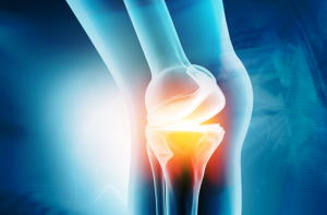
New research from Robert Mauck, Mary Black Ralston Professor in Orthopaedic Surgery and Bioengineering and Director of Penn Medicine’s McKay Orthopaedic Research Laboratory, announces a “new biosealant therapy may help to stabilize injuries that cause cartilage to break down, paving the way for a future fix or – even better – begin working right away with new cells to enhance healing.” Their research was published in Advanced Healthcare Materials. The study’s lead author was Jay Patel, a former postdoctoral fellow in the McKay Lab and now Assistant Professor at Emory University and was contributed to by Claudia Loebel, a postdoctoral research in the Burdick lab and who will begin an appointment as Assistant Professor at the University of Michigan in Fall 2021. In addition, the technology detailed in this publication is at the heart of a new company (Forsagen LLC) spun out of Penn with support from the Penn Center for Innovation (PCI) Ventures Program, which will attempt to spearhead the system’s entry into the clinic. It is co-founded by both Mauck and Patel, along with study co-author Jason Burdick, Professor in Bioengineering, and Ana Peredo, a PhD student in Bioengineering.
Kinases are a class of enzymes that are responsible for transferring the main chemical energy source used by the body’s cells. As such, they play important roles in diverse cellular processes, including signaling, differentiation, proliferation and metabolism. But since they are so ubiquitous, mutated versions of kinases are frequently found in cancers. Many cancer treatments involve targeting these mutant kinases with specific inhibitors.
Understanding the exact genetic mutations that lead to these aberrant kinases can therefore be critical in predicting the progression of a given patient’s cancer and tailoring the appropriate response.
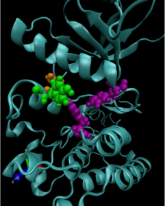
To achieve this understanding on a more fundamental level, a team of researchers from the University of Pennsylvania’s School of Engineering and Applied Science and Perelman School of Medicine, the Children’s Hospital of Philadelphia (CHOP) and researchers at the Yale School of Medicine’s Cancer Biology Institute, have constructed molecular simulations of a mutant kinase implicated in pediatric neuroblastoma, a childhood cancer impacting the central nervous system.
Using their computational model to study the relationship between single-point changes in the kinase’s underlying gene and the altered structure of the protein it ultimately produces, the researchers revealed useful commonalities in the mutations that result in tumor formation and growth. Their findings suggest that such computational approaches could outperform existing profiling methods for other cancers and lead to more personalized treatments.
The study, published in the Proceedings of the National Academy of Sciences, was led by Ravi Radhakrishnan, Professor and chair of Penn Engineering’s Department of Bioengineering and professor in its Department of Chemical and Biomolecular Engineering, and Mark A. Lemmon, Professor of Pharmacology at Yale and co-director of Yale’s Cancer Biology Institute. The study’s first authors were Keshav Patil, a graduate student in Penn Engineering’s Department of Chemical and Biomolecular Engineering, along with Earl Joseph Jordan and Jin H. Park, then members of the Graduate Group in Biochemistry and Molecular Biology in Penn’s Perelman School of Medicine. Krishna Suresh, an undergraduate student in Radhakrishnan’s lab, Courtney M. Smith, a graduate student in Lemmon’s lab, and Abigail A. Lemmon, an undergraduate in Lemmon’s lab, contributed to the study. They collaborated with Yaël P. Mossé, Associate Professor of Pediatrics at Penn Medicine and in the division of oncology at CHOP.
“Some cancers rely on the aberrant activation of a single gene product for tumor initiation and progression,” says Radhakrishnan. “This unique mutational signature may hold the key to understanding which patients suffer from aggressive forms of the disease or for whom a given therapeutic drug may yield short- or long-term benefits. Yet, outside of a few commonly occurring ‘hotspot’ mutations, experimental studies of clinically observed mutations are not commonly pursued.”
The inaugural Joseph Bordogna Forum took place on Wednesday, February 24 and featured a talk from John Brooks Slaughter, Deans’ Professor of Education and Engineering at USC’s Viterbi School of Engineering and Rossier School of Education, entitled a “Call to Action for Racial Justice and Equity in Engineering.”
Dr. Slaughter was joined by panelists Jennifer R. Lukes, Professor in Mechanical Engineering and Applied Mechanics, Oladayo Adewole, an alumnus in Robotics who recently defended his doctoral dissertation in Bioengineering, and CJ Taylor, Raymond S. Markowitz President’s Distinguished Professor in Computer and Information Science and Associate Dean, Diversity, Equity and Inclusion, who moderated the talk.
Dr. Slaughter talked about how microaggressions can often be a barrier to student success and emphasized on the importance of mentorship for underrepresented minorities: “If faculty members seek to improve the retention of underrepresented minorities, often times more has to be done than introducing science and math principles early on in their education, but instead, the unique backgrounds of these students must be understood.”
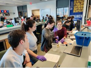
One example is Strella Biotechnology, founded in 2019 by Katherine Sizov (Biology 2019 & President’s Innovation Prize winner). Strella is developing sensors with the ability to reduce the amount of food waste due to going bad in storage. “Having a Bio-MakerSpace that gives you the functionalities of both a wet lab and a traditional electronics lab is extremely helpful in developing novel technologies” says Sizov on the BE Labs Youtube channel.
The Bio-MakerSpace provides students of all academic backgrounds the resources to turn their ideas into realities, including highly knowledgeable lab staff. Seth Fein (BSE ’20, MSE ’21) has worked at the lab since Fall 2020. “Because bioengineering spans many fields, we encourage interdisciplinary work. Students from Mechanical, Electrical, and Chemical Engineering have all found valuable resources in the lab,” says Fein.
The article also discusses the many resources the BioMakerSpace provides to Penn students and their efforts to keep the lab functional, safe, and open for research and education during the current semester.
Penn Health-Tech is an interdisciplinary center launched in 2017 to advance medical device innovation across the Perelman School of Medicine and the School of Engineering and Applied Sciences by forging collaborative connections among Penn researchers and providing seed funding to incubate novel ideas to advance health care.
Continue reading “The Bio-MakerSpace — Fostering Learning and Innovation Across Many Disciplines” at the Penn Health-Tech blog.
We hope you will join us for the 2021 Grace Hopper Distinguished Lecture by Dr. Jennifer Lewis, presented by the Department of Bioengineering. For event links, email ksas@seas.upenn.edu.
Date: Thursday, March 25, 2021
Time: 3:00-4:00 PM EDT
Speaker: Jennifer A. Lewis, Sc.D.
Wyss Professor for Biologically Inspired Engineering
The Wyss Institue
Paulson School of Engineering and Applied Sciences
Harvard University
Title: “Biomanufacturing Vascularized Organoids and Functional Human Tissue”
Following the lecture, join us for a panel discussion “Horizon 2030: Engineering Life & Life in (Bio)Engineering” featuring Dr. Lewis and Penn faculty and moderated by Bioengineering students. Further details here.
Lecture Abstract:
Recent protocols in developmental biology are unlocking the potential for stem cells to undergo differentiation and self-assembly to form “mini-organs”, known as organoids. To bridge the gap from organoid building blocks (OBBs) to therapeutic functional tissues, integrative approaches that combine bottom-up organoid assembly with top-down bioprinting are needed. While it is difficult, if not impossible, to imagine how either organoids or bioprinting alone would fully replicate the complex multiscale features required for organ-specific function – their combination may provide an enabling foundation for de novo tissue manufacturing. My talk will begin by describing our recent efforts to generate organoids in vitro with perfusable microvascular networks that support their viability and maturation. Next, I will describe the generation of 3D vascularized organ-specific tissues by assembling OBBs into a living matrix that supports the embedded printing of macro-vessels by a process known as sacrificial writing in functional tissue (SWIFT). Though broadly applicable, I will highlight our recent work on kidney, cerebral, and cardiac tissue engineering.
Dr. Lewis Bio:
Jennifer A. Lewis is the Jianming Yu Professor of Arts and Sciences, the Wyss Professor for Biologically Inspired Engineering in the Paulson School of Engineering and Applied Sciences, and a core faculty member of the Wyss Institute at Harvard University. Her research focuses on 3D printing of functional, structural, and biological materials that emulate natural systems. Prior to joining Harvard, Lewis was a faculty member in the Materials Science and Engineering Department at the University of Illinois at Urbana-Champaign, where she served as the Director of the Materials Research Laboratory. Currently, she directs the Harvard Materials Research Science and Engineering Center (MRSEC) and serves the NSF Mathematical and Physical Sciences Advisory Committee.
Lewis has received numerous awards, including the Presidential Faculty Fellow Award, the American Chemical Society Langmuir Lecture Award, the Materials Research Society Medal Award, the American Ceramic Society Sosman and Roy Lecture Awards, and the Lush Science Prize. She is an elected member of the National Academy of Sciences, National Academy of Engineering, National Academy of Inventors, and the American Academy of Arts and Sciences. Her research has enjoyed broad coverage in the popular media. To date, she has co-founded two companies, Voxel8 Inc. and Electroninks, that are commercializing technology from her lab.
Information on the GraceHopper Lecture: In support of its educational mission of promoting the role of all engineers in society, the School of Engineering and Applied Science presents the GraceHopper Lecture Series. This series is intended to serve the dual purpose of recognizing successful women in engineering and of inspiring students to achieve at the highest level.
Rear Admiral GraceHopper was a mathematician, computer scientist, systems designer and the inventor of the compiler. Her outstanding contributions to computer science benefited academia, industry and the military. In 1928 she graduated from Vassar College with a B.A. in mathematics and physics and joined the Vassar faculty. While an instructor, she continued her studies in mathematics at Yale University where she earned an M.A. in 1930 and a Ph.D. in 1934. GraceHopper is known worldwide for her work with the first large-scale digital computer, the Navy’s Mark I. In 1949 she joined Philadelphia’s Eckert-Mauchly, founded by the builders of ENIAC, which was building UNIVAC I. Her work on compilers and on making machines understand ordinary language instructions lead ultimately to the development of the business language, COBOL. GraceHopper served on the faculty of the Moore School for 15 years, and in 1974 received an honorary degree from the University. In support of the accomplishments of women in engineering, each department within the School invites a prominent speaker for a one or two-day visit that incorporates a public lecture, various mini-talks and opportunities to interact with undergraduate and graduate students and faculty.
As COVID-19 vaccines roll out, the concept of using mRNA to fend off viruses has become a part of the public dialogue. However, scientists have been researching how mRNA can be used to in life-saving medical treatments well before the pandemic.
The “m” in “mRNA” is for “messenger.” A single-stranded counterpart to DNA, it translates the genetic code into the production of proteins, the building blocks of life. The Moderna and Pfizer COVID-19 vaccines work by introducing mRNA sequences that act as a set of instructions for the body to produce proteins that mimic parts of the virus itself. This prepares the body’s immune response to recognize the real virus and fight it off.
Because it can spur the production of proteins that the body can’t make on its own, mRNA therapies also have the potential to slow or prevent genetic diseases that develop before birth, such as cystic fibrosis and sickle-cell anemia.
However, because mRNA is a relatively unstable molecule that degrades quickly, it needs to be packaged in a way that maintains its integrity as its delivered to the cells of a developing fetus.
To solve this challenge, Michael J. Mitchell, Skirkanich Assistant Professor of Innovation in the Department of Bioengineering, is researching the use of lipid nanoparticles as packages that transport mRNA into the cell. He and William H. Peranteau, an attending surgeon in the Division of General, Thoracic and Fetal Surgery and the Adzick-McCausland Distinguished Chair in Fetal and Pediatric Surgery at Children’s Hospital of Philadelphia, recently co-authored a “proof-of-concept” paper investigating this technique.
In this study, published in Science Advances, Mitchel examined which nanoparticles were optimal in the transport of mRNA to fetal mice. Although no disease or organ was targeted in this study, the ability to administer mRNA to a mouse while still in the womb was demonstrated, and the results are promising for the next stages of targeted disease prevention in humans.
Mitchel spoke with Tom Avril at The Philadelphia Inquirer about the mouse study and its implications for treatment of rare infant diseases through the use of mRNA, ‘the messenger of life.’
Penn bioengineering professor Michael J. Mitchell, the other senior author of the mouse study, tested various combinations of lipids to see which would work best.
The appeal of the fatty substances is that they are biocompatible. In the vaccines, for example, two of the four lipids used to make the delivery spheres are identical to lipids found in the membranes of human cells — including plain old cholesterol.
When injected, the spheres, called nanoparticles, are engulfed by the person’s cells and then deposit their cargo, the RNA molecules, inside. The cells respond by making the proteins, just as they make proteins by following the instructions in the person’s own RNA. (Important reminder: The RNA in the vaccines cannot become part of your DNA.)
Among the different lipid combinations that Mitchell and his lab members tested, some were better at delivering their cargo to specific organs, such as the liver and lungs, meaning they could be a good vehicle for treating disease in those tissues.
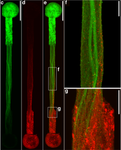
Connecting the human brain to electrical devices is a long-standing goal of neuroscientists, bioengineers, and clinicians, with applications ranging from deep brain stimulation (DBS) to treat Parkinson’s disease to more futuristic endeavors such as Elon Musk’s NeuraLink initiative to record and translate brain activity. However, these approaches currently rely on using implantable metallic electrodes that inherently provoke a lasting immune response due to their non-biological nature, generally complicating the reliability and stability of these interfaces over time. To address these challenges, D. Kacy Cullen, Associate Professor in Neurosurgery and Bioengineering, and Dayo Adewole, a doctoral candidate in Bioengineering, worked with a multi-disciplinary team of collaborators to develop the first “living electrodes” as an implantable, biological bridge between the brain and external devices. In a recent article published in Science Advances, the team demonstrated the fabrication of hair-like microtissue comprised of living neuronal networks and bundled tracts of axons — the signal sending fibers of the nervous system — protected within soft hydrogel cylinders. They showed that these axon-based living electrodes could be fully controlled and monitored with light — thus eliminating the need for electrical contact — and are capable of surviving and forming synapses with the brain as demonstrated in an adult rat model. While further advancements are necessary prior to clinical use, the current findings provide the foundation for a new class of “living electrodes” as a biological intermediary between humans and devices capable of leveraging natural mechanisms to potentially provide a stable interface for clinical applications.
The process of discovery sometimes starts with a hunch. Maria Ovando arrived at Penn Engineering with an affinity for math and science, extensive experience volunteering at her local health clinic and an assumption that she was preparing for a career in medicine. She was drawn to Penn Engineering because of the flexibility in the curriculum and the ability to both tailor her course of study and pursue cross-disciplinary subjects.
As a pre-med student, bioengineering seemed to be the natural choice for a major, but during her freshman year, Ovando found that she genuinely enjoyed bioengineering as a discipline in its own right, and only then did her future goals come into view.
“I’ve discovered that I have a passion for research, working on low-cost devices that can have a direct impact on individuals,” she says.
One of the most important opportunities she’s had at Penn is her work with Dr. Michelle J. Johnson at the Rehabilitation Robotics Lab in the Perelman School of Medicine. There, Ovando has been working to improve aspects of the Community-based Affordable Robot Exercise System, which helps stroke patients with lower extremity impairment. She’s also worked on a project that involved analyzing and reevaluating data in the early detection of cerebral palsy in infants. As an undergraduate, she found it both meaningful and moving to have a role in this groundbreaking research.
Researchers at Children’s Hospital of Philadelphia and the School of Engineering and Applied Science at the University of Pennsylvania have identified ionizable lipid nanoparticles that could be used to deliver mRNA as part of fetal therapy. The proof-of-concept study, published today in Science Advances, engineered and screened a number of lipid nanoparticle formulations for targeting mouse fetal organs and has laid the groundwork for testing potential therapies to treat genetic diseases before birth.
“This is an important first step in identifying nonviral mediated approaches for delivering cutting-edge therapies before birth,” said co-senior author William H. Peranteau, MD, an attending surgeon in the Division of General, Thoracic and Fetal Surgery and the Adzick-McCausland Distinguished Chair in Fetal and Pediatric Surgery at CHOP. “These lipid nanoparticles may provide a platform for in utero mRNA delivery, which would be used in therapies like fetal protein replacement and gene editing.”
Michael J. Mitchell, Skirkanich Assistant Professor of Innovation in Penn Engineering’s Department of Bioengineering, is the other co-senior author of the study. The co-first authors are Mitchell Lab members Rachel Riley, a postdoctoral fellow, and Margaret Billingsley, a graduate student, and Peranteau Lab member Meghana Kashyap, a research fellow.
Recent advances in DNA sequencing technology and prenatal diagnostics have made it possible to diagnose many genetic diseases before birth. Some of these diseases are treated by protein or enzyme replacement therapies after birth, but by then, some of the damaging effects of the disease have taken hold. Thus, applying therapies while the patient is still in the womb has the potential to be more effective for some conditions. The small fetal size allows for maximal therapeutic dosing, and the immature fetal immune system may be more tolerant of replacement therapy.