Carl June on the Boundless Potential of CAR T Cell Therapy

by Meagan Raeke

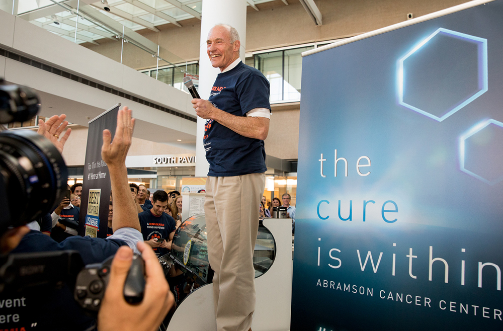
Carl June, at the flash mob celebration of the FDA approval of the CAR T cell therapy he developed, in August 2017. (Image: Courtesy of Penn Medicine Magazine)

For most of modern medicine, cancer drugs have been developed the same way: by designing molecules to treat diseased cells. With the advent of immunotherapy, that changed. For the first time, scientists engineered patients’ own immune systems to recognize and attack diseased cells.

One of the best examples of this pioneering type of medicine is CAR T cell therapy. Invented in the Perelman School of Medicine by Carl June, the Richard W. Vague Professor in Immunotherapy, CAR T cell therapy works by collecting T cells from a patient, modifying those cells in the lab so that they are designed to destroy cancerous cells, and reinfusing them into the patient. June’s research led to the first FDA approval for this type of therapy, in 2017. Six different CAR T cell therapies are now approved to treat various types of blood cancers. Carl June, at the flash mob celebration of the FDA approval of the CAR T cell therapy he developed, in August 2017. (Image: Courtesy of Penn Medicine Magazine)

CAR T cell therapy holds the potential to help millions more patients—if it can be successfully translated to other conditions. June and colleagues, including Daniel Baker, a fourth-year doctoral student in the Cell and Molecular Biology department, discuss this potential in a perspective published in Nature.

In the piece, June and Baker highlight other diseases that CAR T cell therapy could be effective.

“CAR T cell therapy has been remarkably successful for blood cancers like leukemias and lymphomas. There’s a lot of work happening here at Penn and elsewhere to push it to other blood cancers and to earlier stage disease, so patients don’t have to go through chemo first,” June says. “Another big priority is patients with solid tumors because they make up the vast majority of cancer patients. Beyond cancer, we’re seeing early signs that CAR T cell therapy could work in autoimmune diseases, like lupus.”

As for which diseases to pursue as for possible future treatment, June says, “essentially it boils down to two questions: Can we identify a population of cells that are bad? And can we target them specifically? Whether that’s asthma or chronic diseases or lupus, if you can find a bad population of cells and get rid of them, then CAR T cells could be therapeutic in that context.”

“What’s exciting is it’s not just theoretical at this point. There have been clinical reports in other autoimmune diseases, including myasthenia gravis and inflammatory myopathy,” Baker says. “But we are seeing early evidence that CAR T cell therapy will be successful beyond cancer. And it’s really opening the minds of people in the field to think about how else we could use CAR T. For example, there’s some pioneering work at Penn from the Epstein lab for heart failure. The idea is that you could use CAR T cells to get rid of fibrotic tissue after a cardiac injury, and potentially restore the damage following a heart attack.”

Baker adds, “there’s no question that over the last decade, CAR T cell therapy has revolutionized cancer. I’m hoping to play a role in bringing these next generation therapies to patients and make a real impact over the next decade. I think there’s potential for cell therapy to be a new pillar of medicine at large, and not just a new pillar of oncology.”

Read the full story at Penn Medicine Today.

An Improved Delivery System for mRNA Vaccines Provides More Powerful Protection

by Devorah Fischler

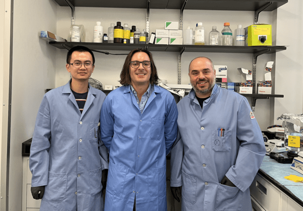
(From left to right) Xuexiang Han, Michael Mitchell and Mohamad-Gabriel Alameh

The COVID-19 vaccine swiftly undercut the worst of the pandemic for hundreds of millions around the world. Available sooner than almost anyone expected, these vaccines were a triumph of resourcefulness and skill.

Messenger RNA vaccines, like the ones manufactured by Moderna or Pfizer/BioNTech, owed their speed and success to decades of research reinforcing the safety and effectiveness of their unique immune-instructive technology.

Now, researchers from the University of Pennsylvania School of Engineering and Applied Science and the Perelman School of Medicine are refining the COVID-19 vaccine, creating an innovative delivery system for even more robust protection against the virus.

In addition to outlining a more flexible and effective COVID-19 vaccine, this work has potential to increase the scope of mRNA vaccines writ large, contributing to prevention and treatment for a range of different illnesses.

Michael Mitchell, associate professor in Penn Engineering’s Department of Bioengineering, Xuexiang Han, postdoctoral fellow in Mitchell’s lab, and Mohamad-Gabriel Alameh, postdoctoral fellow in Drew Weissman’s lab at Penn Medicine and incoming assistant professor in the Department of Pathology and Laboratory Medicine at the Perelman School of Medicine, recently published their findings in Nature Nanotechnology.

mRNA, or messenger ribonucleic acid, is the body’s natural go-between. mRNA contains the instructions our cells need to produce proteins that play important roles in our bodies’ health, including mounting immune responses.

The COVID-19 vaccines follow suit, sending a single strand of RNA to teach our cells how to recognize and fight the virus.

Read the full story in Penn Engineering Today.

The Physics of Fat Droplets Reveal DNA Danger

by Devorah Fischler

Fat is a normal and necessary part of the body. Fat cells store and release energy, as well as play significant roles in hormonal regulation and immunity.

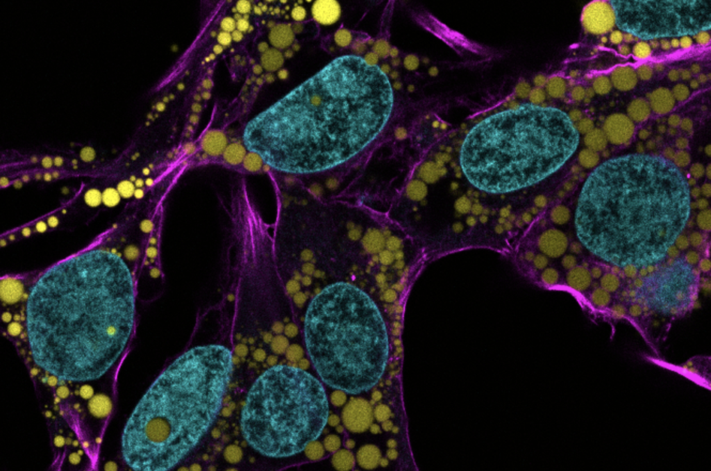
Engineers and scientists at the University of Pennsylvania are the first to discover fat-filled lipid droplets’ (pictured above in green) surprising capability to indent and puncture the nucleus, the organelle which contains and regulates a cell’s DNA.

In recent decades, a concerning rise in metabolic illnesses – such as cardiovascular disease, high blood pressure and diabetes – has focused scientific attention on the biology and chemistry of fat, resulting in a wealth of information about how fat cells work.

But fat cells and their metabolic activities are only part of the story.

Fat-filled lipid droplets, tiny spheres of fat many times smaller than fat cells, are a growing subject of scientific interest. Found inside many different cell types, these lipid particles have long been little understood. Studies have begun to illuminate these droplets’ participation in metabolic functions and cellular protection, but we still know next to nothing about the physical nature of fat.

Now, researchers at the University of Pennsylvania School of Engineering and Applied Science have looked beyond biochemistry to publish groundbreaking work on the physics of these droplets, revealing them to be a potential threat to a cell’s nucleus. In the August issue of the Journal of Cell Biology, they are the first to discover fat-filled lipid droplets’ surprising capability to indent and puncture the nucleus, the organelle which contains and regulates a cell’s DNA.

The stakes of their findings are high: a ruptured nucleus can lead to elevated DNA damage that is characteristic of many diseases, including cancer.

The study was led by Dennis E. Discher, Robert D. Bent Professor in the Department of Chemical and Biomolecular Engineering, Bioengineering, and in Mechanical Engineering and Applied Mechanics, Irena Ivanovska, Ph.D. research associate in Penn’s Molecular and Cell Biophysics Lab, and Michael Tobin, Ph.D. candidate in the Department of Bioengineering.

“Intuitively, people think of fat as soft,” says Discher. “And on a cellular level it is. But at this small size of droplet— measuring just a few microns rather than the hundreds of microns of a mature fat cell—it stops being soft. Its shape has a much higher curvature, bending other objects very sharply. This changes its physics in the cell. It can deform. It can damage. It can rupture.”

Read the full story in Penn Engineering Today.

SCALAR: A Microchip Designed to Transform the Production of mRNA Therapeutics and Vaccines

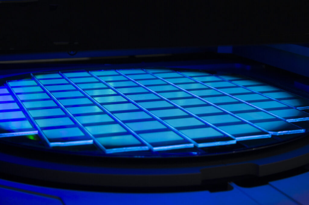
Led by Michael Mitchell and David Issadore of the School of Engineering and Applied Science, a team of researchers has developed a platform that could rapidly accelerate the development of mRNA-based lipid nanoparticle vaccines and therapeutics at both the small and large scale, SCALAR. (Image: iStock / Anatoly Morozov)

Following the global COVID-19 pandemic, the development and rapid deployment of mRNA vaccines highlighted the critical role of lipid nanoparticles (LNPs) in the context of pharmaceuticals. Used as the essential delivery vehicles for fragile RNA-based therapies and vaccines, LNPs protect the RNA from degradation and ensure effective delivery within the body.

Despite their critical importance, the large-scale manufacturing of these LNPs saw numerous bottlenecks during the pandemic, underscoring the need for scalable production techniques that could keep pace with global demand.

Now, in a paper published in the Proceedings of the National Academy of the Sciences, researchers at the University of Pennsylvania describe how the Silicon Scalable Lipid Nanoparticle Generation platform (SCALAR), a reusable silicon- and glass-based platform designed to transform the production landscape of LNPs for RNA therapeutics and vaccines, offers a scalable and efficient solution to the challenges exposed during the COVID-19 crisis.

“We’re excited to create a piece of technology platform that bridges the gap between small-scale discovery and large-scale manufacturing in the realm of RNA lipid nanoparticle vaccines and therapeutics,” says co-author Michael Mitchell, associate professor of bioengineering in the School of Engineering and Applied Science at Penn. “By doing so, we’ve effectively leapfrogged the clunky, time-consuming, and costly barriers that slow down the production ramp-up of promising new RNA medicines and vaccines.”

The intricacies of RNA-based therapies require the RNA to be encased in a delivery system capable of navigating the body’s biological obstacles. LNPs fulfill this role, allowing the RNA to reach the intended cells for maximum therapeutic impact. SCALAR aims to take this a step further, allowing for an unprecedented three orders of magnitude scalability in LNP production rates, addressing the speed and consistency bottlenecks that hinder existing methods.

Sarah Shepherd, the first author of the paper and a recent Ph.D. graduate who worked in the Mitchell Lab, says, “With SCALAR, we’re not just reacting to today’s challenges but proactively preparing for tomorrow’s opportunities and crises. This technology is flexible, uses mixing architectures well-documented in microfluidics, and is scalable enough to meet future demands in real time. That’s an enormous leap forward for the field.”

Shepherd says that SCALAR builds on prior work from the Mitchell lab and is based on a microfluidic chip platform. Akin to a computer chip, wherein a computer’s electrically integrated circuit has numerous little transistors transporting signals as ones or zeroes to produce an output, the SCALAR microchip precisely controls their two key reagents, lipids and RNA, to generate LNPs.

Read the full story in Penn Today.

Penn Engineers Create Low-Cost, Eco-Friendly COVID Test

by Kat Sas

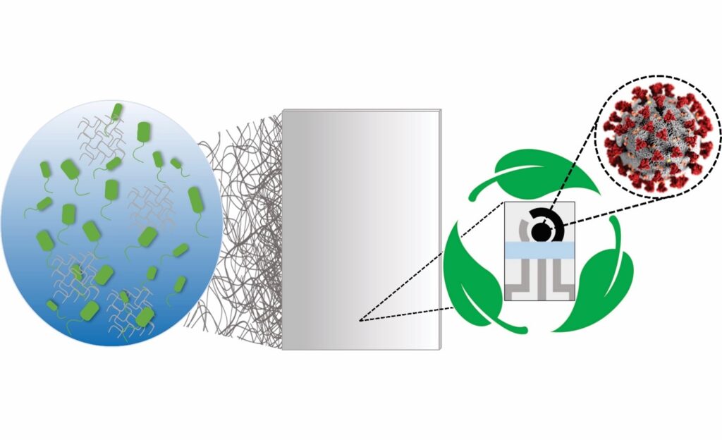
Fabrication steps of the biodegradable BC substrate and the electrochemical devices. (1) Incubation of the bacterium Gluconacetobacter hansenii. (2) BC substrate collected and treated, resulting in a clear sheet. (3) The biodegradable BC sheet is screen-printed, (4) resulting in a device with 3 electrodes, (4) which are cut out using a scissor, (5) resulting in a portable, biodegradable, and inexpensive electrochemical sensor.

The availability of rapid, accessible testing was integral to overcoming the worst surges of the COVID-19 pandemic, and will be necessary to keep up with emerging variants. However, these tests come with unfortunate costs.

Polymerase chain reaction (PCR) tests, the “gold standard” for diagnostic testing, are hampered by waste. They require significant time (results can take up to a day or more) as well as specialized equipment and labor, all of which increase costs. The sophistication of PCR tests makes them harder to tweak, and therefore slower to respond to new variants. They also carry environmental impacts. For example, most biosensor tests developed to date use printed circuit boards, or PCBs, the same materials used in computers. PCBs are difficult to recycle and slow to biodegrade, using large amounts of metal, plastic and non-eco-friendly materials.

In addition, most PCR tests end up in landfills, resulting in material waste and secondary contamination. An analysis by the World Health Organization (WHO) estimated that, as of February 2022, “over 140 million test kits, with a potential to generate 2,600 tonnes of non-infectious waste (mainly plastic) and 731,000 litres of chemical waste (equivalent to one-third of an Olympic-size swimming pool) have been shipped.”

In order to balance the need for fast, affordable and accurate testing while addressing these environmental concerns, César de la Fuente, Presidential Assistant Professor in Bioengineering and Chemical and Biomolecular Engineering in the School of Engineering and Applied Science, with additional primary appointments in Psychiatry and Microbiology within the Perelman School of Medicine, has turned his attention to the urgent need for “green” testing materials.

The de la Fuente lab has been working on creative ways to create faster and cheaper testing for COVID-19 since the outbreak of the pandemic. Utilizing his lab’s focus on machine biology and the treatment of infectious disease, they created RAPID, an aptly named test that generates results in minutes with a high degree of accuracy. An even more cost-effective version, called LEAD, was created using electrodes made from graphite. A third test, called COLOR, was a low-cost optodiagnostic test printed on cotton swabs.

The team’s latest innovation incorporates the speed and cost-effectiveness of previous tests with eco-friendly materials. In a paper published in Cell Reports Physical Science, the group introduces a new test made from Bacterial Cellulose (BC), an organic compound synthesized from several strains of bacteria, as a substitute for PCBs.

Read the full story in Penn Engineering Today.

For a New Generation of Antibiotics, Scientists are Bringing Extinct Molecules Back to Life – and Discovering the Hidden Genetics of Immunity Along the Way

by Devorah Fischler

Marrying artificial intelligence with advanced experimental methods, the Machine Biology Group has mined the ancient past for future medical breakthroughs, bringing extinct molecules back to life. (Image credit: Ella Marushchenko)

“We need to think big in antibiotics research,” says Cesar de la Fuente. “Over one million people die every year from drug-resistant infections, and this is predicted to reach 10 million by 2050. There hasn’t been a truly new class of antibiotics in decades, and there are so few of us tackling this issue that we need to be thinking about more than just new drugs. We need new frameworks.”

De la Fuente is Presidential Assistant Professor in the Department of Bioengineering and the Department of Chemical and Biomolecular Engineering at the University of Pennsylvania School of Engineering and Applied Science. He holds additional primary appointments in Psychiatry and Microbiology in the Perelman School of Medicine.

De la Fuente’s lab, the Machine Biology Group, creates these new frameworks using potent partnerships in engineering and the health sciences, drawing on the “power of machines to accelerate discoveries in biology and medicine.”

Marrying artificial intelligence with advanced experimental methods, the group has mined the ancient past for future medical breakthroughs. In a recent study published in Cell Host and Microbe, the team has launched the field of “molecular de-extinction.”

Our genomes – our genetic material – and the genomes of our ancient ancestors, express proteins with natural antimicrobial properties. “Molecular de-extinction” hypothesizes that these molecules could be prime candidates for safe new drugs. Naturally produced and selected through evolution, these molecules offer promising advantages over molecular discovery using AI alone.

In this paper, the team explored the proteomic expressions of two extinct organisms –Neanderthals and Denisovans, archaic precursors to the human species – and found dozens of small protein sequences with antibiotic qualities. Their lab then worked to synthesize these molecules, bringing these long-since-vanished chemistries back to life.

“The computer gives us a sequence of amino acids,” says de la Fuente. “These are the building blocks of a peptide, a small protein. Then we can make these molecules using a method called ‘solid-phase chemical synthesis.’ We translate the recipe of amino acids into an actual molecule and then build it.”

The team next applied these molecules to pathogens in a dish and in mice to test the veracity and efficacy of their computational predictions.

“The ones that worked, worked quite well,” continues de la Fuente. “In two cases, the peptides were comparable – if not better – than the standard of care. The ones that didn’t work helped us learn what needed to be improved in our AI tools. We think this research opens the door to new ways of thinking about antibiotics and drug discovery, and this first step will allow scientists to explore it with increasing creativity and precision.”

Read the full story in Penn Engineering Today.

“QR Code for Cancer Cells” – Uncovering Why Some Cells Become Resistant to Anti-Cancer Therapies

by Win Reynolds

QR codeA research team led by engineers at the University of Pennsylvania and Northwestern University scientists has created a new synthetic biology approach, or a “QR code for cancer cells,” to follow tumor cells over time, finding there are meaningful differences in why a cancer cell dies or survives in response to anti-cancer therapies.
Remarkably, what fate cancer cells choose after months of therapy is “entirely predictable” based on seemingly small, yet important, differences that appear even before treatment begins. The researchers also discovered the reason is not genetics, contrary to beliefs held in the field.

The findings were recently published in Nature.

The study outlined the team’s new technology platform that developed a QR code for each of the millions of cells for scientists to find and use later — much like tagging swans in a pond. The QR code directs researchers to a genome-wide molecular makeup of these cells and provides information about how they’ve reacted to cancer treatment.

“We think this work stands to really change how we think about therapy resistance,” said Arjun Raj, co-senior author and Professor in Bioengineering in the School of Engineering and Applied Science at the University of Pennsylvania. “Rather than drug-resistant cells coming in just one flavor, we show that even in highly controlled conditions, different ‘flavors’ can emerge, raising the possibility that each of these flavors may need to be treated individually.”

In the study, the lab and collaborators sought to apply synthetic biology tools to answer a key question in cancer research: What makes certain tumors come back a few months or years after therapy? In other words, could the lab understand what causes some rare cells to develop therapeutic resistance to a drug?

“There are many ways cells become different from each other,” said Yogesh Goyal, the co-senior author at Northwestern University. “Our lab asks, how do individual cells make decisions? Understanding this in the context of cancer is all the more exciting because there’s a clinically relevant dichotomy: A cell dies or becomes resistant when faced with therapies.”

Using the interdisciplinary team, the scientists put the before-and-after cloned cells through a whole genome sequencing pipeline to compare the populations and found no systematic underlying genetic mutations to investigate the hypothesis. Raj and Goyal  helped develop the QR code framework, FateMap, that could identify each unique cell that seemed to develop resistance to drug therapy. “Fate” refers to whether a cell dies or survives (and if so, how), and the scientists “map” the cells across their lifespan, prior to and following anti-cancer therapy. FateMap is the result of work from several research institutions, and it applies an amalgamation of concepts spanning several disciplines, including synthetic biology, genome engineering, bioinformatics, machine learning and thermodynamics.

“Some are different by chance — just as not all leaves on a tree look the same — but we wanted to determine if that matters,” Goyal said. “The cell biology field has a hard time defining if differences have meaning.”

Read the full story in Penn Engineering Today.

AI-guided Brain Stimulation Aids Memory in Traumatic Brain Injury

by Erica Moser

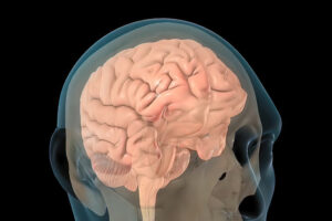
Illustration of a human brain
Image: iStock/Ogzu Arslan

Traumatic brain injury (TBI) has disabled 1 to 2% of the population, and one of their most common disabilities is problems with short-term memory. Electrical stimulation has emerged as a viable tool to improve brain function in people with other neurological disorders.

Now, a new study in the journal Brain Stimulation shows that targeted electrical stimulation in patients with traumatic brain injury led to an average 19% boost in recalling words.

Led by University of Pennsylvania psychology professor Michael Jacob Kahana, a team of neuroscientists studied TBI patients with implanted electrodes, analyzed neural data as patients studied words, and used a machine learning algorithm to predict momentary memory lapses. Other lead authors included Wesleyan University psychology professor Youssef Ezzyat and Penn research scientist Paul Wanda.

“The last decade has seen tremendous advances in the use of brain stimulation as a therapy for several neurological and psychiatric disorders including epilepsy, Parkinson’s disease, and depression,” Kahana says. “Memory loss, however, represents a huge burden on society. We lack effective therapies for the 27 million Americans suffering.”

Read the full story in Penn Today.

Michael Kahana is the Edmund J. and Louise W. Kahn Term Professor of Psychology at the University of Pennsylvania. He is a member of the Penn Bioengineering Graduate Group.

Artificial Intelligence is Leveling Up the Fight Against Infectious Diseases

by

Image credit: NIAID

Artificial intelligence is a new addition to the infectious disease researcher’s toolbox. Yet in merely half a decade, AI has accelerated progress on some of the most urgent issues in medical science and public health. Researchers in this field blend knowledge of life sciences with skill in computation, chemistry and design, satisfying decades-long appeals for interdisciplinary tactics to treat these disorders and stop their spread.

Diseases are “infectious” when they are caused by organisms, including parasites, viruses, bacteria and fungi. People and animals can contract infectious diseases from their environments or food, or through interactions with one another. Some, but not all, are contagious.

Infectious diseases are an intractable global challenge, posing problems that continue to grow in severity even as science has offered a steady pace of solutions. The world continues to become more interconnected, bringing people into new kinds and levels of relation, and the climate crisis is throwing environmental and ecological networks out of balance. Diseases that were once treatable by drugs have become resistant, and new drug discovery is more costly than ever. Uneven resource distribution means that certain parts of the world are perennial hotspots for diseases that others never fear.

Cesar de la Fuente brings an expert eye to how AI has transformed infectious disease research in a recently published piece in Science with co-authors Felix Wong and James J. Collins from MIT.

Presidential Assistant Professor in the Department of Bioengineering and the Department of Chemical and Biomolecular Engineering at the University of Pennsylvania School of Engineering and Applied Science, with additional primary appointments in Psychiatry and Microbiology within the Perelman School of Medicine, de la Fuente brings a multifaceted perspective to his survey of the field.

In the paper, de la Fuente and co-authors assess the progress, limitations and promise of research in AI and infectious diseases in three major areas of inquiry: anti-infective drug discovery, infection biology, and diagnostics for infectious diseases.

Read more in Penn Engineering Today.

The Art and Science of Living-Like Architecture

by

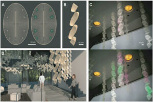
Collaborators from Penn Engineering and the Stuart Weitzman School of Design have created “living-like” bioactive interior architecture designed to one day protect us from hidden airborne threats. The figure above demonstrates (A) design for support lattices for the team’s innovative bioactive sites, (B) a ribbon-like geometry for hanging and (C, D) how these structures may be integrated into indoor environments to biologically sense and react to air.

“This technology is not alive,” says Laia Mogas-Soldevila. “It is living-like.”

The distinction is an important one for the assistant professor at the Stuart Weitzman School of Design, for reasons both scientific and artistic. With a doctorate in biomedical engineering, several degrees in architecture, and a devotion to sustainable design, Mogas-Soldevila brings biology to everyday life, creating materials for a future built halfway between nature and artifice.

The architectural technology she describes is unassuming at first look: A freeze-dried pellet, small enough to get lost in your pocket. But this tiny lump of matter, the result of more than a year’s collaboration between designers, engineers and biologists, is a biomaterial that contains a “living-like” system.

When touched by water, the pellet activates and expresses a glowing protein, its fluorescence demonstrating that life and art can harmonize into a third and very different thing, as ready to please as to protect. Woven into lattices made of flexible natural materials promoting air and moisture flow, the pellets form striking interior design elements that could one day keep us healthy.

“We envision them as sensors,” explains Mogas-Soldevila. “They may detect pathogens, such as bacteria or viruses, or alert people to toxins inside their home. The pellets are designed to interact with air. With development, they could monitor or even clean it.”

For now, they glow, a triumphant first stop on the team’s roadmap to the future. The fluorescence establishes that the lab’s biomaterial manufacturing process is compatible with the leading-edge cell-free engineering that gives the pellets their life-like properties.

A rapidly expanding technology, cell-free protein expression systems allow researchers to manufacture proteins without the use of living cells.

Gabrielle Ho, Ph.D. candidate in the Department of Bioengineering and co-leader of the project, explains how the team’s design work came to be cell-free, a technique rarely explored outside of lab study or medical applications.

“Typically, we’d use living E. coli cells to make a protein,” says Ho. “E. coli is a biological workhorse, accessible and very productive. We’d introduce DNA to the cell to encourage expression of specific proteins. But this traditional method was not an option for this project. You can’t have engineered E. coli hanging on your walls.”

Cell-free systems contain all the components a living cell requires to manufacture protein —energy, enzymes and amino acids — and not much else. These systems are therefore not alive. They do not replicate, and neither can they cause infection. They are “living-like,” designed to take in DNA and push out protein in ways that previously were only possible using living cells.

“One of the nicest things about these materials not being alive,” says Mogas-Soldevila, “is that we don’t need to worry about keeping them that way.”

Unlike living cells, cell-free materials don’t need a wet environment or constant monitoring in a lab. The team’s research has established a process for making these dry pellets that preserves bioactivity throughout manufacturing, storage and use.

Bioactive, expressive and programmable, this technology is designed to capitalize on the unique properties of organic materials.

Mogas-Soldevila, whose lab focuses exclusively on biodegradable architecture, understands the value of biomaterials as both environmentally responsible and aesthetically rich.

“Architects are coming to the realization that conventional materials — concrete, steel, glass, ceramic, etc. — are environmentally damaging and they are becoming more and more interested in alternatives to replace at least some of them. Because we use so much, even being able to replace a small percentage would result in a significant reduction in waste and pollution.”

Her lab’s signature materials — biopolymers made from shrimp shells, wood pulp, sand and soil, silk cocoons, and algae gums — lend qualities over and above their sustainable advantages.

“My obsession is diagnostic, but my passion is playfulness,” says Mogas-Soldevila. “Biomaterials are the only materials that can encapsulate this double function observed in nature.”

This multivalent approach benefited from the help of Penn Engineering’s George H. Stephenson Foundation Educational Laboratory & Bio-MakerSpace, and the support of its director, Sevile Mannickarottu. In addition to contributing essential equipment and research infrastructure to the team, Mannickarottu was instrumental in enabling the interdisciplinary relationships that led the team to success, introducing Ho to the DumoLab Research team collaborators. These include Mogas-Soldevila, Camila Irabien, a Penn Biology major who provided crucial contributions to experimental work, and Fulbright design fellow Vlasta Kubušová, who co-led the project during her time at Penn and who will continue fueling the project’s next steps.

Read the full story in Penn Engineering Today.