Carl June, MD, Professor in the Perelman School of Medicine and member of the Penn Bioengineering Graduate Group, was quoted in a recent press release announcing a new international partnership between Penn Medicine (PSOM), the Children’s Hospital of Pennsylvania (CHOP), and Costa Rica’s CCSS, or the Caja Costarricense de Seguro Social (Social Security Program), to develop CAR T research in Costa Rica. June is a world renowned cancer cell therapy pioneer whose research led to the initial development and FDA approval of CAR T cell therapy:
“‘At least 15,000 patients across the world have received CAR T cells, and dozens more clinical trials using this approach are in progress, for almost every major tumor type, but people in many parts of the globe still do not have access to treatment with these transformative therapies,’ said Carl H. June, MD, the Richard W. Vague Professor in Immunotherapy and director of the Center for Cellular Immunotherapies in Penn’s Perelman School of Medicine. “We are honored to work with our colleagues in Costa Rica in hopes of building a path for patients in underserved areas to have the opportunity to benefit from clinical research programs offering this personalized therapy.’”
Yale E. Cohen, Professor of Otorhinolaryngology, with secondary appointments in Neuroscience and Bioengineering, was appointed Assistant Dean of Research Facilities and Resources at the Perelman School of Medicine at the University of Pennsylvania, effective April 1, 2022. Cohen is currently Chair of the Penn Bioengineering Graduate Group, and Director of the Hearing Sciences Center:
“Many of you are already quite familiar with Dr. Cohen, as his leadership roles in research training and education at PSOM and the University are far-reaching and impactful. Dr. Cohen is a Professor of Otorhinolaryngology with secondary appointments in the Department of Neuroscience and Engineering’s Department of Bioengineering. Recognized widely for his deep commitment to our teaching and training community, Dr. Cohen chairs the Bioengineering Graduate Group, and in 2020 received the prestigious Jane M. Glick Graduate Student Teaching Award, which honors clinicians and scientists who exemplify outstanding quality of patient care, mentoring, research, and teaching.”
Kevin B. Johnson, David L. Cohen University Professor in Biostatistics, Epidemiology and Informatics and in Computer and Information Science, has been elected to the 2022 Class of the American Institute for Medical and Biological Engineering (AIMBE) Fellows. Johnson joined the Penn faculty in 2021. He also holds secondary appointments in Bioengineering, in Pediatrics, and in the Annenberg School for Communication, and is the Vice President for Applied Informatics for the University of Pennsylvania Health System.
Election to the AIMBE College of Fellows is among the highest professional distinctions accorded to a medical and biological engineer. College membership honors those who have made outstanding contributions to “engineering and medicine research, practice, or education” and to “the pioneering of new and developing fields of technology, making major advancements in traditional fields of medical and biological engineering, or developing/implementing innovative approaches to bioengineering education.”
Johnson was nominated, reviewed, and elected by peers and members of the AIMBE College of Fellows for his pioneering discoveries in clinical informatics, leading to advances in data acquisition, medication management, and information aggregation in medical settings.
A formal induction ceremony was held during AIMBE’s 2022 Annual Event on March 25, 2022. Johnson was inducted along with 152 colleagues who make up the AIMBE Fellow Class of 2022. For more information about the AIMBE Annual Event, please visit www.aimbe.org.
Read Johnson’s AIMBE election press release here. Find the full list of 2022 Fellows here.
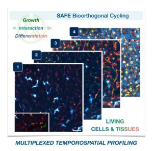
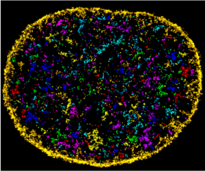
Cells in complex organisms undergo frequent changes, and researchers have struggled to monitor these changes and create a comprehensive profile for living cells and tissues. Historically researchers have been limited to only 3-5 markers due to spectral overlaps in fluorescence microscopy, an essential tool required for imaging cells. With only this small handful of markers, it is difficult to monitor protein expressions of live cells and a comprehensive profile of cellular dynamics cannot be created. However, a new study in Nature Biotechnology addresses these limitations by demonstrating a new method for comprehensive profiling of living cells.
Jina Ko, PhD
Jina Ko, Assistant Professor in Bioengineering in the School of Engineering and Applied Science and in Pathology and Laboratory Medicine in the Perelman School of Medicine, conducted postdoctoral research at Massachusetts General Hospital (MGH) and the Wyss Institute at Harvard University, and the work for this study was done under the supervision of Jonathan Carlson M.D., Ph.D. and Ralph Weissleder M.D., Ph.D. of MGH. Ko’s lab at Penn develops novel technologies using bioengineering, molecular biology, and chemistry to address diagnostic challenges for precision medicine.
To address these limitations in microscopy, the team developed a new chemistry tool which was highly gentle to cells. This “scission-accelerated fluorophore exchange (or SAFE)” method utilizes “click” chemistry, a type of chemistry that follows examples found in nature to create fast and simple reactions. This new SAFE method functions with non-toxic conditions to living cells and tissues, whereas previous methods have used harsh chemicals that would strip off fluorophores and consequently would not work with living cells and tissues.
With the development of SAFE, the authors demonstrated that researchers can now effectively perform multiple cycles of cell profiling and can monitor cellular changes over the course of their observations. Instead of the previous limitation of 3-5 markers total, SAFE allows for many more cycles and can keep track of almost as many markers as the researcher wants. One can now stain cells and quench/release fluorophores and repeat the cycle multiple times for multiplexing on living cells. Each cycle can profile 3 markers, and so someone interested in profiling 15 markers could easily perform 5 cycles to achieve this much more comprehensive cell profile. With this breakthrough in more detailed imaging of cells, SAFE demonstrates broad applicability for allowing researchers to better investigate the physiologic dynamics in living systems.
This study was supported by the Schmidt Science Fellows in Partnership with the Rhodes Trust and National Institutes of Health, National Cancer Institute (K99CA256353).
She joins 28 early-career scientists from around the world in this year’s cohort, with each receiving support for one to two years, $100,000 in salary support per year, individualized mentoring, and a series of professional development sessions as they pivot to the next stages of their research agendas.
The fellowship is a program of Schmidt Futures, the philanthropic initiative of Eric and Wendy Schmidt that aims to tackle society’s toughest challenges by supporting interdisciplinary researchers at the start of their careers.
“Our latest group of Schmidt Science Fellows embodies our vision for this Program at its inception five years ago,” says Eric Schmidt, co-founder of Schmidt Futures and former CEO and Chairman of Google. “We find the most talented next-generation leaders from around the world and back these impressive young adults with the resources and networks they need to realize their full potential while addressing some of the big scientific questions facing the world. Congratulations to the 2022 Schmidt Science Fellows, I am excited to see where your science takes you and what you will achieve.”
Working at the intersection of materials science, biology, and applied clinical research, Zlotnick’s postdoctoral work will involve developing advanced bioprinting techniques for regenerative medicine. Such advances are necessary to recreate the multi-cellular composition of orthopedic tissues, such as those found in the knee joint. Lab-grown tissue models can then be used to broaden our understanding of how degenerative diseases progress after injury, limit the need for animal models, and serve as a platform for therapeutic discovery.
The U.S. Food and Drug Administration has expanded its approval for Kymriah, a personalized cellular therapy developed at the Abramson Cancer Center, this time for the treatment of adults with relapsed/refractory follicular lymphoma who have received at least two lines of systemic therapy. “Patients with follicular lymphoma who relapse or don’t respond to treatment have a poor prognosis and may face a series of treatment options without a meaningful, lasting response,” said Stephen J. Schuster, the Robert and Margarita Louis-Dreyfus Professor in Chronic Lymphocytic Leukemia and Lymphoma in the Division of Hematology Oncology. It’s the third FDAapproval for the “living drug,” which was the first of its kind to be approved, in 2017, and remains the only CAR T cell therapy approved for both adult and pediatric patients.
“In just over a decade, we have moved from treating the very first patients with CAR T cell therapy and seeing them live healthy lives beyond cancer to having three FDA-approved uses of these living drugs which have helped thousands of patients across the globe,” said Carl June, MD, the Richard W. Vague Professor in Immunotherapy in the department of Pathology and Laboratory Medicine in Penn’s Perelman School of Medicine and director of the Center for Cellular Immunotherapies in the Abramson Cancer Center and director of the Parker Institute for Cancer Immunotherapy at Penn. “Today’s news is new fuel for our work to define the future of cell therapy and set new standards in harnessing the immune system to treat cancer.”
Research from June, a member of the Penn Bioengineering Graduate Group, led to the initial FDA approval for the CAR T therapy (sold by Novartis as Kymriah) for treating acute lymphoblastic leukemia (ALL), one of the most common childhood cancers.
The dynamics governing mechanointelligence vary greatly along time- and length-scales, so detailed models of individual cells and their components are necessary to connect the effects of their physical environments to the downstream effects those forces have on biological processes.
The National Science Foundation’s Science and Technology Center (STC) program is its flagship funding mechanism for organizing interdisciplinary research on cutting-edge topics. Penn’s Center for Engineering MechanoBiology (CEMB) is one of the 18 active STCs, bringing together dozens of researchers from Penn Engineering and the Perelman School of Medicine, as well as others spread across campus and at partner institutions around the world.
With its NSF funding now renewed for another five years, the Center is entering into a new phase of its mission, centered on the nascent concept of “mechanointelligence.”
Mechanobiology is the study of the physical forces that govern the behavior of cells and their communication with their neighbors. Mechanointelligence adds another layer of complexity, attempting to understand the forces that allow cells to sense, remember and adapt to their environments.
Ultimately, harnessing these forces would allow researchers to help multicellular organisms — plants, animals and humans — better adapt to their environments as well.
“Mechanointelligence is a key element of a cell’s ability to survive and reproduce,” says CEMB Director and Eduardo D. Glandt President’s Distinguished Professor Vivek Shenoy. “Just like with complex organisms, a cell’s ‘fitness’ depends on its environment, and adapting means rewiring how its genes are expressed.”
Vivek Shenoy is Eduardo D. Glandt President’s Distinguished Professor in Materials Science and Engineering, Bioengineering and Mechanical Engineering and Applied Mechanics.
César de la Fuente, Presidential Assistant Professor in Psychiatry, Bioengineering, Microbiology, and in Chemical and Biomolecular Engineering has been honored with a 2022 Young Investigator Award by the Royal Spanish Society of Chemistry (RSEQ) for his pioneering research efforts to combine the power of machines and biology to help prevent, detect, and treat infectious diseases.
A new feature in Chemistry World explores the history of CAR (chimeric antigen receptor)-T cell therapy, a revolutionary type of therapeutic treatment for certain types of cancer. One of the pioneers of CAR-T cell therapy is Carl June, Richard W. Vague Professor in Immunotherapy in the Perelman School of Medicine and member of the Penn Bioengineering Graduate Group. His groundbreaking research opened the door for FDA approval of the CAR T therapy called Kymriah, which treats acute lymphoblastic leukemia (ALL), one of the most common childhood cancers.
The Solomon R. Pollack Award for Excellence in Graduate Bioengineering Research is given annually to the most deserving Bioengineering graduate students who have successfully completed research that is original and recognized as being at the forefront of their field. This year Penn Bioengineering recognizes the outstanding work of two graduate students in Bioengineering: Erin Berlew and Rhea Chitalia.
Erin Berlew, Ph.D. candidate in Bioengineering
Erin Berlew is a Ph.D. candidate in the lab of Brian Chow, Associate Professor in Bioengineering. She successfully defended her thesis, titled “Single-component optogenetic tools for cytoskeletal rearrangements,” in December 2021. In her research, she used the BcLOV4 optogenetic platform discovered/developed in the Chow lab to control RhoGTPase signaling. Erin earned a B.S. in Chemistry from Haverford College in 2015 and was an Americorps member with City Year Philadelphia from 2015-2016. “Erin is a world-class bioengineering with an uncommon record of productivity gained through her complementary expertise in molecular, cellular, and computational biology,” says Chow. “She embodies everything wonderful, both academically and culturally, about our graduate program and its distinguished history.” Erin’s hobbies outside the lab include spending time with family, reading mystery novels, enjoying Philadelphia, and crossword puzzles. In the future, she hopes to continue to teach for the BE department (she has already taught ENGR 105 and served as a TA for undergraduate and graduate courses) and to conduct further research at Penn.
Rhea Chitalia, Ph.D. candidate in Bioengineering
Rhea Chitalia is a Ph.D. candidate in Bioengineering and a member of the Computational Biomarker Imaging Group (CBIG), advised by Despina Kontos, Matthew J. Wilson Associate Professor of Research Radiology II in the Perelman School of Medicine. Rhea completed her B.S.E. in Biomedical Engineering at Duke University in 2015. Her doctoral research concerns leveraging machine learning, bioinformatics, and computer vision to develop computational imaging biomarkers for improved precision cancer care. In December 2021 she successfully defended her thesis titled “Computational imaging biomarkers for precision medicine: characterizing intratumor heterogeneity in breast cancer.” “It has been such a privilege to mentor Rhea on her dissertation research,” says Kontos. “Rhea has been a star graduate student. Her work has made fundamental contributions in developing computational methods that will allow us to gain important insight into tumor heterogeneity by utilizing a multi-modality imaging approach.” David Mankoff, Matthew J. Wilson Professor of Research Radiology in the Perelman School of Medicine, served as Rhea’s second thesis advisor. “It was a true pleasure for me to work with Rhea and to Chair her BE Thesis Committee,” Mankoff adds. “Rhea’s Ph.D. thesis and thesis presentation was one of the best I have had the chance to be involved with in my graduate mentoring career.” After graduation, Rhea hopes to further precision medicine initiatives through the use of real world, multi-omic data in translational industry settings. She will be joining Invicro as an Imaging Scientist. In her spare time, Rhea enjoys trying new restaurants, reading, and spending time with friends and family.