Kathleen J. Stebe and Michel Koo urge “the academic community to adopt a coordinated approach uniting dental medicine and engineering to support research, training and entrepreneurship to address unmet needs and spur oral health care innovations.” (Image: Min Jun Oh and Seokyoung Yoon)
Penn’s Center for Innovation & Precision Dentistry (CiPD) is the first cross-disciplinary initiative in the nation to unite oral-craniofacial health sciences and engineering.
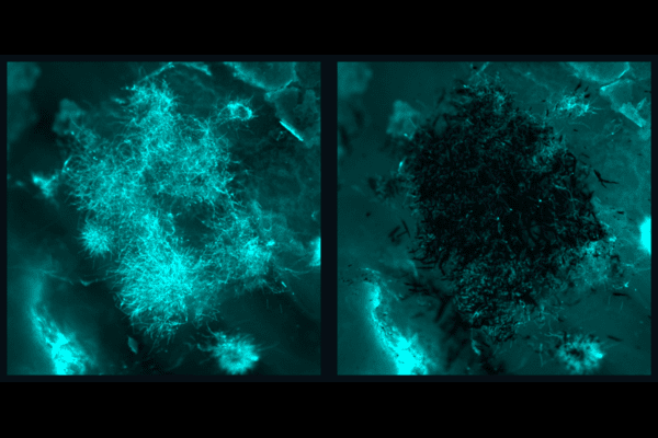
In just two years since CiPD was founded, the outcomes of this newly conceived research partnership have proven its value: microrobots that clean teeth for people with limited mobility, a completely new understanding of bacterial physics in tooth decay, enzymes from plant chloroplasts that degrade plaque, promising futures for lipid nanoparticles in oral cancer treatment and new techniques and materials to restore nerves in facial reconstructive surgery.
In addition, CiPD is training the next generation of dentists, scientists and engineers through an NIH/NIDCR-sponsored postdoctoral training program as well as fellowships from industry.
The center’s Founding Co-Directors, Kathleen J. Stebe, Richer & Elizabeth Goodwin Professor in Chemical and Biomolecular Engineering, and Michel Koo, Professor of Orthodontics in Penn Dental Medicine, published an editorial in the Journal of Dental Research, planting a flag for CiPD’s mission and encouraging others to mirror its method.
The two urge “the academic community to adopt a coordinated approach uniting dental medicine and engineering to support research, training and entrepreneurship to address unmet needs and spur oral health care innovations.”
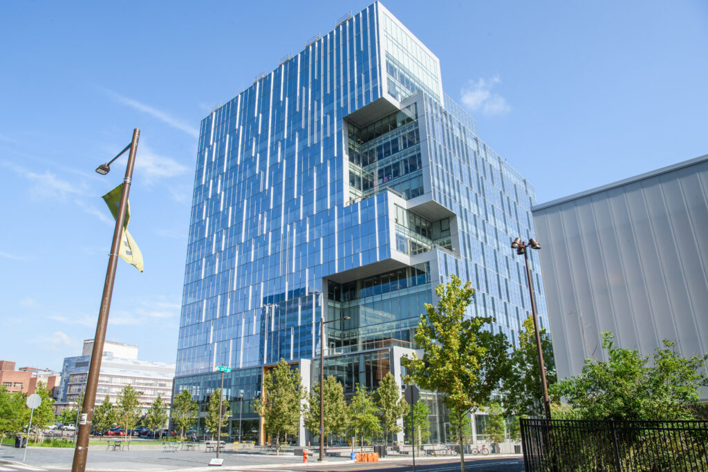
On September 14, Wexford Science & Technology, LLC and the University of Pennsylvania announced that the University has signed a lease for new laboratory space that will usher in a wave of novel vaccine, therapeutics, and engineered diagnostics research to West Philadelphia. Research teams from Penn are poised to move into 115,000 square feet of space at One uCity Square, the 13-story, 400,000 square foot purpose-built lab and office building within the vibrant uCity Square Knowledge Community being developed by Wexford. This is the largest lease in the building, encompassing four floors, and bringing the building to over 90% leased. The building currently includes industry tenants Century Therapeutics (NASDAQ: IPSC), Integral Molecular, Exponent (NASDAQ: EXPO), and Charles River Laboratories (NYSE: CRL).
The new University space will house Penn Medicine’s Institute for RNA Innovation and Penn Engineering’s Center for Precision Engineering for Health, underscoring the University’s commitment to a multi-disciplinary and collaborative approach to research that will attract and retain the best talent and engage partners from across the region. Penn’s decision to locate at One uCity Square reinforces uCity Square’s evolution as a central cluster of academic, clinical, commercial, entrepreneurial, and amenity spaces for the area’s innovation ecosystem, and further cements Philadelphia’s position as a top life sciences market.
Jonathan Epstein, MD, Executive Vice Dean and Chief Scientific Officer of Penn Medicine, shared his anticipation for the opportunities that lie ahead: “Penn Medicine is proud to build on its existing clinical presence in uCity Square and establish an innovative and collaborative research presence at the heart of uCity Square’s multidisciplinary innovation ecosystem. This strategic move underscores our commitment to accelerating advancements in biomedical research, industry collaboration, and equipping our talented teams with the resources they need to shape the future of healthcare.”
Locating the Penn Institute for RNA Innovation in the heart of the uCity Square community brings together researchers across disciplines who are already pursuing new vaccines and treatments, and better ways to deliver them. Their shared work will help to power the next phase of vaccine discovery and development.
Likewise, anchoring the work of Penn Engineering’s Center in the One uCity Square space will allow the School’s multi-disciplinary researchers and their collaborators to advance new clinical and diagnostic methods that will focus on intelligent therapeutics, genome design, diagnostics for discovery of human biology, and engineering the human immune shield.
“Penn Engineering has made a substantial commitment to precision engineering for health, an area that is not only important and relevant to engineering, but also critical to the future of humanity,” said Vijay Kumar, Nemirovsky Family Dean of Penn Engineering. “The space in One uCity Square will add another 30,000 square feet of space for our engineers to develop technologies that will fight future pandemics, cure incurable diseases, and extend healthy life spans around the world.”
Spearheading the Penn Institute for RNA Innovation will be Drew Weissman, MD, PhD, the Roberts Family Professor for Vaccine Research, who along with Katalin Karikó, PhD, adjunct professor of Neurosurgery, discovered foundational mRNA technology that enabled the creation of vital vaccine technology, including the FDA-approved mRNA-based COVID-19 vaccines developed by Pfizer-BioNTech and Moderna.
In this new space at One uCity Square, Weissman and his research team and collaborators will further pursue their groundbreaking research efforts with a goal to develop new therapeutics and vaccines and initiate clinical trials for other devastating diseases.
In addition, two established researchers will join the Institute at One uCity Square: Harvey Friedman, MD, a professor of Infectious Diseases, who leads a team researching various vaccines. He will be joined by Vladimir Muzykantov, MD, PhD, Founders Professor in Nanoparticle Research, who focuses on several projects related to targeting the delivery of drugs, including mRNA, to create more effective, targeted pathways to deliver drugs to the vascular system, treating a wide range of diseases that impact the brain, lung, heart, and blood.
Dan Hammer, Alfred G. and Meta A. Ennis Professor in the Departments of Bioengineering and Chemical and Biomolecular Engineering in Penn Engineering and Director of the Center for Precision Engineering for Health, will oversee the Center’s innovations in diagnostics and delivery, cellular and tissue engineering, and the development of new devices that integrate novel materials with human tissues. The Center will bring together scholars from all departments within Penn Engineering and will help to foster increased collaboration with campus colleagues at Penn’s Perelman School of Medicine and with industry partners.
Joining the Center researchers in One uCity Square are Noor Momin, Sherry Gao, and Michael Mitchell. Noor Momin, who will join Penn Engineering in early 2024 as an assistant professor in Bioengineering, will leverage her lab’s expertise in cardiovascular immunology, protein engineering and pharmacokinetic modeling to develop next-generation treatments and diagnostics for cardiovascular diseases.
The award recognizes faculty who are conducting some of the most innovative and impactful studies in the field of biomedical engineering. Recipients will present their research and be officially recognized at the BMES Annual Meeting in October.
Mitchell is being honored for creating an RNA nanoparticle therapy that stops the spread of the deadly bone marrow cancer multiple myeloma and helps to eliminate it altogether. Known for being difficult to treat, the disease kills over 100,000 people every year.
“We urgently need innovative, effective therapies against this cancer,” Mitchell says. “The nanotechnology we developed can potentially serve as a platform to treat multiple myeloma and other bone marrow-based malignancies.”
Mitchell, along with Christian Figuerora-Espada, a doctoral student in Bioengineering, previously published a study in PNAS describing how their RNA nanoparticle therapy stops multiple myeloma from moving through the blood vessels and mutating. In their current paper in Cellular and Molecular Bioengineering, which expands upon this RNA nanoparticle platform, they show that inhibition of both multiple myeloma migration and adhesion to bone marrow blood vessels, combined with an FDA-approved multiple myeloma therapeutic, extends survival in a mouse model of multiple myeloma.
Gabriella Daltoso, Sophie Ishiwari, Gabriela Cano, Caroline Amanda Magro, and Tifara Eliana Boyce
A team of recent Penn Bioengineering graduates have been included in list of prominent young Philadelphia innovators as chosen by The Philadelphia Business Journal and PHL Inno.
Gabriella Daltoso, Sophie Ishiwari, Gabriela Cano, Caroline Amanda Magro, and Tifara Eliana Boyce founded Sonura as their Senior Design Project in Bioengineering. The team, who all graduated in 2023, picked up a competitive President’s Innovation Prize for their beanie that promotes the cognitive and socioemotional development of newborns in the NICU by protecting them from the auditory hazards of their environments while fostering parental connection. Now, they have been included in the list of fourteen Inno Under 25 honorees for 2023.
“To determine this year’s list, the Philadelphia Business Journal and PHL Inno sought nominations from the public and considered candidates put forth by our editorial team. To be considered, nominees must be 25 years of age or younger and work for a company based in Greater Philadelphia and/or reside in the region.
Honorees span a wide range of industries, including consumer goods, biotechnology and environmental solutions. Many are products of the region’s colleges and universities, though some studied farther afield before setting up shop locally.”
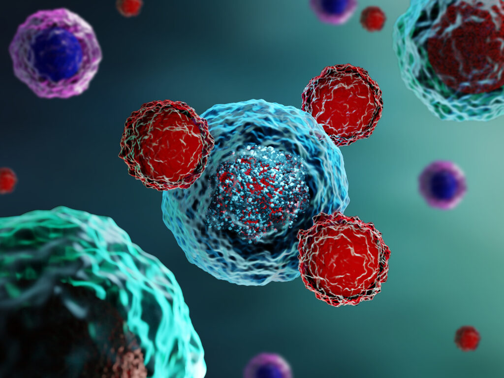
Chimeric antigen receptor T cell (CAR T) therapy has delivered promising results, transforming the fight against various forms of cancer, but for many, the therapy comes with severe and potentially lethal side effects. Now, a research team led by Michael Mitchell of the School of Engineering and Applied Science has found a solution that could help CAR T therapies reach their full potential while minimizing severe side effects. (Image: iStock / Meletios Verras)
In recent years, cancer researchers have hailed the arrival of chimeric antigen receptor T cell (CAR T) therapy, which has delivered promising results, transforming the fight against various forms of cancer. The process involves modifying patients’ T-cells to target cancer cells, resulting in remarkable success rates for previously intractable forms of cancer.
Six CAR T cell therapies have secured FDA approval, and several more are in the pipeline. However, these therapies come with severe and potentially lethal side effects, namely cytokine release syndrome (CRS) and neurotoxicity. These drawbacks manifest as a range of symptoms—from high fever and vomiting to multiple organ failure and patient death—posing significant challenges to broader clinical application.
“Addressing CRS and neurotoxicity without compromising the therapeutic effectiveness of CAR T cells has been a complex challenge,” says Mitchell.
He says that unwanted interactions between CAR T and immune cells called macrophages drive the overactivation of macrophages, which in turn result in the release of toxic cytokines that lead to CRS and neurotoxicity.
“Controlling CAR T-macrophage interactions in vivo is difficult,” Mitchell says. “So, our study introduces a materials engineering-based strategy that involves incorporating a sugar molecule onto the surface of CAR T cells. These sugars are then used as a reactive handle to create a biomaterial coating around these cells directly in the body, which acts as a ‘suit of armor,’ preventing dangerous interactions with macrophages.”
First author Ningqiang Gong, a postdoctoral researcher in the Mitchell Lab, elaborates on the technique, “We attached this sugar molecule to the CAR T cells using metabolic labeling. This modification enables the CAR T cells to attack cancer cells without any hindrance.”
“When symptoms of CRS begin to manifest, we introduce another molecule—polyethylene glycol (PEG)—to create the suit of armor, which effectively blocks dangerous interactions between these engineered T cells, macrophages, and the tumor cells themselves,” Gong says.
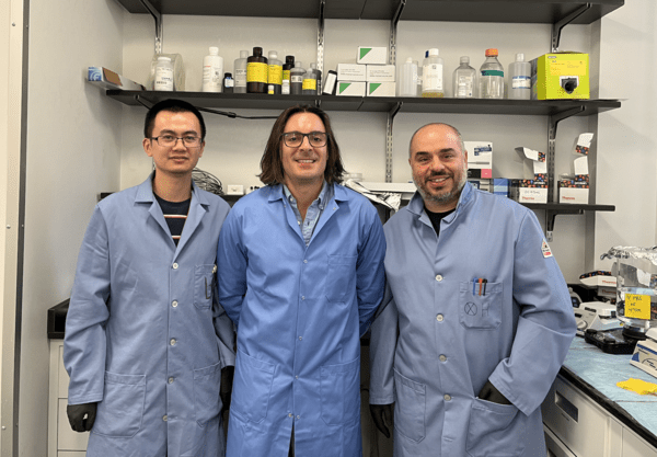
(From left to right) Xuexiang Han, Michael Mitchell and Mohamad-Gabriel Alameh
The COVID-19 vaccine swiftly undercut the worst of the pandemic for hundreds of millions around the world. Available sooner than almost anyone expected, these vaccines were a triumph of resourcefulness and skill.
Messenger RNA vaccines, like the ones manufactured by Moderna or Pfizer/BioNTech, owed their speed and success to decades of research reinforcing the safety and effectiveness of their unique immune-instructive technology.
In addition to outlining a more flexible and effective COVID-19 vaccine, this work has potential to increase the scope of mRNA vaccines writ large, contributing to prevention and treatment for a range of different illnesses.
Michael Mitchell, associate professor in Penn Engineering’s Department of Bioengineering, Xuexiang Han, postdoctoral fellow in Mitchell’s lab, and Mohamad-Gabriel Alameh, postdoctoral fellow in Drew Weissman’s lab at Penn Medicine and incoming assistant professor in the Department of Pathology and Laboratory Medicine at the Perelman School of Medicine, recently published their findings in Nature Nanotechnology.
mRNA, or messenger ribonucleic acid, is the body’s natural go-between. mRNA contains the instructions our cells need to produce proteins that play important roles in our bodies’ health, including mounting immune responses.
The COVID-19 vaccines follow suit, sending a single strand of RNA to teach our cells how to recognize and fight the virus.
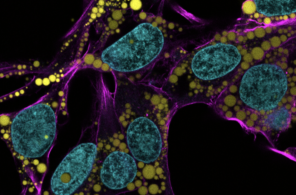
Fat is a normal and necessary part of the body. Fat cells store and release energy, as well as play significant roles in hormonal regulation and immunity.
Engineers and scientists at the University of Pennsylvania are the first to discover fat-filled lipid droplets’ (pictured above in green) surprising capability to indent and puncture the nucleus, the organelle which contains and regulates a cell’s DNA.
In recent decades, a concerning rise in metabolic illnesses – such as cardiovascular disease, high blood pressure and diabetes – has focused scientific attention on the biology and chemistry of fat, resulting in a wealth of information about how fat cells work.
But fat cells and their metabolic activities are only part of the story.
Fat-filled lipid droplets, tiny spheres of fat many times smaller than fat cells, are a growing subject of scientific interest. Found inside many different cell types, these lipid particles have long been little understood. Studies have begun to illuminate these droplets’ participation in metabolic functions and cellular protection, but we still know next to nothing about the physical nature of fat.
Now, researchers at the University of Pennsylvania School of Engineering and Applied Science have looked beyond biochemistry to publish groundbreaking work on the physics of these droplets, revealing them to be a potential threat to a cell’s nucleus. In the August issue of the Journal of Cell Biology, they are the first to discover fat-filled lipid droplets’ surprising capability to indent and puncture the nucleus, the organelle which contains and regulates a cell’s DNA.
The stakes of their findings are high: a ruptured nucleus can lead to elevated DNA damage that is characteristic of many diseases, including cancer.
“Intuitively, people think of fat as soft,” says Discher. “And on a cellular level it is. But at this small size of droplet— measuring just a few microns rather than the hundreds of microns of a mature fat cell—it stops being soft. Its shape has a much higher curvature, bending other objects very sharply. This changes its physics in the cell. It can deform. It can damage. It can rupture.”
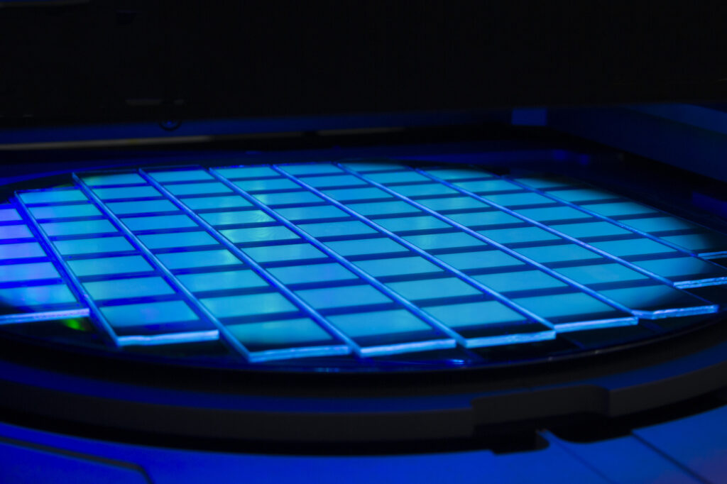
Led by Michael Mitchell and David Issadore of the School of Engineering and Applied Science, a team of researchers has developed a platform that could rapidly accelerate the development of mRNA-based lipid nanoparticle vaccines and therapeutics at both the small and large scale, SCALAR. (Image: iStock / Anatoly Morozov)
Following the global COVID-19 pandemic, the development and rapid deployment of mRNA vaccines highlighted the critical role of lipid nanoparticles (LNPs) in the context of pharmaceuticals. Used as the essential delivery vehicles for fragile RNA-based therapies and vaccines, LNPs protect the RNA from degradation and ensure effective delivery within the body.
Despite their critical importance, the large-scale manufacturing of these LNPs saw numerous bottlenecks during the pandemic, underscoring the need for scalable production techniques that could keep pace with global demand.
Now, in a paper published in the Proceedings of the National Academy of the Sciences, researchers at the University of Pennsylvania describe how the Silicon Scalable Lipid Nanoparticle Generation platform (SCALAR), a reusable silicon- and glass-based platform designed to transform the production landscape of LNPs for RNA therapeutics and vaccines, offers a scalable and efficient solution to the challenges exposed during the COVID-19 crisis.
“We’re excited to create a piece of technology platform that bridges the gap between small-scale discovery and large-scale manufacturing in the realm of RNA lipid nanoparticle vaccines and therapeutics,” says co-author Michael Mitchell, associate professor of bioengineering in the School of Engineering and Applied Science at Penn. “By doing so, we’ve effectively leapfrogged the clunky, time-consuming, and costly barriers that slow down the production ramp-up of promising new RNA medicines and vaccines.”
The intricacies of RNA-based therapies require the RNA to be encased in a delivery system capable of navigating the body’s biological obstacles. LNPs fulfill this role, allowing the RNA to reach the intended cells for maximum therapeutic impact. SCALAR aims to take this a step further, allowing for an unprecedented three orders of magnitude scalability in LNP production rates, addressing the speed and consistency bottlenecks that hinder existing methods.
Sarah Shepherd, the first author of the paper and a recent Ph.D. graduate who worked in the Mitchell Lab, says, “With SCALAR, we’re not just reacting to today’s challenges but proactively preparing for tomorrow’s opportunities and crises. This technology is flexible, uses mixing architectures well-documented in microfluidics, and is scalable enough to meet future demands in real time. That’s an enormous leap forward for the field.”
Shepherd says that SCALAR builds on prior work from the Mitchell lab and is based on a microfluidic chip platform. Akin to a computer chip, wherein a computer’s electrically integrated circuit has numerous little transistors transporting signals as ones or zeroes to produce an output, the SCALAR microchip precisely controls their two key reagents, lipids and RNA, to generate LNPs.
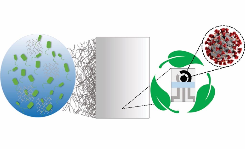
Fabrication steps of the biodegradable BC substrate and the electrochemical devices. (1) Incubation of the bacterium Gluconacetobacter hansenii. (2) BC substrate collected and treated, resulting in a clear sheet. (3) The biodegradable BC sheet is screen-printed, (4) resulting in a device with 3 electrodes, (4) which are cut out using a scissor, (5) resulting in a portable, biodegradable, and inexpensive electrochemical sensor.
The availability of rapid, accessible testing was integral to overcoming the worst surges of the COVID-19 pandemic, and will be necessary to keep up with emerging variants. However, these tests come with unfortunate costs.
Polymerase chain reaction (PCR) tests, the “gold standard” for diagnostic testing, are hampered by waste. They require significant time (results can take up to a day or more) as well as specialized equipment and labor, all of which increase costs. The sophistication of PCR tests makes them harder to tweak, and therefore slower to respond to new variants. They also carry environmental impacts. For example, most biosensor tests developed to date use printed circuit boards, or PCBs, the same materials used in computers. PCBs are difficult to recycle and slow to biodegrade, using large amounts of metal, plastic and non-eco-friendly materials.
In addition, most PCR tests end up in landfills, resulting in material waste and secondary contamination. An analysis by the World Health Organization (WHO) estimated that, as of February 2022, “over 140 million test kits, with a potential to generate 2,600 tonnes of non-infectious waste (mainly plastic) and 731,000 litres of chemical waste (equivalent to one-third of an Olympic-size swimming pool) have been shipped.”
In order to balance the need for fast, affordable and accurate testing while addressing these environmental concerns, César de la Fuente, Presidential Assistant Professor in Bioengineering and Chemical and Biomolecular Engineering in the School of Engineering and Applied Science, with additional primary appointments in Psychiatry and Microbiology within the Perelman School of Medicine, has turned his attention to the urgent need for “green” testing materials.
The de la Fuente lab has been working on creative ways to create faster and cheaper testing for COVID-19 since the outbreak of the pandemic. Utilizing his lab’s focus on machine biology and the treatment of infectious disease, they created RAPID, an aptly named test that generates results in minutes with a high degree of accuracy. An even more cost-effective version, called LEAD, was created using electrodes made from graphite. A third test, called COLOR, was a low-cost optodiagnostic test printed on cotton swabs.
The team’s latest innovation incorporates the speed and cost-effectiveness of previous tests with eco-friendly materials. In a paper published in Cell Reports Physical Science, the group introduces a new test made from Bacterial Cellulose (BC), an organic compound synthesized from several strains of bacteria, as a substitute for PCBs.
Genetic diseases that involve the central nervous system (CNS) often impact children before birth, meaning that once a child is born, irreversible damage has already been done. Given that many of these conditions result from a mutation in a single gene, there has been growing interest in using gene editing tools to correct these mutations before birth.
However, identifying the appropriate vehicle to deliver these gene editing tools to the CNS and brain has been a challenge. Viral vectors used to deliver gene therapies have some potential drawbacks, including pre-existing viral immunity and vector-related adverse events, and other options like lipid nanoparticles (LNPs) have not been investigated extensively in the perinatal brain.
Now, researchers in the Center for Fetal Research at Children’s Hospital of Philadelphia (CHOP) and Penn Engineering have identified an ionizable LNP that can deliver mRNA base editing tools to the brain and have shown it can mitigate CNS disease in perinatal mouse models. The findings, published in ACS Nano, open the door to mRNA therapies that could be delivered pre- or postnatally to treat genetic CNS diseases.
The research team began by screening a library of ionizable LNPs – microscopic fat bubbles that have a positive charge at low pH but neutral charge at physiological conditions in the body. After identifying which LNPs were best able to penetrate the blood-brain barrier in fetal and newborn mice, they optimized their top-performing LNP to be able to deliver base editing tools. The LNPs were then used to deliver mRNA for an adenine base editor, which would correct a disease-causing mutation in the lysosomal storage disease, MPSI, by changing the errant adenine to guanine.
The researchers showed that their LNP was able to improve the symptoms of the lysosomal storage disease in the neonatal mouse brain, as well as deliver mRNA base editing tools to the brain of other animal models. They also showed the LNP was stable in human cerebrospinal fluid and could deliver mRNA base editing tools to patient-derived brain tissue.
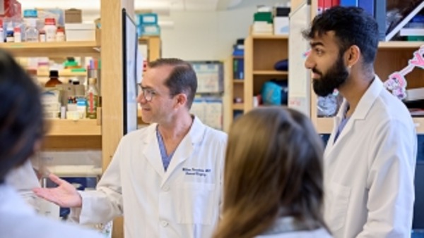
“This proof-of-concept study – co-led by Rohan Palanki, an MD/PhD student in my lab, and Michael Mitchell’s lab at Penn Bioengineering – supports the safety and efficacy of LNPs for the delivery of mRNA-based therapies to the central nervous system,” said co-senior author William H. Peranteau, MD, an attending surgeon in the Division of General, Thoracic and Fetal Surgery at CHOP and the Adzick-McCausland Distinguished Chair in Fetal and Pediatric Surgery. “Taken together, these experiments provide the foundation for additional translational studies and demonstrate base editing facilitated by a nonviral delivery carrier in the NHP fetal brain and primary human brain tissue.”
This story was written by Dana Bate. It originally appeared on CHOP’s website.