Illuminating the Unseen: Former Penn iGEM Team Publishes Award-Winning Optogenetic Device

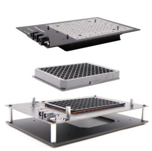
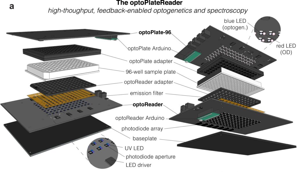
Diagram of the optoPlateReader, a high-throughput, feedback-enabled optogenetics and spectroscopy device initially developed by Penn 2021 iGEM team.

For bioengineers today, light does more than illuminate microscopes. Stimulating cells with light waves, a field known as optogenetics, has opened new doors to understanding the molecular activity within cells, with potential applications in drug discovery and more.

Thanks to recent advances in optogenetic technology, much of which is cheap and open-source, more researchers than ever before can construct arrays capable of running multiple experiments at once, using different wavelengths of light. Computing languages like Python allow researchers to manipulate light sources and precisely control what happens in the many “wells” containing cells in a typical optogenetic experiment.

However, researchers have struggled to simultaneously gather data on all these experiments in real time. Collecting data manually comes with multiple disadvantages: transferring cells to a microscope may expose them to other, non-experimental sources of light. The time it takes to collect the data also makes it difficult to adjust metabolic conditions quickly and precisely in sample cells.

Now, a team of Penn Engineers has published a paper in Communications Biology, an open access journal in the Nature portfolio, outlining the first low-cost solution to this problem. The paper describes the development of optoPlateReader (or oPR), an open-source device that addresses the need for instrumentation to monitor optogenetic experiments in real time. The oPR could make possible features such as automated reading, writing and feedback in microwell plates for optogenetic experiments.
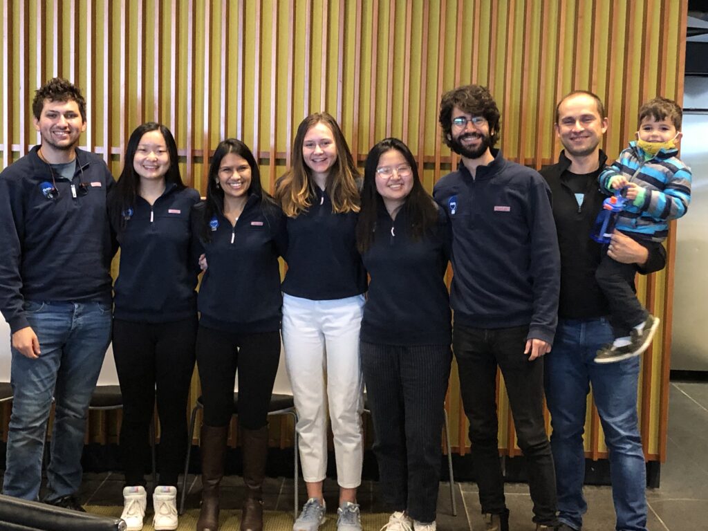
Left to right: Will Benman, Gloria Lee, Saachi Datta, Juliette Hooper, Grace Qian, David Gonzalez-Martinez, and Lukasz Bugaj (with Max).

The paper follows up on the award-winning work of six University of Pennsylvania alumni — Saachi Datta, M.D. Candidate at Stanford School of Medicine; Juliette Hooper, Programmer Analyst in Penn’s Perelman School of Medicine; Gabrielle Leavitt, M.D. Candidate at Temple University; Gloria Lee, graduate student at Oxford University; Grace Qian, Drug Excipient and Residual Analysis Research Co-op at GSK; and Lana Salloum, M.D. Candidate at Albert Einstein College of Medicine — who claimed multiple prizes at the 2021 International Genetically Engineered Machine Competition (iGEM) as Penn undergraduates.

The International Genetically Engineered Machine Competition (or iGEM) is the largest synthetic biology community and the premiere synthetic biology competition for both university and high school students from around the world. Hundreds of interdisciplinary teams of students compete annually, combining molecular biology techniques and engineering concepts to create novel biological systems and compete for prizes and awards through oral presentations and poster sessions.

The optoPlateReader was initially developed by Penn’s 2021 iGEM team, combining a light-stimulation device with a plate reader. At the iGEM competition, the invention took home Best Foundational Advance (best in track), Best Hardware (best from all undergraduate teams), and Best Presentation (best from all undergraduate teams), as well as a Gold Medal Distinction and inclusion in the Top 10 Overall and Top 10 Websites lists. (Read more about the 2021 iGEM team on the BE Blog.)

The original iGEM project focused on the design, construction, and testing of the hardware and software that make up the oPR, the focus of the new paper. After iGEM concluded, the team showed that the oPR could be used with real biological samples, such as cultures of bacteria. This work demonstrated that the oPR could be applied to real research questions, a necessary precursor to publication, and that the device could simultaneously monitor and manipulate living samples. 

The main application for the oPR is in metabolic production (such as the creation of pharmaceuticals and bio-fuels). The oPR is able to issue commands to cells via light but can also take live readings about their current state. In the oPR, certain colors of light cause cells to carry out different tasks, and optical measurements give information on growth rates and protein production rates.

In this way, the new device is able to support production processes that can adapt in real time to what cells need, altering their behavior to maximize yield. For example, if an experiment produces a product that is toxic to cells, the oPR could instruct those cells to “turn on” only when the population of cells is dense and “turn off” when the concentration of that product becomes toxic and the cellular population needs to recover. This ability to pivot in real time could assist industries that rely on bioproduction.

The main challenges in developing this device were in incorporating the many light emitting diodes (LEDs) and sensors into a tiny space, as well as insulating the sensors from the nearby LEDs to ensure that the measured light came from the sample and not from the instrument itself. The team also had to create software that could coordinate the function of nearly 100 different sets of LEDs and sensors. Going forward, the team hopes to spread the word about the open-source oPR to other researchers studying metabolic production to enable more efficient research.

Lukasz Bugaj, Assistant Professor in Bioengineering and senior author of the paper, served as the team’s mentor along with Brian Chow, formerly an Associate Professor in Bioengineering and a founding member of the iGEM program at MIT, and Jose Avalos, Associate Professor of Chemical and Biological Engineering at Princeton University.

Key to the project’s development was the guidance of Bioengineering graduate students Will Benman, David Gonzalez Martinez, and Gabrielle Ho, as well as that of Saurabh Malani, a graduate student at Princeton University.

Much of the original work was conducted in Penn Bioengineering’s Stephenson Foundation Educational Laboratory & Bio-MakerSpace, with important contributions made by Michael Patterson, Director of Educational Laboratories in Bioengineering, and Sevile Mannickarottu, Director of Technological Innovation and Entrepreneurship in Penn Engineering’s Entrepreneurship Program.

Read “High-throughput feedback-enabled optogenetic stimulation and spectroscopy in microwell plates” in Communications Biology.

This project was supported by the Department of Bioengineering, the School of Engineering and Applied Science, and the Office of the Vice Provost for Research (OVPR), and by funding from the National Institute of Health (NIH), the National Science Foundation (NSF), and the Department of Energy (DOE).

The iGEM program was created at the Massachusetts Institute of Technology in 2003. Read stories in the BE Blog featuring recent Penn iGEM teams here.

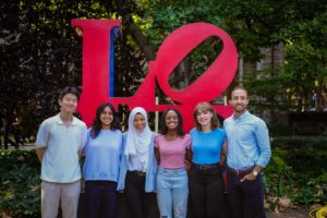
2022 Penn iGEM Team Wins Gold Medal in Grand Jamboree

The 2022 iGEM team from left to right: June Ahn, Shreya Villimanalan, Adiva Daniar, Wangari Mbuthia, Cristina Perez and Moses Zeidan.

Congratulations to the 2022 University of Pennsylvania iGEM Team who took home a gold medal in the iGEM Grand Jamboree. This international competition of multidisciplinary teams of graduate and undergraduate students presenting original projects in synthetic biology culminated in the in-person Jamboree event held in Paris, France in October 2022. Over 370 judges awarded prizes and medals to the 350+ teams representing over 40 countries.

The 2022 Penn team was awarded a Gold Medal for their project “Photocreate,” a toolbox to control intercellular communication using optogenetics. Their plasmid constructs are designed to control protein secretion, display and shedding using a photocleavable protein, Phocl. The full abstract reads:

Intercellular communication is primarily studied using synthetic protein-level circuits. These circuits currently lack the spatial and temporal control necessary for targeted and time-sensitive applications. To address this gap, we developed Photocrete, a toolbox of protein constructs for light-inducible control of protein display, secretion, and shedding. We expanded upon RELEASE (Vlahos et al.), a modular and generalizable protein circuit which utilizes an ER retention motif and an exogenous protease to control protein secretion. We optogenetically modified RELEASE by replacing different components with the photocleavable protein PhoCl, allowing us to control the mammalian secretion pathway at distinct nodes with finely-tuned light administration regimens. Preliminary results indicate integration of Photocrete into the secretion pathway, but more research is necessary to determine optimal light administration settings. The potential for high spatial and temporal control of Photocrete could allow researchers to perform various signaling studies and develop therapeutics at a new level of precision.

The 2022 iGEM team includes undergraduates June Ahn (B.S. in Biochemistry, Physics and Nutrition), Adiva Daniar (B.S.E. in Bioengineering, minor in Engineering Entrepreneurship), Wangari Mbuthia (B.S.E. in Bioengineering), Cristina Perez (B.S.E. in Bioengineering, minor in Physics), Shreya Vallimanalan (B.S.E. in Bioengineering, minor in Computational Neuroscience), an d Moses Zeidan (B.S.E. in Bioengineering, minor in Chemistry and Spanish). They were mentored by graduate students David Gonzalez-Martinez, Gabrielle Ho, Zikang Huang, and Will Benman. Their faculty advisor is Lukasz Bugaj, Assistant Professor in Bioengineering.

Read the full results of the 2022 iGEM Competition here.

Work for the annual iGEM competition is conducted in the George H. Stephenson Foundation Educational Laboratory & Bio-MakerSpace.

We acknowledge financial support from the Bradley Gabel Memorial Fund.

Erin Berlew and Rhea Chitalia Receive Solomon R. Pollack Awards for Excellence in Graduate Bioengineering Research

The Solomon R. Pollack Award for Excellence in Graduate Bioengineering Research is given annually to the most deserving Bioengineering graduate students who have successfully completed research that is original and recognized as being at the forefront of their field. This year Penn Bioengineering recognizes the outstanding work of two graduate students in Bioengineering: Erin Berlew and Rhea Chitalia.

Erin Berlew, Ph.D. candidate in Bioengineering

Erin Berlew is a Ph.D. candidate in the lab of Brian Chow, Associate Professor in Bioengineering. She successfully defended her thesis, titled “Single-component optogenetic tools for cytoskeletal rearrangements,” in December 2021. In her research, she used the BcLOV4 optogenetic platform discovered/developed in the Chow lab to control RhoGTPase signaling. Erin earned a B.S. in Chemistry from Haverford College in 2015 and was an Americorps member with City Year Philadelphia from 2015-2016. “Erin is a world-class bioengineering with an uncommon record of productivity gained through her complementary expertise in molecular, cellular, and computational biology,” says Chow. “She embodies everything wonderful, both academically and culturally, about our graduate program and its distinguished history.” Erin’s hobbies outside the lab include spending time with family, reading mystery novels, enjoying Philadelphia, and crossword puzzles. In the future, she hopes to continue to teach for the BE department (she has already taught ENGR 105 and served as a TA for undergraduate and graduate courses) and to conduct further research at Penn.

Rhea Chitalia, Ph.D. candidate in Bioengineering

Rhea Chitalia is a Ph.D. candidate in Bioengineering and a member of the Computational Biomarker Imaging Group (CBIG), advised by Despina Kontos, Matthew J. Wilson Associate Professor of Research Radiology II in the Perelman School of Medicine. Rhea completed her B.S.E. in Biomedical Engineering at Duke University in 2015. Her doctoral research concerns leveraging machine learning, bioinformatics, and computer vision to develop computational imaging biomarkers for improved precision cancer care. In December 2021 she successfully defended her thesis titled “Computational imaging biomarkers for precision medicine: characterizing intratumor heterogeneity in breast cancer.” “It has been such a privilege to mentor Rhea on her dissertation research,” says Kontos. “Rhea has been a star graduate student. Her work has made fundamental contributions in developing computational methods that will allow us to gain important insight into tumor heterogeneity by utilizing a multi-modality imaging approach.” David Mankoff, Matthew J. Wilson Professor of Research Radiology in the Perelman School of Medicine, served as Rhea’s second thesis advisor. “It was a true pleasure for me to work with Rhea and to Chair her BE Thesis Committee,” Mankoff adds. “Rhea’s Ph.D. thesis and thesis presentation was one of the best I have had the chance to be involved with in my graduate mentoring career.” After graduation, Rhea hopes to further precision medicine initiatives through the use of real world, multi-omic data in translational industry settings. She will be joining Invicro as an Imaging Scientist. In her spare time, Rhea enjoys trying new restaurants, reading, and spending time with friends and family.


A Protein Controlled by both Light and Temperature May Open Doors to Understanding Disease-related Cell Signal Pathways

by Melissa Pappas

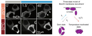
The brighter edges of the cells in the middle and upper right panels show the optogenetic proteins collecting at the membrane after light exposure. At higher temperatures, however, the proteins become rapidly inactivated and thus do not stay at the membrane, resulting in the duller edges seen in the bottom right panel.

Most organisms have proteins that react to light. Even creatures that don’t have eyes or other visual organs use these proteins to regulate many cellular processes, such as transcription, translation, cell growth and cell survival.

The field of optogenetics relies on such proteins to better understand and manipulate these processes. Using lasers and genetically engineered versions of these naturally occurring proteins, known as probes, researchers can precisely activate and deactivate a variety of cellular pathways, just like flipping a switch.

Now, Penn Engineering researchers have described a new type of optogenetic protein that can be controlled not only by light, but also by temperature, allowing for a higher degree of control in the manipulation of cellular pathways. The research will open new horizons for both basic science and translational research.

Lukasz Bugaj, Bomyi Lim, and Brian Chow

Lukasz Bugaj, Assistant Professor in Bioengineering (BE), Bomyi Lim, Assistant Professor in Chemical and Biomolecular Engineering, Brian Chow, Associate Professor in BE, and graduate students William Benman in Bugaj’s lab, Hao Deng in Lim’s lab, and Erin Berlew and Ivan Kuznetsov in Chow’s lab, published their study in Nature Chemical Biology. Arndt Siekmann, Associate Professor of Cell and Developmental Biology at the Perelman School of Medicine, and Caitlyn Parker, a research technician in his lab, also contributed to this research.

The team’s original aim was to develop a single-component probe that would be able to manipulate specific cellular pathways more efficiently. The model for their probe was a protein called BcLOV4, and through further investigation of this protein’s function, they made a fortuitous discovery: that the protein is controlled by both light and temperature.

Read more in Penn Engineering Today.

Single-cell Cancer Detection Project Wins 2021 NEMO Prize

This scProteome-seq array shows separated protein biomarkers (green and magenta spots) from thousands of single cells.

Penn Health-Tech’s Nemirovsky Engineering and Medicine Opportunity (NEMO) Prize awards $80,000 to support early-stage ideas joining engineering and medicine. The goal of the prize is to encourage collaboration between the University of Pennsylvania’s Perelman School of Medicine and the School of Engineering and Applied Science by supporting innovative ideas that might not receive funding from traditional sources.

This year, the NEMO Prize has been awarded to a team of researchers from Penn Engineering’s Department of Bioengineering. Their project aims to develop a technology that can detect multiple cancer biomarkers in single cells from tumor biopsy samples.

As cancer cells grow in the body, one of the characteristics that influences tumor growth and response to treatment is cancer cell state heterogeneity, or differences in cell states. Methods that rapidly catalogue cell heterogeneity may be able to detect rare cells responsible for tumor growth and drug resistance.

Single-cell transcriptomics (scRNA-seq) is the standard method for studying cell states; by amplifying and analyzing the cell’s complement of RNA sequences at a given time, researchers can get a snapshot of what proteins the cell is in the process of making. However, this method does not fully capture the function of the cell. The field of proteomics, which captures the actual protein content of cells along with post-translational modifications, provides a better picture of the cell’s function, but single-cell proteomic methods with the same sensitivity as scRNA-seq do not currently exist.

Alex Hughes, Lukasz Bugaj and Andrew Tsourkas

This collaborative project, which joins Assistant Professors Alex Hughes and Lukasz Bugaj, as well as Professor Andrew Tsourkas, aims to change that by developing multiplexed, sensitive and highly specific single-cell proteomics technologies to advance our understanding of cancer, its detection and its treatment.

This new technology, called scProteome-seq, builds from Hughes’s previous work.

“My specific expertise here is as an inventor of single-cell western blotting, which is the core technology that our team is building on,” says Hughes. “Single-cell proteomics technologies of this type have a track-record of commercial translation for applications in basic science and clinical automation, so our approach has a high potential for real-world impact.”

The current technology from Hughes’ lab separates proteins in cells by their molecular weight and “blots” them on a piece of paper. Improvements to this technology included in this project will remove the limitation of using light-emitting dyes to detect different proteins and instead use DNA barcodes to differentiate them.

Read the full story in Penn Engineering Today.

Penn’s 2021 iGEM Team Takes Home Multiple Prizes

Four of Penn’s 2021 iGEM team (left to right): Juliette Hooper, Grace Qian, Saachi Datta, and Gloria Lee.

The University of Pennsylvania’s 2021 iGEM team has been awarded several distinctions in this year’s highly competitive iGEM Competition. The International Genetically Engineered Machine Competition is the largest synthetic biology community and the premiere synthetic biology competition for both university and high school level students from around the world. Each year, hundreds of interdisciplinary teams of students combine molecular biology techniques and engineering concepts to create novel biological systems and compete for prizes and awards through oral presentations and poster sessions.

The Penn team’s project, “OptoReader,” is a combined light-simulation device and plate reader, which makes optogenetic experiments more powerful and accessible. The abstract reads:

“Metabolic engineering has the potential to change the world, and optogenetic tools can make metabolic engineering research easier by providing spatiotemporal control over cells. However, current optogenetic experiments are low-throughput, expensive, and laborious, which makes them inaccessible to many. To tackle this problem, we combined a light-stimulation device with a plate reader, creating our OptoReader. This device allows us to automate ~100 complex optogenetic experiments at the same time. Because it is open source and inexpensive, our device would make optogenetic experiments more efficient and available to all.”

Watch the team’s presentation on OptoReader here.

This year’s Penn team was mentored by Lukasz Bugaj, Assistant Professor in Bioengineering. In addition, the team was supported by Brian Chow, Associate Professor in Bioengineering. Chow has supported previous undergraduate iGEM teams at Penn, and was involved in the creation of the iGEM program during his time as a graduate student at MIT.

OptoReader took home the top prizes in three of the four categories in which it was nominated. These prizes include:

  • Best Foundational Advance (best in track)
  • Best Hardware (best from all undergraduate teams)
  • Best Presentation (best from all undergraduate teams)

They were also awarded a Gold Medal Distinction and were included in the Top 10 Overall (from all undergraduate teams, and the only team from the United States to make the top 10) and Top 10 Websites (from all undergraduate teams).

The awards were announced during iGEM’s online Jamboree Award Ceremony on November 14, 2021 (watch the full award ceremony here).

In addition to the outstanding awards recognition, OptoReader was also selected for an iGEM Impact Grant which awards teams $2,500 to continue development of their projects. This new initiative from the iGEM Foundation was announced earlier this year, and with the support of the Frederick Gardner Cottrell Foundation, is distributing a total of $225,000 in grant funds to 90 iGEM teams during the 2021 competition season. Learn more about the Impact Grant and read the full list of winning teams here.

Penn’s 2021 iGEM team was made up of an interdisciplinary group of women undergraduates from the School of Engineering and Applied Science (SEAS) and the School of Arts and Sciences (SAS):

  • Saachi Datta (B.A. in Biology and Religious Studies 2021)
  • Juliette Hooper (B.S.E. and M.S.E. in Bioengineering 2022)
  • Gabrielle Leavitt (B.S.E. in Bioengineering 2021 and current Master’s student in Bioengineering)
  • Gloria Lee (B.A. in Physics and B.S.E. in Bioengineering 2023)
  • Grace Qian (B.S.E. in Bioengineering 2023)
  • Lana Salloum (B.A. in Neuroscience 2022)

They were mentored by three doctoral students in Bioengineering: Will Benman (Bugaj Lab), David Gonzalez Martinez (Bugaj Lab), Gabrielle Ho (Chow Lab). Saurabh Malani, a graduate student in the Avalos Lab at Prince University, was also very involved in mentoring the team.

OptoReader

The graduate mentors were instrumental in quickly bringing the undergraduates up to speed on a diverse array of skills needed to accomplish this project including circuit design, optics, optogenetics, programming, and additive manufacturing. They then coached the team through building and testing prototypes, as well as accomplishing other objectives required for success at iGEM. These other objectives included establishing collaborations with other iGEM teams, performing outreach, and effectively communicating their project through a website and online presentations.

“This team and their work is outstanding,” said William Benman. “Not only did they sweep several awards, but they did it all with a small team and while working with technology they had no prior experience with. They created a device that not only increases accessibility to optogenetics but also allows optogenetic systems to interface directly with computer programs, allowing for completely new research avenues within the field. They are truly a remarkable group.”

Due to the COVID pandemic, the team operated virtually through the summer of 2020, and then continued in person in the summer of 2021 as the project progressed and more students returned to Penn’s campus. Upon return to campus, the work was conducted in both the Bugaj lab in the Stephenson Foundation Educational Laboratory & Bio-MakerSpace, the primary teaching laboratory in Penn Bioengineering and an interdisciplinary makerspace open to anyone at Penn. The team also collaborated with the Avalos Lab at Princeton University, which conducts research in the application of optogenetics to optimize production of valuable  chemicals in microbes.

“I’m beyond excited about this phenomenal showing from team Penn at the iGEM Jamboree awards ceremony,” said faculty mentor Lukasz Bugaj. “This is truly outstanding recognition for what the team has accomplished, and it wouldn’t have happened without essential contributions from everyone on the team.”

Brian Chow added that this achievement is “no small feat,” especially for a hardware project. “The iGEM competition leans toward genetic strain engineering, but the advances in the field made by these incredible students were undeniable,” he said.

Going forward, the team plans to publish a scientific article and file a patent application describing their device. “It’s clear that there is excitement in the scientific community for what our students created, and we’re excited to share the details and designs of their work,” said Bugaj.

Congratulations to all the team members and mentors of OptoReader on this incredible achievement! Check out the OptoReader project website and Instagram to learn more about their project.

This project was supported by the Department of Bioengineering, the School of Engineering and Applied Science, and the Office of the Vice Provost for Research (OVPR). 

Penn Engineers’ ‘LADL’ Uses Light to Serve Up On-demand Genome Folding

Every cell in your body has a copy of your genome, tightly coiled and packed into its nucleus. Since every copy is effectively identical, the difference between cell types and their biological functions comes down to which, how and when the individual genes in the genome are expressed, or translated into proteins.

Scientists are increasingly understanding the role that genome folding plays in this process. The way in which that linear sequence of genes are packed into the nucleus determines which genes come into physical contact with each other, which in turn influences gene expression.

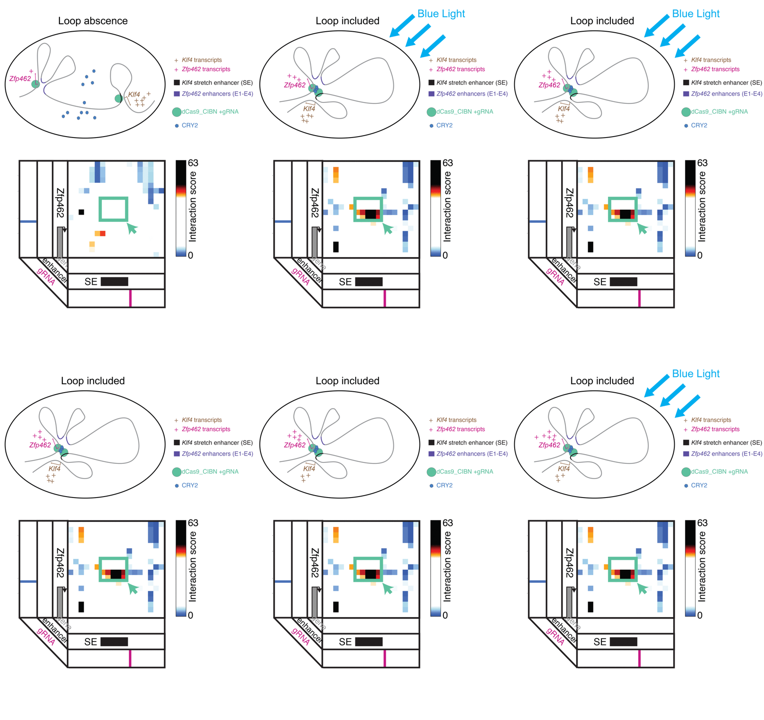
LADL combines CRISPR/Cas9 and optogenetics to bring two distant points in a linear gene sequence into physical contact, forming a folding pattern known as a “loop.” Looping interactions influence gene expression, so the researchers envision LADL as being a powerful tool for studying these dynamics.

Jennifer Phillips-Cremins, assistant professor in Penn Engineering’s Department of Bioengineering, is a pioneer in this field, known as “3-D Epigenetics.” She and her colleagues have now demonstrated a new technique for quickly creating specific folding patterns on demand, using light as a trigger.

The technique, known as LADL or light-activated dynamic looping, combines aspects of two other powerful biotechnological tools: CRISPR/Cas9 and optogenetics. By using the former to target the ends of a specific genome fold, or loop, and then using the latter to snap the ends together like a magnet, the researchers can temporarily create loops between exact genomic segments in a matter of hours.

The ability to make these genome folds, and undo them, on such a short timeframe makes LADL a promising tool for studying 3D-epigenetic mechanisms in more detail. With previous research from the Phillips-Cremins lab implicating these mechanisms in a variety of neurodevelopmental diseases, they hope LADL will eventually play a role in future studies, or even treatments.

Jennifer Phillips-Cremins, Ji Hun Kim and Mayuri Rege

Alongside Phillips-Cremins, lab members Ji Hun Kim and Mayuri Rege led the study, and Jacqueline Valeri, Aryeh Metzger, Katelyn R. Titus, Thomas G. Gilgenast, Wanfeng Gong and Jonathan A. Beagan contributed to it. They collaborated with associate professor of Bioengineering Arjun Raj and Margaret C. Dunagin, a member of his lab.

The study was published in the journal Nature Methods.

“In recent years,” Phillips-Cremins says, “scientists in our fields have overcome technical and experimental challenges in order to create ultra-high resolution maps of how the DNA folds into intricate 3D patterns within the nucleus. Although we are now capable of visualizing the topological structures, such as loops, there is a critical gap in knowledge in how genome structure configurations contribute to genome function.”

In order to conduct experiments on these relationships, researchers studying these 3D patterns were in need of tools that could manipulate specific loops on command. Beyond the intrinsic physical challenges — putting two distant parts of the linear genome in physical contact is quite literally like threading a needle with a thread that is only a few atoms thick — such a technique would need to be rapid, reversible and work on the target regions with a minimum of disturbance to neighboring sequences.

The advent of CRISPR/Cas9 solved the targeting problem. A modification of the gene editing tool allowed researchers to home in on the desired sequences of DNA on either end of the loop they wanted to form. If those sequences could be engineered to seek one another out and snap together under the other necessary conditions, the loop could be formed on demand.

Cremins Lab members then sought out biological mechanisms that could bind the ends of the loops together, and found an ideal one in the toolkit of optogenetics. The proteins CIB1 and CRY2, found in Arabidopsis, a flowering plant that’s a common model organism for geneticists, are known to bind together when exposed to blue light.

“Once we turn the light on, these mechanisms begin working in a matter of milliseconds and make loops within four hours,” says Rege. “And when we turn the light off, the proteins disassociate, meaning that we expect the loop to fall apart.”

“There are tens of thousands of DNA loops formed in a cell,” Kim says. “Some are formed slowly, but many are fast, occurring within the span of a second. If we want to study those faster looping mechanisms, we need tools that can act on a comparable time scales.”

As shown in a 2013 Nature Methods paper by fellow Penn bioengineer Lukasz Bugaj, the optical response of the CRY2 protein is a key component of LADL. When the blue light is turned on, CRY2 proteins in cell immediately find one another and bind together into clumps large enough to be seen under magnification. When the light is turned off, the clumps begin to dissolve away.”

Fast acting folding mechanisms also have an advantage in that they lead to fewer perturbations of the surrounding genome, reducing the potential for unintended effects that would add noise to an experiment’s results.

The researchers tested LADL’s ability to create the desired loops using their high-definition 3D genome mapping techniques. With the help of Arjun Raj, an expert in measuring the activity of transcriptional RNA sequences, they also were able to demonstrate that the newly created loops were impacting gene expression.

The promise of the field of 3D-epigenetics is in investigating the relationships between these long-range loops and mechanisms that determine the timing and quantity of the proteins they code for. Being able to engineer those loops means researchers will be able to mimic those mechanisms in experimental conditions, making LADL a critical tool for studying the role of genome folding on a variety of diseases and disorders.

“It is critical to understand the genome structure-function relationship on short timescales because the spatiotemporal regulation of gene expression is essential to faithful human development and because the mis-expression of genes often goes wrong in human disease,” Phillips-Cremins says. “The engineering of genome topology with light opens up new possibilities to understanding the cause-and-effect of this relationship. Moreover we anticipate that, over the long term, the use of light will allow us to target specific human tissues and even to control looping in specific neuron subtypes in the brain.”

The research was supported by the New York Stem Cell Foundation; Alfred P. Sloan Foundation; the National Institutes of Health through its Director’s New Innovator Award from the National Institute of Mental Health, grant no. 1DP2MH11024701, and a 4D Nucleome Common Fund, grant no. 1U01HL1299980; and the National Science Foundation through a joint NSF-National Institute of General Medical Sciences grant to support research at the interface of the biological and mathematical sciences, grant no. 1562665, and a Graduate Research Fellowship, grant no. DGE-1321851.

Originally published on the Penn Engineering Medium blog.

New Faculty: Interview With Lukasz Bugaj

Bugaj
Lukasz Bugaj, Ph.D.

This week, we present our interview with incoming faculty member Lukasz Bugaj, who starts as an assistant professor at Penn BE in January. Lukasz and Andrew Mathis discuss tennis and crew, Lukasz’s subfield of optogenetics, and life as the child of a statistician.

Please note: This was our first interview recorded by telephone. We will try to improve the quality of the audio, but for now, be advised that the questions are at a far lower volume than the responses, so set your volume, accordingly, particularly if you are listening on headphones.

PlayPlay