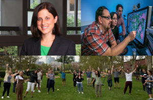
Jennifer E. Phillips-Cremins (upper left) and members of her lab.
Each year, the National Institutes of Health (NIH) recognizes exceptionally creative scientists through its High-Risk, High-Reward Research Program. The four awards granted by this program are designed to support researchers whose “out of the box” and “trailblazing” ideas have the potential for broad impact.
Jennifer E. Phillips-Cremins, Associate Professor and Dean’s Faculty Fellow in Penn Engineering’s Department of Bioengineering and the Perelman School of Medicine’s Department of Genetics, is one such researcher. As a recipient of an NIH Director’s Pioneer Award, she will receive $3.5 million over five years to support her work on the role that the physical folding of chromatin plays in the encoding of neural circuit and synapse properties contributing to long-term memory.
Phillips-Cremins’ award is one of 106 grants made through the High-Risk, High-Reward program this year, though she is only one of 10 to receive the Pioneer Award, which is the program’s largest funding opportunity.
“The science put forward by this cohort is exceptionally novel and creative and is sure to push at the boundaries of what is known,” said NIH Director Francis S. Collins.
Phillips-Cremins’ research is in the general field of epigenetics, the molecular and structural modifications that allow the genome — an identical copy of which is found in each cell — to express genes differently at different times and in different parts of the body. Within this field, her lab focuses on higher-order folding patterns of the DNA sequence, which bring distant sets of genes and regulatory elements into close proximity with one another as they are compressed inside the cell’s nucleus.
Previous work from the Cremins lab has investigated severe genome misfolding patterns common across a class of genetic neurological disorders, including fragile X syndrome, Huntington’s disease, ALS and Friedreich’s ataxia.
With the support of the Pioneer Award, she and the members of her lab will extend that research to a more fundamental question of neuroscience: how memory is encoded over decades, despite the rapid turnover of the relevant proteins and RNA sequences within the brain’s synapses.
“Our long-term goals are to understand how, when and why pathologic genome misfolding leads to synaptic dysfunction by way of disrupted gene expression,” said Phillips-Cremins, “as well as to engineer the genome’s structure-function relationship to reverse pathologic synaptic defects in debilitating neurological diseases.”
Our next Penn Bioengineering seminar will be held on zoom next Thursday.
Kenneth Yamada, MD, PhD
Speaker: Kenneth Yamada, M.D., Ph.D.
NIH Distinguished Investigator
Cell Biology Section
National Institute of Dental and Craniofacial Research, National Institutes of Health (NIH)
Date: Thursday, September 9, 2021
Time: 3:30-4:30 PM EDT
Zoom – check email for link or contact ksas@seas.upenn.edu
Location: Moore Room 216, 200 S. 33rd Street
Abstract: Real-time microscopy of the dynamics of cells and tissues in 3D environments is opening new windows to understanding the biophysical mechanisms of complex biological processes. Direct visualization is allowing us to explore fundamental questions in more depth that include: How do cells migrate in 3D? How do cancer cells invade? How is the extracellular matrix assembled? How are organs formed? Visualizing how cells move and organize into tissues is not only providing descriptive insights, but is also leading to the identification of novel, unexpected physical and mechanical mechanisms relevant to tissue engineering. Cells can use varying combinations of cell adhesion to adjacent cells and to the surrounding extracellular matrix with localized cellular contractility to migrate, invade, and produce the complex tissue architecture needed for organ formation.
Kenneth Yamada Bio: Kenneth Yamada has been an NIH Distinguished Investigator since 2011. He received MD and PhD degrees from Stanford. He was a Section Chief at the National Cancer Institute for 10 years and has been a Section Chief at NIDCR since 1990. He is an elected Fellow of the AAAS and American Society for Cell Biology. His research focuses on discovering novel mechanisms and regulators of cell interactions with the extracellular matrix and their roles in embryonic development and cancer. His research group focuses on the mechanisms by which three-dimensional (3D) extracellular matrix mediates key biological events, including cell migration, tissue morphogenesis, and cancer cell invasion. His research places particular emphasis on characterizing the dynamic movements of cells and their extracellular matrix as tissues are remodeled in 3D in real time. The biological systems they study include human primary cells migrating in 3D, human tumor cells and tissues, and mouse organ development. He places particularly high priority on developing future independent research leaders.
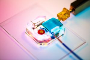
To combat the COVID-19 pandemic caused by the SARS-CoV2 virus, Dr. Andrew Tsourkas’s Targeted Imaging Therapeutics and Nanomedicine (Titan) Lab in Penn Bioengineering, in collaboration with the Penn-based startup, AlphaThera, was recently awarded a $667,000 SBIR Phase II Grant Extension to support its efforts in commercializing COVID-19 detection technology. The grant supports work to address the growing need for anti-viral antibody testing. Specifically, the Tsourkas Lab and AlphaThera hope to leverage their expertise with antibody conjugation technologies to reduce the steps and complexity of existing detection assays to enable greater production and higher sensitivity tests. AlphaThera was founded in 2016 by Andrew Tsourkas, PhD, Professor of Bioengineering and James Hui, MD, PhD, a graduate of the Perelman School of Medicine and Penn Bioengineering’s doctoral program.
During this pandemic it is crucial to characterize disease prevalence among populations, understand immunity, test vaccine efficacy and monitor disease resurgence. Projections have indicated that millions of daily tests will be needed to effectively control the virus spread. One important testing method is the serological assay: These tests detect the presence of SARS-CoV2 antibodies in a person’s blood produced by the body’s immune system responding to infection. Serological tests not only diagnose active infections, but also establish prior infection in an individual, which can greatly aid in forecasting disease spread and contact tracing. To perform the serological assays for antibody detection, well-established immunoassay methods are used such as ELISA.
A variety of issues have slowed the distribution of these serological assays for antibody testing. The surge in demand for testing has caused shortages in materials and reagents that are crucial for the assays. Furthermore, complexity in some of the assay formats can slow both production and affect the sensitivity of test results. Recognizing these problems, AlphaThera is leveraging its novel conjugation technology to greatly improve upon traditional assay formats.
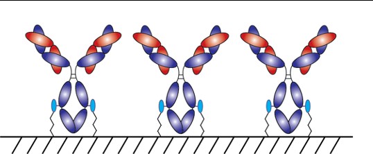
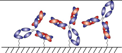
With AlphaThera’s conjugation technology, the orientation of antibodies can be precisely controlled so that they are aligned and uniformly immobilized on assay detection plates. This is crucial as traditional serological assays often bind antibodies to plates in a non-uniform manner, which increases variability of results and reduces sensitivity. See Fig 1 below. With AlphaThera’s uniform antibody immobilization, assay specificity could increase by as much as 1000- fold for detection of a patient’s SaRS-CoV2 antibodies.
Fig 1: Uniform vs Non-Uniform Immobilized Antibodies on Surface: Top is AlphaThera improvement, showing how antibodies would be uniformly immobilized and oriented on a plate for detection. Bottom is how many traditional serological assays immobilize antibodies, resulting in variability of results and lower specificity.
Furthermore, AlphaThera is addressing the shortage of assay reagents, specifically secondary antibody reagents, by removing certain steps from traditional serological assays. Rather than relying on secondary antibodies for detection of the patient antibodies, AlphaThera’s technology can label the patient SaRS-CoV2 primary antibodies directly in serum with a detection reagent. This eliminates several processing steps, reducing the time of the assay by as much as 50%, as well as the costs.
The Tsourkas Lab and AlphaThera have initiated their COVID-19 project, expanding into the Pennovation Center and onboarding new lab staff. Other antibody labeling products have also become available and are currently being prepared for commercialization. Check out the AlphaThera website to learn more about their technology at https://www.alphathera.com.
NIH SBIR Phase II Grant Extension— 5-R44-EB023750-03 (PI: Yu) — 10/07/2020 – 10/07/2021
We would like to congratulate Assistant Professor in Bioengineering Alex Hughes, Ph.D., on receiving the Maximizing Investigators’ Research Award (MIRA) from the National Institutes of Health (NIH), which funds investigators to create flexible and forward-thinking research programs. Hughes is the first recipient of this award in Penn’s School of Engineering and Applied Science, marking a major accomplishment for him and his lab.
The award recognizes Hughes’ efforts to create new tools used for tissue engineering, in particular by fusing concepts from developmental biology into tissue construction efforts. Hughes believes this approach will have impacts on fundamental understanding human disease, leading to new strategies to combat them. Hughes and his lab specifically focus on kidney disease. As Hughes says, “defects in the kidney and urinary tract account for up to a third of all birth defects.” Furthermore, because kidney development involves many different kinds of cell interactions, there’s a gap in understanding exactly how these defects occur.
Unlike other grants that focus on funding projects, the MIRA prioritizes the people behind the research, giving them funding as a sign of faith in the future work they’ll choose to do. “The MIRA has allowed us significant leeway to integrate several complementary approaches here,” Hughes says. Because of this flexibility, Hughes and his lab thinks it will allow them to reach for more innovative and risky approaches in their research, in the hopes that this will lead to a better understanding of kidney defects and modes of treatment for them.
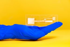
Rachel Young, a graduate student in Huh’s lab, holds up the new eye-on-a-chip device. The latest iteration of the lab’s eye-on-a-chip has a mechanical eyelid to simulate blinking, and was used to test an experimental drug for dry eye disease. By incorporating human cells into an engineered scaffolding, the eye-on-a-chip has many of the benefits of testing on living subjects, while minimizing risks and ethical concerns.
People who spend eight or more hours a day staring at a computer screen may notice their eyes becoming tired or dry, and, if those conditions are severe enough, they may eventually develop dry eye disease (DED). DED is a common disease with shockingly few FDA-approved drug options, partially because of the difficulties of modeling the complex pathophysiology in human eyes. Enter the blinking eye-on-a-chip: an artificial human eye replica constructed in the laboratory of Penn Engineering researchers.
This eye-on-a-chip, complete with a blinking eyelid, is helping scientists and drug developers to improve their understanding and treatment of DED, among other potential uses. The research, published in Nature Medicine, outlines the accuracy of the eye-on-a-chip as an organ stand-in and demonstrates its utility as a drug testing platform.
They collaborated with Vivian Lee, Vatinee Bunya and Mina Massaro-Giordano from the Department of Ophthalmology in Penn’s Perelman School of Medicine, as well as with Vivek Shenoy, Eduardo D. Glandt President’s Distinguished Professor in Penn Engineering’s Department of Materials Science and Engineering. Other collaborators included Woo Byun, Andrei Georgescu and Yoon-suk Yi, members of Huh’s lab, and Farid Alisafaei, a member of Shenoy’s lab.
Huh’s lab specializes in creating organs-on-a-chip that provide microengineered in vitro platforms to mimic their in vivo counterparts, including lung and bone marrow proxies launched into space this May to study astronaut illness. The lab has spent years fine-tuning its eye-on-a-chip, which earned them the 2018 Lush Prize for its promise in animal-free testing of drugs, chemicals, and cosmetics.
In this study, Huh and Seo focused on engineering an eye model that could imitate a healthy eye and an eye with DED, allowing them to test an experimental drug without risk of human harm.
The Huh lab’s eye-on-a-chip attached to a motorized, gelatin-based eyelid. Blinking spreads tears over the corneal surface, and so was a critical aspect to replicate in the researchers’ model of dry eye disease. cells. The cells of the cornea grow on the inner circle of scaffolding, dyed yellow, and the cells of the conjunctiva grow on the surrounding red circle. Artificial tears are supplied by a tear duct, dyed blue.
To construct their eye-on-a-chip, Huh’s team starts with a porous scaffold engineered with 3D printing, about the size of a dime and the shape of a contact lens, on which they grow human eye cells. The cells of the cornea grow on the inner circle of scaffolding, dyed yellow, and the cells of the conjunctiva, the specialized tissue covering the white part of human eyes, grow on the surrounding red circle. A slab of gelatin acts as the eyelid, mechanically sliding over the eye at the same rate as human blinking. Fed by a tear duct, dyed blue, the eyelid spreads artificial tear secretions over the eye to form what is called a tear film.
“From an engineering standpoint, we found it interesting to think about the possibility of mimicking the dynamic environment of a blinking human eye. Blinking serves to spread tears and generate a thin film that keeps the ocular surface hydrated. It also helps form a smooth refractive surface for light transmission. This was a key feature of the ocular surface that we wanted to recapitulate in our device,” says Huh.
For people with DED, that tear film evaporates faster than it’s replenished, resulting in inflammation and irritation. A common cause of DED is the reduced blinking that occurs during excessive computer usage, but people can develop the disease for other reasons as well. DED affects about 14 percent of the world’s population but has been notably difficult to develop new treatments for, with 200 failed clinical drug trials since 2010 and only two currently available FDA-approved drugs for treatment.
Huh’s lab has been considering the drug-testing potential of organs-on-a-chip since their initial conceptualization, and, because of its surface-level area of impact, DED seemed the perfect place to start putting their eye model to the test. But before they started a drug trial, the team had to ensure their model could really imitate the conditions of DED.
“Initially, we thought modeling DED would be as simple as just keeping the culture environment dry. But as it turns out, it’s an incredibly complex multifactorial disease with a variety of sub-types,” Huh says. “Regardless of type, however, there are two core mechanisms that underlie the development and progression of DED. First, as water evaporates from the tear film, salt concentration increases dramatically, resulting in hyperosmolarity of tears. And second, with increased tear evaporation, the tear film becomes thinner more rapidly and often ruptures prematurely, which is referred to as tear film instability. The question was: Is our model capable of modeling these core mechanisms of dry eye?”
The answer, after much experimentation, was yes. The team evoked DED conditions in their eye-on-a-chip by cutting their device’s artificial blinking in half and carefully creating an enclosed environment that simulated the humidity of real-life conditions. When put to the test against real human eyes, both healthy and with DED, the corresponding eye-on-a-chip models proved their similarity to the actual organ on multiple clinical measures. The eyes-on-a-chip mimicked actual eyes’ performance in a Schirmer strip, which tests liquid production; in an osmolarity test, which looks at tear film salt content; and in a keratography test, which evaluates the time it takes for a tear film to break up.
Having confirmed their eye-on-a-chip’s ability to mirror the performance of a human eye in normal and DED-inducing settings, Huh’s team turned to the pharmaceutical industry to find a promising DED drug candidate to test-drive their model. They landed on an upcoming drug based on lubricin, a protein primarily found in the lubricating fluid that protects joints.
“When people think of DED, they normally treat it as a chronic disease driven by inflammation,” says Huh, “but there’s now increasing evidence suggesting that mechanical forces are important for understanding the pathophysiology of DED. As the tear film becomes thinner and more unstable, friction between the eyelids and the ocular surface increases, and this can damage the epithelial surface and also trigger adverse biological responses such as inflammation. Based on these observations, there is emerging interest in developing ophthalmic lubricants as a topical treatment for dry eye. In our study, we used an lubricin-based drug that is currently undergoing clinical trials. When we tested this drug in our device, we were able to demonstrate its friction-lowering effects, but, more importantly, using this model we discovered its previously unknown capacity to suppress inflammation of the ocular surface.”
By comparing the testing results of their models of a healthy eye, an eye with DED, and an eye with DED plus lubricin, Huh and Seo were able to further scientists’ understanding of how lubricin works and show the drug’s promise as a DED treatment.
Similarly, the process of building a blinking eye-on-a-chip pushed forward scientists’ understanding of the eye itself, providing insights into the role of mechanics in biology. Collaborating with Shenoy, director of the Center for Engineering MechanoBiology, the team’s attention was drawn to how the physical blinking action was affecting the cells they cultivated to engineer an artificial eye on top of their scaffolding.
“Initially, the corneal cells start off as a single layer, but they become stratified and form multiple layers as a result of differentiation, which happens when these cells are cultured at the air-liquid interface. They also form tight cell-cell junctions and express a set of markers during differentiation,” Huh says. “Interestingly, we found out that mechanical forces due to blinking actually help the cells differentiate more rapidly and more efficiently. When the corneal cells were cultured under air in the presence of blinking, the rate and extent of differentiation increased significantly in comparison to static models without blinking. Based on this result, we speculate that blink-induced physiological forces may contribute to differentiation and maintenance of the cornea.”
In other words, human cornea cells growing on the scientists’ scaffold more quickly became specialized and efficient at their particular jobs when the artificial eyelid was blinking on top of them, suggesting that mechanical forces like blinking contribute significantly to how cells function. These types of conceptual advances, coupled with drug discovery applications, highlight the multifaceted value that engineered organs-on-a-chip can contribute to science.
Huh and Seo’s eye-on-a-chip is still just dipping its toes into the field of drug testing, but this first step is a victory that represents years of work refining their artificial eye to reach this level of accuracy and utility.
“Although we have just demonstrated proof-of-concept,” says Seo, “I hope our eye-on-a-chip platform is further advanced and used for a variety of applications besides drug screening, such as testing of contact lenses and eye surgeries in the future.”
“We are particularly proud of the fact that our work offers a great and rare example of interdisciplinary efforts encompassing a broad spectrum of research activities from design and fabrication of novel bioengineering systems to in vitro modeling of complex human disease to drug testing,” says Huh. “I think this is what makes our study unique and representative of innovation that can be brought about by organ-on-a-chip technology.”
This work was supported by the National Institutes of Health through grants 1DP2HL127720–0, R01EY026972 and K08EY025742–01, the National Science Foundation through grants CMMI:15–48571, and Research to Prevent Blindness.
New 3D Tumor Models Could Improve Cancer Treatment
New ways of testing cancer treatments may now be possible thanks to researchers at the University of Akron who developed three-dimensional tumor models of triple-negative breast cancer. Led by Dr. Hossein Tavana, Ph. D., an associate professor of biomedical engineering at the university, the Tissue Engineering Microtechnologies Lab recently received a $1.13 million grant from the prestigious National Cancer Institute (NCI) of the National Institute of Health (NIH) to continue improving these tumor models. Tumors are difficult to fully replicate in vitro, as they are comprised of cancerous cells, connective tissue, and matrix proteins, among several other components. With this new grant, Tavana sees creating a high-throughput system that uses many identical copies of the tumor model for drug testing and better understanding of the way tumors operate. This high-throughput method would allow Tavana and his lab to isolate and test several different approaches at once, which they hope will help change the way tumors are studied and treated everywhere.
Noise-Induced Hearing Loss Poses Greater Threat to Neural Processing
Even though we all know we probably shouldn’t listen to music at high volumes, most of us typically do it anyway. But researchers at Purdue University recently found that noise-induced hearing loss could cause significant changes in neural processing of more complex sound inputs. Led by Kenneth Henry, Ph.D., an assistant professor of otolaryngology at the University of Rochester Medical Center, and Michael Heinz, Ph.D., a professor of biomedical engineering at Purdue University, the study shows that when compared with age-related hearing loss, noise-induced hearing loss will result in a greater decrease in hearing perception even when the two kinds of hearing loss appear to be of the same degree on an audiogram. This is because noise-induced hearing loss occurs because of physical trauma to the ear, rather than the long-term electrochemical degradation of some components that come happen with age. The evidence of this research is yet another reason why we should be more careful about exposing our ears to louder volumes, as they pose a greater risk of serious damage.
Increasing the Patient Populations for Research in Cartilage Therapy and Regenration
Despite the great progress in research of knee cartilage therapy and regeneration, there are still issues with the patient populations that most studies consider. Researchers often want to test new methods on patients that have the greatest chance of injury recovery without complications – often referred to as “green knees” – but this leaves out those patient populations who suffer from conditions or defects that have the potential to cause complications – often referred to as “red knees.” In a new paper published in Regenerative Medicine, the Mary Black Ralston Professor for Education and Research in Orthopaedic Surgery and secondary faculty in the Department of Bioengineering at Penn, Robert Mauck, Ph.D., discusses some cartilage therapies that may be suitable for red knee populations.
Working with James Carey, M.D., the Director of the Penn Center for Cartilage Repair and Osteochondritis, Mauck and his research team realized that even those with common knee cartilage conditions such as the presence of lesions or osteoarthritis were liable to be excluded from most regeneration studies. In discussing alternatives methods and structures of studying cartilage repair and regeneration, Mauck and Carey hope that future therapies will be applicable to a wider range of patient populations, and that there will soon be more options beyond full joint replacement for those with red knee conditions.
Plant-Like Superhydrophobicity Has Applications in Biomedical Engineering
Researchers in the Department of Biomedical Engineering at Texas A&M University recently found ways of incorporating the superhydrophobic properties of some plant leaves into biomedical applications through what they’re calling a “lotus effect.” The Gaharwar Lab, led by principal investigator and assistant professor of biomedical engineering Akhilesh Gaharwar, Ph.D., developed an assembly of two-dimensional atomic layers that they describe as a “nanoflower” to help control surface wetting in a biomedical setting. A recent paper published in Chemical Communications describes Gaharwar and his team’s work as expanding the use of superhydrophobic surface properties in biomedical devices by demonstrating the important role that atomic vacancies play in the wetting characteristic. While Gaharwar hopes to research the impact that controlling superhydrophobicity could have in stem cell applications, his work already allows for innovations in self-cleaning and surface properties of devices involving labs-on-a-chip and biosensing.
People and Places
Nader Engheta, H. Nedwill Ramsey Professor in Electrical and Systems Engineering, Bioengineering and Materials Science and Engineering, has been inducted into the Canadian Academy of Engineering (CAE) as an International Fellow. The CAE comprises many of Canada’s most accomplished engineers and Engheta was among the five international fellows that were inducted this year.
The Academy’s President Eddy Isaacs remarked: “Over our past 32 years, Fellows of Academy have provided insights in the fields of education, infrastructure, and innovation, and we are expecting the new Fellows to expand upon these contributions to public policy considerably.”
We would like to congratulate Anthony Lowman, Ph.D., on his appointment as the Provost and Senior Vice President for Academic Affairs at Rowan University. Formerly the Dean of Rowan’s College of Engineering, Lowman helped the college double in size, and helped foster a stronger research community. Lowman also helped to launch a Ph.D. program for the school, and added two new departments of Biomedical Engineering and Experiential Engineering Education in his tenure as the dean. Widely recognized for his research on hydrogels and drug delivery, Lowman was also formerly a professor of bioengineering at Temple University and Drexel University.
Lastly, we would like to congratulate Daniel Lemons, Ph.D., on his appointment as the Interim President of Lehman College of the City University of New York. Lemons, a professor in the Department of Biology at City College, specializes in cardiovascular and comparative physiology, and was also one of the original faculty members of the New York Center for Biomedical Engineering. With prior research funded by both the National Institute of Health (NIH) and the National Science Foundation (NSF), Lemons also holds patents in biomechanics teaching models and mechanical heart simulators.
Tulane Researchers Use Cancer Imaging Technique to Help Detect Preeclampsia
Preeclampsia is potentially life-threatening pregnancy disorder that typically occurs in about 200,000 expectant mothers every year. With symptoms of high blood pressure, swelling of the hands and feet, and protein presence in urine, preeclampsia is usually treatable if diagnosed early enough. However, current methods for diagnosis involve invasive procedures like cordocentesis, a procedure which takes a sample of fetal blood.
Researchers at Tulane School of Medicine led by assistant professor of bioengineering Carolyn Bayer, Ph.D., hope to improve diagnostics for preeclampsia with the use of spectral photoacoustic imaging. Using this technique, Bayer’s team noticed a nearly 12 percent decrease in placental oxygenation in rats with induced preeclampsia when compared to normal pregnant rats after only two days. If success in using this imaging technology continues at the clinical level, Bayer plans to find more applications of it in the detection and diagnosis of breast and ovarian cancers as well.
New CRISPR-powered device detects genetic mutations in minutes
This new chip eliminates the long and expensive amplification process involved in the typical polymerase chain reaction (PCR) used to read DNA sequences. In doing so, the CRISPR-Chip is much more of a point-of-care diagnostic, having the ability to quickly detect a given mutation or sequence when given a pure DNA sample. Led by Kiana Aran, Ph.D., the research team behind the CRISPR-Chip hopes that this new combination of nanoelectronics and modern biology will allow for a new world of possibilities in personalized medicine.
New Method of Brain Stimulation Might Alleviate Symptoms of Depression
Depression is one of the most common mental health disorders in the United States, with nearly 3 million cases every year. For most patients suffering from depression, treatment involves prolonged psychotherapy, antidepressant medication, or even electroconvulsive therapy in extreme cases. Now, scientists at the University of North Carolina School of Medicine study the use of transcranial alternating current stimulation (tACS) to alleviate symptoms of depression.
Led by Flavio Frohlich, Ph.D., who has an adjunct appointment in biomedical engineering, this team of researchers based this new solution on information from each patient’s specific alpha oscillations, which are a kind of wave that can be detected by an electroencephalogram (EEG). Those who suffer from depression tend to have imbalanced alpha oscillations, so Frohlich and his team sought to use tACS to restore this balance in those patients. After seeing positive results from data collected two weeks after patients in a clinical trial receives the tACS treatment, Frohlich hopes that future applications will include treatment for even more mental health disorders and psychiatric illnesses.
University of Utah Researchers Receive Grant to Improve Hearing Devices for Deaf Patients
Engineers at the University of Utah are part of team that recently received a $9.7 million grant from the National Institute of Health (NIH) to design new implantable hearing devices for deaf patients, with the hope to improve beyond the sound quality of existing devices. The work will build upon a previous project at the University of Utah called the Utah Electrode Array, a brain-computer interface originally developed by Richard Normann, Ph.D., that can send and receive neural impulses to and from the brain. This new device will differ from a typical cochlear implant because the Utah Electrode Array assembly will be attached directly to the auditory nerve instead of the cochlea, providing the patient with a much higher resolution of sound.
People & Places
Vivek Shenoy, Eduardo D. Glandt President’s Distinguished Scholar in the Department of Materials Science and Engineering and Secondary Faculty in Bioengineering, has been named the recipient of the 2018–19 George H. Heilmeier Faculty Award for Excellence in Research for “for pioneering multi-scale models of nanomaterials and biological systems.”
The Heilmeier Award honors a Penn Engineering faculty member whose work is scientifically meritorious and has high technological impact and visibility. It is named for George H. Heilmeier, a Penn Engineering alumnus and advisor whose technological contributions include the development of liquid crystal displays and whose honors include the National Medal of Science and Kyoto Prize.
We would also like to congratulate Jay Goldberg, Ph.D., from Marquette University on his election as a fellow to the National Academy of Inventors. Nominated largely for his six patents involving medical devices, Goldberg also brings this innovation to his courses. One in particular called Clinical Issues in Biomedical Engineering Design allows junior and senior undergraduates to observe the use of technology in clinical settings like the operating room, in an effort to get students thinking about how to improve the use of medical devices in these areas.
Louisiana Tech Sends First All-Female Team to RockOn
A team of faculty and students from Louisiana Tech University will participate in RockOn, a NASA-sponsored workshop on rocketry and engineering. Mechanical Engineering Lecturer Krystal Corbett, Ph.D., and Assistant Professor of bioengineering Mary Caldorera-Moore, Ph.D., will work together to lead the university’s first team of three all-female students at the event. At the program, they will have the chance to work on projects involving components of spacecraft systems, increasing students’ experience in hands-on activities and real-world engineering.
Refining Autism Treatments Using Big Data
Though treatments like therapy and medication exist for patients with autism, one of the biggest challenges that those caring for these patients face is in measuring their effects over time. Many of the markers of progress are qualitative, and based on a given professional’s opinion on a case-by-case basis. But now, a team of researchers from Rensselaer Polytechnic Institute (RPI) hopes to change that with the use of big data.
Juergen Hahn, Ph. D., and his lab recently published a paper in Frontiers in Cellular Neuroscience discussing their findings in connecting metabolic changes with behavioral improvements in autistic patients. Their analysis looks for multiple chemical and medical markers simultaneously in data from three distinct clinical trials involving metabolic treatment for patients. Being able to quantitatively describe the effects of current autism treatments would revolutionize clinical trials in the field, and lead to overall better patient care.
Penn Engineers Can Detect Ultra Rare Proteins in Blood Using a Cellphone Camera
One of the frontiers of medical diagnostics is the race for more sensitive blood tests. The ability to detect extremely rare proteins could make a life-saving difference for many conditions, such as the early detection of certain cancers or the diagnosis of traumatic brain injury, where the relevant biomarkers only appear in vanishingly small quantities. Commercial approaches to ultrasensitive protein detection are starting to become available, but they are based on expensive optics and fluid handlers, which make them relatively bulky and expensive and constrain their use to laboratory settings.
Knowing that having this sort of diagnostic system available as a point-of-care device would be critical for many conditions — especially traumatic brain injury — a team of engineers led by Assistant Professor in the Department of Bioengineering, David Issadore, Ph.D., at the University of Pennsylvania have developed a test that uses off-the-shelf components and can detect single proteins with results in a matter of minutes, compared to the traditional workflow, which can take days.
Treating Cerebral Palsy with Battery-Powered Exoskeletons
Cerebral palsy is one of the most common movement disorders in the United States. The disorder affects a patient’s control over even basic movements like walking, so treatments for cerebral palsy often involve the use of assistive devices in an effort to give patients better command over their muscles. Zach Lerner, Ph.D., is an Assistant Professor of Mechanical Engineering and faculty in Northern Arizona University’s Center for Bioengineering Innovation whose research looks to improve these kinds of assistive devices through the use of battery-powered exoskeletons.
Lerner and his lab recently received three grants, one each from the National Institute of Health (NIH), the National Science Foundation (NSF), and the Arabidopsis Biological Resource Center, to continue their research in developing these exoskeletons. Their goal is to create devices with powered assistance at joints like the ankle or knee to help improve patient gait patterns in rehabilitating the neuromuscular systems associated with walking. The team hopes that their work under these new grants will help further advance treatment for children with cerebral palsy, and improve overall patient care.
People & Places
David Aguilar, a 19-year-old bioengineering student at Universitat Internacional de Catalunya made headlines recently for a robotic prosthetic arm that he built for himself using Lego pieces. Due to a rare genetic condition, Aguilar was born without a right forearm, a disability that inspired him to play with the idea of creating his own prosthetic arm from age nine. His design includes a working elbow joint and grabber that functions like a hand. In the future, Aguilar hopes to continue improving his own prosthetic designs, and to help create similar versions of affordable devices for other patients who need them.
This week, we would like to congratulate two recipients of the National Science Foundation’s Career Awards, given to junior faculty that exemplify the role of teacher-scholars in their research. The first recipient we’d like to acknowledge is the University of Arkansas’ Kyle Quinn, Ph.D., who received the award for his work in developing new image analysis methods and models using the fluorescence of two metabolic cofactors. Dr. Quinn completed his Ph.D. here at Penn in Dr. Beth Winkelstein’s lab, and received the Solomon R. Pollack Award for Excellence in Graduate Bioengineering Dissertation Research for his work.
The second recipient of the award we wish to congratulate is Reuben Kraft, Ph.D., who is an Assistant Professor in Mechanical and Biomedical Engineering at Penn State. Dr. Kraft’s research centers around developing computational models of the brain through linking neuroimaging and biomechanical assessments. Dr. Kraft also collaborates with Kacy Cullen, Ph.D., who is a secondary faculty member in Penn’s bioengineering department and a member of the BE Graduate Group faculty.
Finally, we’d like to congratulate Dawn Elliott, Ph.D., on being awarded the Orthopaedic Research Society’s Adele L. Boskey, PhD Award, awarded annually to a member of the Society with a commitment to both mentorship and innovative research. Dr. Elliott’s spent 12 years here at Penn as a member of the orthopaedic surgery and bioengineering faculty before joining the University of Delaware in 2011 to become the founding director of the bioengineering department there. Her research focuses primarily on the biomechanics of fibrous tissue in tendons and the spine.
Detecting Infectious Diseases with Paper-Based Devices
Dr. Linnes’ paper device. Image used courtesy of Erin Easterling, Purdue College of Engineering.
Despite great advancements in diagnostics technology over the past few decades, patient accessibility to these technologies remains one of the biggest challenges of the field today. Particularly in low-resource areas, even simple processes can end up taking weeks or months to return results from tests that are normally completed in days. But what if these tests could be simplified to smaller, at-home tests based on properties of microfluidics – something like a pregnancy test but for infectious diseases like HIV?
Jacqueline Linnes, Ph.D., and her team of researchers at Purdue University are working towards finding a way to do just that by creating paper-based devices that use microfluidics to help carry out the necessary diagnostic tests. Specifically, her lab designed such a paper-based system that can detect HIV nucleic acids within 90 minutes of receiving a drop of patient blood. The success of this design shows promise for producing devices for diseases whose diagnostics process involve similar pathways of pathogen detection, opening the door to more applications of at-home tests based in the properties of paper microfluidics.
Here at Penn, undergraduate bioengineering students enrolled in the two-semester laboratory course Bioengineering Modeling, Analysis, and Design (BE 309 & BE 310) have the chance to create their own models of paper microfluidics delivery systems based on given time constraints in a multi-step process. Though the students’ challenge only involves water as a substrate, Linnes’ research demonstrates the later implications of studying fluid flow through a medium as cheap and accessible as paper.
Watch the video below demonstrating Dr. Linnes’ device:
Funding for Cancer Research in Tumor Mimicry and Imaging
Two of the deadliest forms of cancer today are breast cancer and pancreatic cancer, with the latter having a five-year survival rate of only about 8%. Because cancer treatments are often adjusted according to a unique patient-to-patient basis, learning how to improve predictions of tumor behavior could help determine proper therapies sooner.
Chien-Chi Lin, Ph.D., an associate professor of biomedical engineering at Indiana University – Purdue University Indianapolis, recently received a grant from the National Institute of Health to advance his research in pancreatic cancer treatment. His project under the grant involves the development of bio-inspired, responsive, and viscoelastic (BRAVE) cell-laden hydrogels to help understand cell interactions in pancreatic ductal adenocarcinoma, which is the most common form of malignancy in the pancreas. These hydrogels mimic tumor tissue, as well as model tumor development over time, helping to eventually find better ways of treating pancreatic cancer.
In other news surrounding cancer-related research, a team of researchers led by Kenneth Tichauer, Ph.D., at the Illinois Institute of Technology won the university’s Nayar Prize for their development of the Agent-Dependent Early Photon Tomography (ADEPT) Cancer Imager, a machine designed to find early tumor development in the lymph nodes of breast cancer patients. Through the use of a special dyeing process that now dyes the entire lymph node, providing a sharper image that allows for a quicker discovery of smaller tumors.
Penn’s Women in Computer Science (WiCS) Hosts FemmeHacks
Penn President Amy Gutmann and Penn Engineering Dean Vijay Kumar stopped by FemmeHacks at the Pennovation Center Feb. 9. The annual event is a beginner-friendly collegiate hackathon for women-identifying people with an interest in computer programming, and featured a day of all-levels workshops Feb. 8. The event is sponsored by Penn’s Women in Computer Science student organization.
Though the event is not specifically tailored towards applications in bioengineering, skills relating to coding and software development are increasingly important for those interested in pursuing a career in medical device design. In fact, in the evaluation of new medical devices, the FDA often focuses more on software over hardware, as the former is associated with more security liabilities, due to its relative novelty.
Case Western Reserve University and Cleveland Clinic announced the launch of an alliance last year with the goal of creating better synergy across the two renowned institutions, hoping to provide more opportunities for students with interest in medicine at all levels, from high school to postdoctoral education. Though researchers from both institutions frequently partner on projects, this new alliance will create a more structured platform for future collaborations.
We would like to commend Steven George, M.D./Ph.D., on his new position as the chair of the Department of Biomedical Engineering at the University of California at Davis. His research involves the development of “organ-on-a-chip” technologies using stem cells and microfluidics to mimic human organ functions of vascularized cardiac, tumor, and pancreatic tissues.
Finally, we want to congratulate Paul Yock, M.D., on his being chosen to receive the National Academy of Engineering’s 2019 Fritz J. and Dolores H. Russ Prize. The prize honors two of Dr. Yock’s inventions from his research in interventional cardiology, one of which is Rapid Exchange, which is a kind of stenting and balloon angioplasty system. Dr. Yock is the Martha Meier Weiland Professor in the School of Medicine and Professor of Bioengineering.
Zhiliang Cheng, Ph.D., a research assistant professor in the Department of Bioengineering at the University of Pennsylvania, has received an R01 grant from the National Institute of Neurological Disorders and Stroke to study chronic pain. The grant, which provides nearly $1.7 million over the next five years, will support the work of Dr. Cheng, Bioengineering Professor Andrew Tsourkas, and Vice Provost for Education and Professor Beth Winkelstein, in developing a novel nanotechnology platform for greater effectiveness in radiculopathy treatment.
Based on the idea that phospholipase-A2 (PLA2) enzymes, which modulate inflammation, play an important role in pain due to nerve damage, the group’s research seeks to develop PLA2-responsive multifunctional nanoparticles (PRMNs) that could both deliver anti-inflammatory drugs and magnetic resonance contrast agents to sites of pain so that the molecular mechanisms at work in producing chronic pain can be imaged, as well as allowing for the closer monitoring of treatment.
This research builds on previous findings by Drs. Cheng, Tsourkas, and Winkelstein. In a 2011 paper, Drs. Tsourkas and Winkelstein used superparamagnetic iron oxide nanoparticles to enhance magnetic resonance imaging of neurological injury in a rat model. Based on the theory of reactive oxygen species playing a role in pain following neural trauma, a subsequent paper published in July with Sonia Kartha as first author and Dr. Cheng as a coauthor found that a type of nanoparticle called polymersomes could be used to deploy superoxide dismutase, an antioxidant, to sites of neuropathic pain. The current grant-supported study combines the technologies developed in the previous studies.
“To the best of our knowledge, no studies have sought to combine and/or leverage this aspect of the inflammatory and PLA2 response for developing effective pain treatment. We hypothesize that this theranostic agent, which integrates both diagnostic and therapeutic functions into a single system, offers a unique opportunity and tremendous potential for monitoring and treating patients with direct, clinically translational impact,” Dr. Cheng said.


 New ways of testing cancer treatments may now be possible thanks to researchers at the University of Akron who developed three-dimensional tumor models of triple-negative breast cancer. Led by
  Preeclampsia is potentially life-threatening pregnancy disorder that typically occurs in about 200,000 expectant mothers every year. With symptoms of high blood pressure, swelling of the hands and feet, and protein presence in urine, preeclampsia is usually treatable if diagnosed early enough. However, current methods for diagnosis involve invasive procedures like cordocentesis, a procedure which takes a sample of fetal blood.