Riccardo Gottardi, Assistant Professor in Pediatrics and in Bioengineering and leader of the Bioengineering and Biomaterials Laboratory at the Children’s Hospital of Philadelphia (CHOP), received the Rising Star Award from the Biomedical Engineering Society-Cellular and Molecular Bioengineering (BMES-CMBE). The Rising Star Award recognizes a BMES-CMBE member who is at the early independent career stage and has made an outstanding impact on the field of cellular and molecular bioengineering. Awardees will give an oral presentation on their research at the BMES-CMBE conference in Puerto Rico in January and be recognized at the conference Gala dinner.
Dr. Gottardi’s research focuses on engineering solutions for pediatric health, primarily for airway disorders. He has previously received awards for work to create a biomaterial patch to repair the tympanic membrane and for work to develop cartilage implants to treat severe subglottic stenosis. He received grant support from the National Institutes of Health to further his work in subglottic stenosis.
The Heilmeier Award honors a Penn Engineering faculty member whose work is scientifically meritorious and has high technological impact and visibility. It is named for the late George H. Heilmeier, a Penn Engineering alumnus and member of the School’s Board of Advisors, whose technological contributions include the development of liquid crystal displays and whose honors include the National Medal of Science and Kyoto Prize.
Raj, who also holds an appointment in Genetics in the Perelman School of Medicine, is a pioneer in the burgeoning field of single-cell engineering and biology. Powered by innovative techniques he has developed for molecular profiling of single cells, his scientific discoveries range from the molecular underpinnings of cellular variability to the behavior of single cells across biology, including in diseases such as cancer.
Raj will deliver the 2023-24 Heilmeier Lecture at Penn Engineering during the spring 2024 semester.
This story originally appeared in Penn Engineering Today.
The award recognizes faculty who are conducting some of the most innovative and impactful studies in the field of biomedical engineering. Recipients will present their research and be officially recognized at the BMES Annual Meeting in October.
Mitchell is being honored for creating an RNA nanoparticle therapy that stops the spread of the deadly bone marrow cancer multiple myeloma and helps to eliminate it altogether. Known for being difficult to treat, the disease kills over 100,000 people every year.
“We urgently need innovative, effective therapies against this cancer,” Mitchell says. “The nanotechnology we developed can potentially serve as a platform to treat multiple myeloma and other bone marrow-based malignancies.”
Mitchell, along with Christian Figuerora-Espada, a doctoral student in Bioengineering, previously published a study in PNAS describing how their RNA nanoparticle therapy stops multiple myeloma from moving through the blood vessels and mutating. In their current paper in Cellular and Molecular Bioengineering, which expands upon this RNA nanoparticle platform, they show that inhibition of both multiple myeloma migration and adhesion to bone marrow blood vessels, combined with an FDA-approved multiple myeloma therapeutic, extends survival in a mouse model of multiple myeloma.
Carl June, at the flash mob celebration of the FDA approval of the CAR T cell therapy he developed, in August 2017. (Image: Courtesy of Penn Medicine Magazine)
For most of modern medicine, cancer drugs have been developed the same way: by designing molecules to treat diseased cells. With the advent of immunotherapy, that changed. For the first time, scientists engineered patients’ own immune systems to recognize and attack diseased cells.
One of the best examples of this pioneering type of medicine is CAR T cell therapy. Invented in the Perelman School of Medicine by Carl June, the Richard W. Vague Professor in Immunotherapy, CAR T cell therapy works by collecting T cells from a patient, modifying those cells in the lab so that they are designed to destroy cancerous cells, and reinfusing them into the patient. June’s research led to the first FDA approval for this type of therapy, in 2017. Six different CAR T cell therapies are now approved to treat various types of blood cancers. Carl June, at the flash mob celebration of the FDA approval of the CAR T cell therapy he developed, in August 2017. (Image: Courtesy of Penn Medicine Magazine)
CAR T cell therapy holds the potential to help millions more patients—if it can be successfully translated to other conditions. June and colleagues, including Daniel Baker, a fourth-year doctoral student in the Cell and Molecular Biology department, discuss this potential in a perspective published in Nature.
In the piece, June and Baker highlight other diseases that CAR T cell therapy could be effective.
“CAR T cell therapy has been remarkably successful for blood cancers like leukemias and lymphomas. There’s a lot of work happening here at Penn and elsewhere to push it to other blood cancers and to earlier stage disease, so patients don’t have to go through chemo first,” June says. “Another big priority is patients with solid tumors because they make up the vast majority of cancer patients. Beyond cancer, we’re seeing early signs that CAR T cell therapy could work in autoimmune diseases, like lupus.”
As for which diseases to pursue as for possible future treatment, June says, “essentially it boils down to two questions: Can we identify a population of cells that are bad? And can we target them specifically? Whether that’s asthma or chronic diseases or lupus, if you can find a bad population of cells and get rid of them, then CAR T cells could be therapeutic in that context.”
“What’s exciting is it’s not just theoretical at this point. There have been clinical reports in other autoimmune diseases, including myasthenia gravis and inflammatory myopathy,” Baker says. “But we are seeing early evidence that CAR T cell therapy will be successful beyond cancer. And it’s really opening the minds of people in the field to think about how else we could use CAR T. For example, there’s some pioneering work at Penn from the Epstein lab for heart failure. The idea is that you could use CAR T cells to get rid of fibrotic tissue after a cardiac injury, and potentially restore the damage following a heart attack.”
Baker adds, “there’s no question that over the last decade, CAR T cell therapy has revolutionized cancer. I’m hoping to play a role in bringing these next generation therapies to patients and make a real impact over the next decade. I think there’s potential for cell therapy to be a new pillar of medicine at large, and not just a new pillar of oncology.”
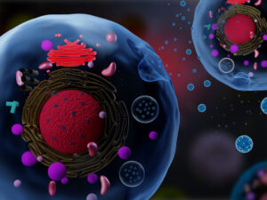
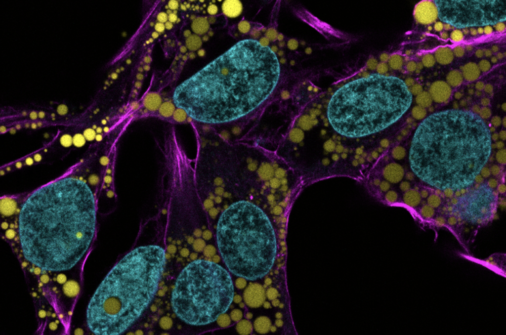
Fat is a normal and necessary part of the body. Fat cells store and release energy, as well as play significant roles in hormonal regulation and immunity.
Engineers and scientists at the University of Pennsylvania are the first to discover fat-filled lipid droplets’ (pictured above in green) surprising capability to indent and puncture the nucleus, the organelle which contains and regulates a cell’s DNA.
In recent decades, a concerning rise in metabolic illnesses – such as cardiovascular disease, high blood pressure and diabetes – has focused scientific attention on the biology and chemistry of fat, resulting in a wealth of information about how fat cells work.
But fat cells and their metabolic activities are only part of the story.
Fat-filled lipid droplets, tiny spheres of fat many times smaller than fat cells, are a growing subject of scientific interest. Found inside many different cell types, these lipid particles have long been little understood. Studies have begun to illuminate these droplets’ participation in metabolic functions and cellular protection, but we still know next to nothing about the physical nature of fat.
Now, researchers at the University of Pennsylvania School of Engineering and Applied Science have looked beyond biochemistry to publish groundbreaking work on the physics of these droplets, revealing them to be a potential threat to a cell’s nucleus. In the August issue of the Journal of Cell Biology, they are the first to discover fat-filled lipid droplets’ surprising capability to indent and puncture the nucleus, the organelle which contains and regulates a cell’s DNA.
The stakes of their findings are high: a ruptured nucleus can lead to elevated DNA damage that is characteristic of many diseases, including cancer.
“Intuitively, people think of fat as soft,” says Discher. “And on a cellular level it is. But at this small size of droplet— measuring just a few microns rather than the hundreds of microns of a mature fat cell—it stops being soft. Its shape has a much higher curvature, bending other objects very sharply. This changes its physics in the cell. It can deform. It can damage. It can rupture.”
They join 120 members and 23 international members elected by their peers this year to NAS. Recognized for “distinguished and continuing achievements in original research,” this new class brings the total number of active members to 2,565 and of international members to 526.
David Brainard is the RRL Professor of Psychology, director of the Vision Research Center, and associate dean for the natural sciences in the School of Arts & Sciences. His research focuses on human vision, using both experiments and computer modeling of visual processing, to understand how the visual system deciphers information about objects from light entering the eye. Specifically, he and his lab are interested in color vision, conducting psychophysical experiments to investigate how the appearance of color is affected by an object’s surface properties and ambient light, and how color perception aids in identifying objects. Brainard is the recipient of many honors, including the Macbeth Award from the Inter-Society Color Council, Stein Innovation Award from Research to Prevent Blindness, and Edgard D. Tillyer Award from Optica. He is an elected member of the Society of Experimental Psychologists, a Silver Fellow of the Association for Research in Vision and Ophthalmology, and a Fellow of the Association for Psychological Science.
Kenneth Zaret
Kenneth S. Zaret is the Joseph Leidy Professor in the Department of Cell and Developmental Biology at the Perelman School of Medicine, director of the Institute for Regenerative Medicine, and a member of the Cell and Molecular Biology Graduate Program. His research focuses on gene regulation, cell differentiation, and chromatin structure, with a goal of elucidating these phenomena in the context of embryonic development and tissue regeneration. Pinpointing these aspects of development at the cellular level can serve as the basis for developing future therapeutics and experimental models that further scientists’ ability to understand and cure disease. Zaret has been the recipient of many honors, including a MERIT Award from the National Institutes of Health, the Stanley N. Cohen Biomedical Research Award, and election as a fellow of the American Association for the Advancement of Science.
A team of researchers led by the School of Arts & Science’s Wei Guo offers new insights into a mechanism that promotes tumor growth. “This information could be used to help clinicians diagnose cancers earlier in the future,” says Guo.
In many instances, the physical manifestation of cancers and the ways they are subsequently diagnosed is via a tumor, tissue masses of mutated cells and structures that grow excessively. One of the major mysteries in understanding what goes awry in cancers relates to the environments within which these structures grow, commonly known as the tumor microenvironment.
These microenvironments play a role in facilitating tumor survival, growth, and spread. Tumors can help generate their own infrastructure in the form of vasculature, immune cells, signaling molecules, and extracellular matrices (ECMs), three-dimensional networks of collagen-rich support scaffolding for a cell. ECMs also help regulate cellular communications, and in the tumor microenvironment ECMs can be a key promoter of tumor growth by providing structural support for cancerous cells and in modulating signaling pathways that promote growth.
Now, new research led by the School of Arts & Science’sWei Guo and published in the journal Nature Cell Biology has bridged the complex structural interactions within the tumor microenvironment to the signals that trigger tumor growth. The researchers studied cancerous liver cells grown on ECMs of varying stiffness and discovered that the stiffening associated with tumor growth can initiate a cascade that increases the production of small lipid-encapsulated vesicles known as exosomes.
“Think of these exosomes as packages that each cell couriers out, and, depending on the address, they get directed to other cells,” says Ravi Radhakrishnan, professor of bioengineering in the School of Engineering and Applied Science and a co-author of the paper.
“By recording the number of packages sent, the addresses on these packages, their contents, and most importantly, how they’re regulated and generated, we can better understand the relationship between a patient’s tumor microenvironment and their unique molecular signaling signatures, hinting at more robust personalized cancer therapies,” Radhakrishnan says.
While studying exosomes in relation to tumor growth and metastasis has been well-documented in recent years, researchers have mostly focused on cataloging their characteristics rather than investigating the many processes that govern the creation and shuttling of exosomes between cells. As members of Penn’s Physical Sciences Oncology Center (PSOC), Guo and Radhakrishnan have long collaborated on projects concerning tissue stiffness. For this paper, they sought to elucidate how stiffening promotes exosome trafficking in cancerous intracellular signaling.
“Our lab previously found that high stiffness promotes the secretion of exosomes,” says Di-Ao Liu, co-first author of the paper and a graduate student in the Guo Lab. “Now, we were able to model the stiffening processes through experiments and identify molecular pathways and protein networks that cause this, which better links ECM stiffening to cancerous signaling.”
The model of tubule packing developed by the Hughes Lab shows the tubules repelling each other and shifting around.
A recent study by Penn Bioengineering researchers sheds new light on the role of physics in kidney development. The kidney uses structures called nephrons and tubules to filter blood and pass urine to the bladder. Nephron number is set at birth and can vary over an order of magnitude (anywhere from 100,000 to over a million nephrons in an individual kidney). While the reasons for this variability remain unclear, low numbers of nephrons predispose patients to hypertension and chronic kidney disease.
Now, research published in Developmental Cell led by Alex J. Hughes, Assistant Professor in the Department of Bioengineering, demonstrates a new physics-driven approach to better visualize and understand how a healthy kidney develops to avoid organizational defects that would impair its function. While previous efforts have typically approached this problem using molecular genetics and mouse models, the Hughes Lab’s physics-based approach could link particular types of defects to this genetic information and possibly highlight new treatments to prevent or fix congenital defects.
Alex J. Hughes, Assistant Professor in Bioengineering
Louis Prahl, NIH F32 Postodctoral Fellow
During embryonic development, kidney tubules grow and the tips divide to make a branched tree with clusters of nephron stem cells surrounding each branch tip. In order to build more nephrons, the tree needs to grow more branches. To keep the branches from overlapping, the kidney’s surface grows more crowded as the number of branches increase. “At this point, it’s like adding more people to a crowded elevator,” says Louis Prahl, first author of the paper and Postdoctoral Fellow in the Hughes Lab. “The branches need to keep rearranging to accommodate more until organ growth stops.”
To understand this process, Hughes, Prahl and their team investigated branch organization in mouse kidneys as well as using computer models and a 3D printed model of tubules. Their results show that tubules have to actively restructure – essentially divide at narrower angles – to accommodate more tubules. Computer simulations also identified ‘defective’ packing, in which the simulation parameters caused tubules to either overlap or be forced beneath the kidney surface. The team’s experimentation and analysis of published studies of genetic mouse models of kidney disease confirmed that these defects do occur.
This study represents a unique synthesis of different fields to understand congenital kidney disease. Mathematicians have studied geometric packing problems for decades in other contexts, but the structural features of the kidney present new applications for these models. Previous models of kidney branching have approached these problems from the perspective of individual branches or using purely geometric models that don’t account for tissue mechanics. By contrast, The Hughes Lab’s computer model demonstrates the physics of how tubule families interact with each other, allowing them to identify ‘phases’ of kidney organization that either relate to normal kidney development or organizational defects. Their 3D printed model of tubules shows that these effects can occur even when one sets the biology aside.
Hughes has been widely recognized for his research in the understanding of kidney development. This new publication is the first fruit of his 2021 CAREER Award from the National Science Foundation (NSF) and he was recently named a 2023 Rising Star by the Cellular and Molecular Bioengineering (CMBE) Special Interest Group. In 2020 he became the first Penn Engineering faculty member to receive the Maximizing Investigators’ Research Award (MIRA) from the National Institutes of Health (NIH) for his forward-thinking work in the creation of new tools for tissue engineering.
Pediatric nephrologists have long worked to understand the cause of these childhood kidney defects. These efforts are often confounded by a lack of evidence for a single causative mutation. The Hughes Lab’s approach presents a new and different application of the packing problem and could help answer some of these unsolved questions and open doors to prevention of these diseases. Following this study, Hughes and his lab members will continue to explore the physics of kidney tubule packing, looking for interesting connections between packing organization, mechanical stresses between neighboring tubule tips, and nephron formation while attempting to copy these principles to build stem cell derived tissues to replace damaged or diseased kidney tissue. Mechanical forces play an important role in developmental biology and there is much scope for Hughes, Prahl and their colleagues to learn about these properties in relation to the kidney.
Other authors include Bioengineering Ph.D. students and Hughes Lab members John Viola and Jiageng Liu.
This work was supported by NSF CAREER 2047271, NIH MIRA R35GM133380, Predoctoral Training Program in Developmental Biology T32HD083185, and NIH F32 fellowship DK126385.
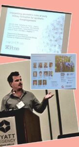
Alex J. Hughes presents at the BMES CMBE conference in January 2023. (Image credit: Riccardo Gottardi, Assistant Professor in Pediatrics and Bioengineering)
Alex J. Hughes, Assistant Professor in the Department of Bioengineering, was one of thirteen recipients of the 2023 Rising Star Award for Junior Faculty by the Cellular and Molecular Bioengineering (CMBE) Special Interest Group. The Rising Star Award recognizes a CMBE member in their early independent career stage that has made an outstanding impact on the field of cellular and molecular bioengineering. CMBE is a special interest group of the Biomedical Engineering Society (BMES), the premier professional organization of bioengineers.
The Hughes Lab in Penn Bioengineering works to “bring developmental processes that operate in vertebrate embryos and regenerating organs under an engineering control framework” in order to “build better tissues.” Hughes’s research interest is in harnessing the developmental principles of organs, allowing him to design medically relevant scaffolds and machines. In 2020 he became the first Penn Engineering faculty member to receive the Maximizing Investigators’ Research Award (MIRA) from the National Institutes of Health (NIH), and he was awarded a prestigious CAREER Award from the National Science Foundation (NSF) in 2021. Most recently, Hughes’s work has focused on understanding the development of cells and tissues in the human kidney via the creation of “organoids”: miniscule organ models that can mimic the biochemical and mechanical properties of the developing kidney. Understanding and engineering how the kidney functions could open doors to more successful regenerative medicine strategies to address highly prevalent congenital and adult diseases.
Hughes and his fellow award recipients were recognized at the annual BMES CBME conference in Indian Wells, CA in January 2023.
Penn Medicine researchers laud the early results for CAR T therapy in lupus patients, which point to broader horizons for the use of personalized cellular therapies.
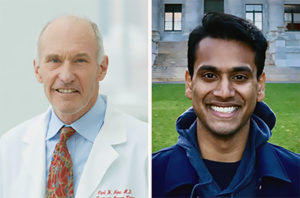
Penn Medicine’s Carl June and Daniel Baker.
Engineered immune cells, known as CAR T cells, have shown the world what personalized immunotherapies can do to fight blood cancers. Now, investigators have reported highly promising early results for CAR T therapy in a small set of patients with the autoimmune disease lupus. Penn Medicine CAR T pioneer Carl June and Daniel Baker, a doctoral student in cell and molecular biology in the Perelman School of Medicine, discuss this development in a commentary published in Cell.
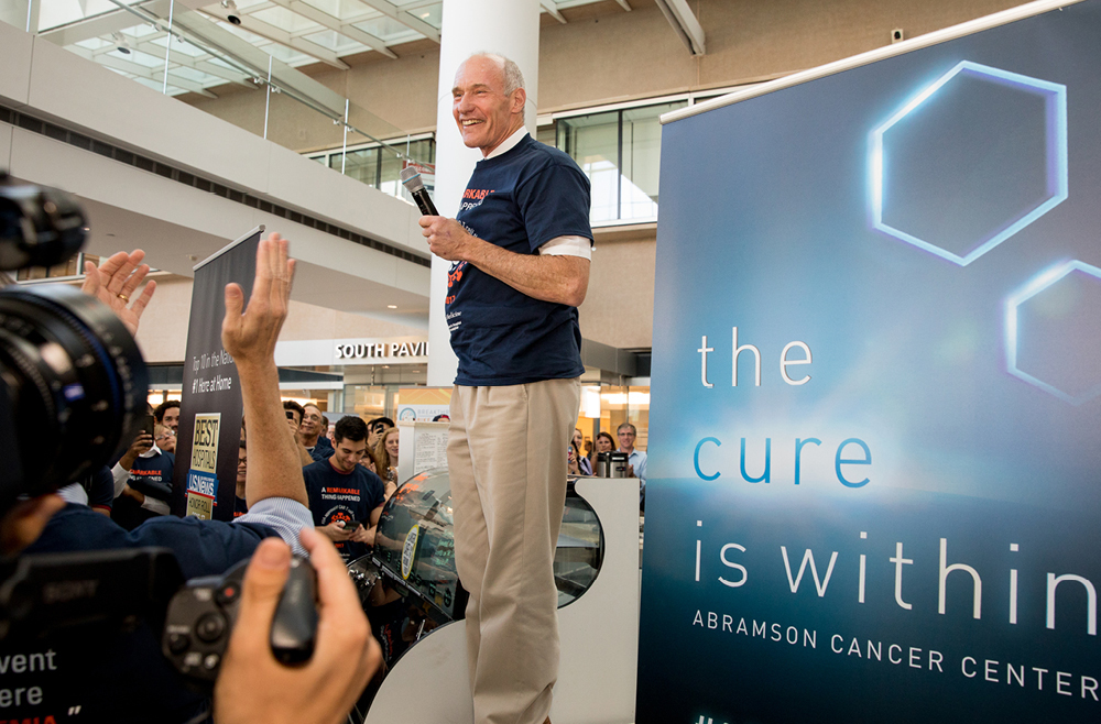
“We’ve always known that in principle, CAR T therapies could have broad applications, and it’s very encouraging to see early evidence that this promise is now being realized,” says June, who is the Richard W. Vague Professor in Immunotherapy in the department of Pathology and Laboratory Medicine at Penn Medicine and director of the Center for Cellular Immunotherapies at the Abramson Cancer Center.
T cells are among the immune system’s most powerful weapons. They can bind to, and kill, other cells they recognize as valid targets, including virus-infected cells. CAR T cells are T cells that have been redirected, through genetic engineering, to efficiently kill specifically defined cell types.
CAR T therapies are created out of each patient’s own cells—collected from the patient’s blood, and then engineered and multiplied in the lab before being reinfused into the patient as a “living drug.” The first CAR T therapy, Kymriah, was developed by June and his team at Penn Medicine, and received Food & Drug Administration approval in 2017. There are now six FDA-approved CAR T cell therapies in the United States, for six different cancers.
From the start of CAR T research, experts believed that T cells could be engineered to fight many conditions other than B cell cancers. Dozens of research teams around the world, including teams at Penn Medicine and biotech spinoffs who are working to develop effective treatments from Penn-developed personalized cellular therapy constructs, are examining these potential new applications. Researchers say lupus is an obvious choice for CAR T therapy because it too is driven by B cells, and thus experimental CAR T therapies against it can employ existing anti-B-cell designs. B cells are the immune system’s antibody-producing cells, and, in lupus, B cells arise that attack the patient’s own organs and tissues.


 David Brainard is the RRL Professor of Psychology, director of the