Week in BioE (March 15, 2019)

by Sophie Burkholder

Synthetic Spinal Discs from a Penn Research Team Might Be the Solution to Chronic Back Pain

Spinal discs, the concentric circles of collagen fiber found between each vertebra of the spine, can be the source of immense back pain when ruptured. Especially for truck and bus drivers, veterans, and cigarette smokers, there is an increased risk in spinal disc rupture due to overuse or deterioration over time. But these patients aren’t alone. In fact, spinal discs erode over time for almost everyone, and are one of the sources of back pain in older patients, especially when the discs erode so much that they allow direct bone-to-bone contact between two or more vertebrae.

Robert Mauck, Ph.D.

Robert Mauck, Ph.D., who is the director of the McKay Orthopaedic Research Laboratory here at Penn and a member of the Bioengineering Graduate Group Faculty, led a research team in creating artificial spinal discs, with an outer layer made from biodegradable polymer and an inner layer made with a sugar-like gel. Their findings appear in Science Translational Medicine. These synthetic discs are also seeded with stem cells that produce collagen over time, meant to replace the polymer as it degrades in vivo over time. Though Mauck and his time are still far from human clinical trials for the discs, they’ve shown some success in goat models so far. If successful, these biodegradable discs could lead to a solution for back pain that integrates itself into the human body over time, potentially eliminating the need of multiple invasive procedures that current solutions require. Mauck’s work was recently featured in Philly.com.

An Untethered, Light-Activated Electrode for Innovations in Neurostimulation

Neurostimulation, a process by which nervous system activity can be purposefully modulated, is a common treatment for patients with some form of paralysis or neurological disorders like Parkinson’s disease. This procedure is typically invasive, and because of the brain’s extreme sensitivity, even the slightest involuntary movement of the cables, electrodes, and other components involved can lead to further brain damage through inflammation and scarring. In an effort to solve this common problem, researchers from the B.I.O.N.I.C. Lab run by Takashi D.Y. Kozai, Ph. D., at the University of Pittsburgh replaced long cables with long wavelength light and a formerly tethered electrode with a smaller, untethered one.

The research team, which includes Pitt senior bioengineering and computer engineering student Kaylene Stocking, centered the device on the principle of the photoelectric effect – a concept first described in a publication by Einstein as the local change in electric potential for an object when hit with a photon. Their design, which includes a 7-8 micron diameter carbon fiber implant, is now patent pending, and Kozai hopes that it will lead to safer and more precise advancements in neurostimulation for patients in the future.

A New Microfluidic Chip Can Detect Cancer in a Drop of Blood

Many forms of cancer cannot be detected until the disease has progressed past the point of optimum treatment time, increasing the risk for patients who receive late diagnoses of these kinds of cancer. But what if the diagnostic process could be simplified and made more efficient so that even a single drop of blood could be enough input to detect the presence of cancer in a patient? Yong Zeng, Ph.D., and his team of researchers at the University of Kansas in Lawrence sought to answer that question.

They designed a self-assembled 3D-nanopatterned microfluidic chip to increase typical microfluidic chip sensitivity so that it can now detect lower levels of tumor-associated exosomes in patient blood plasma. This is in large part due to the nanopatterns in the structure of the chip, which promote mass transfer and increase surface area, which in turn promotes surface-particle interactions in the device. The team applied the device to their studies of ovarian cancer, one of the notoriously more difficult kinds of cancer to detect early on in patients.

A Wearable Respiration Monitor Made from Shrinky Dinks

Michelle Khine, Ph. D., a professor of biomedical engineering at the University of California, Irvine incorporates Shrinky Dinks into her research. After using them once before in a medical device involving microfluidics, her lab recently worked to incorporate them into a wearable respiration monitor – a device that would be useful for patients with asthma, cystic fibrosis, and other chronic pulmonary diseases. The device has the capability to track the rate and volume of its user’s respiration based on measurements of the strain at the locations where the device makes contact with the user’s abdomen.

Paired with Bluetooth technology, this sensor can feed live readings to a smartphone app, giving constant updates to users and doctors, as opposed to the typical pulmonary function test, which only provides information from the time at which the test takes a reading. Though Khine and her team have only tested the device on healthy patients so far, they look forward to testing with patients who have pulmonary disorders, in hopes that the device will provide more comprehensive and accessible data on their respiration.

People and Places

Ashley Kimbel, a high school senior from Grissom High School in Huntsville, Alabama, designed a lightweight prosthetic leg for local Marine, Kendall Bane, after an attack in Afghanistan led him to amputate one of his legs below the knee. Bane, who likes to keep as active as possible, said the new lighter design is more ideal for activities like hiking and mountain biking, especially as any added weight makes balance during these activities more difficult. Kimbel used a CAD-modeling software produced by Siemens called Solid Edge, which the company hopes to continue improving in accessibility so that more students can start projects like Kimbel’s.

This week, we would like to congratulate Angela Belcher, Ph.D., on being named the new head of the Department of Biological Engineering at the Massachusetts Institute of Technology (MIT). With her appointment to this role, now half of the MIT engineering department heads are women. Belcher’s research is in the overlap of materials science and biological engineering, with a particular focus on creating nanostructures based on the evolution of ancient organisms for applications in medical diagnostics, batteries, solar cells, and more.

We would also like to congratulate Eva Dyer, Ph.D., and Chethan Pandarinath, Ph.D., both of whom are faculty members at the Walter H. Coulter Department of Biomedical Engineering at Georgia Tech and Emory University, on receiving research fellowships from the Alfred P. Sloan Foundation. Dr. Dyer, who formerly worked with Penn bioengineering faculty member Dr. Konrad Kording while he was at Northwestern University, leads research in the field of using data analysis methods to quantify neuroanatomy. Dr. Pandarinth leads the Emory and Georgia Tech Systems Neural Engineering Lab, where he works with a team of researchers to use properties of artificial intelligence and machine learning to better understand large neural networks in the brain.


BE Seminar Series: March 14th

The BE Seminar Series continues this week. We hope to see you there!

Shuichi Takayama, Ph.D.

Speaker: Shuichi Takayama, Ph.D.
Professor, GRA Eminent Scholar, Price Gilbert, Jr. Chair in Regenerative Engineering and Medicine
Wallace H. Coulter Department of Biomedical Engineering
Georgia Institute of Technology and Emory University

Date: Thursday, March 14th, 2019
Time: 12:00 pm
Location: Room 337, Towne Building

“Microfluidics and Immuno-Materials for Organs-on-a-Chip”

This presentation will describe microfluidic technologies to conveniently produce life-like pulsatile flows along with applications to study of lung injury, enhancement of in vitro fertilization, and analysis of frequency-dependent cellular responses. The microfluidic technologies range from adaptation of piezo-electric actuator arrays from Braille displays to design of microfluidic circuits that can be designed to switch fluid flow on and off periodically on their own. The presentation will also describe engineered materials to mimic an aspect of the innate immune system to combat bacterial infection. More specifically, reconstituted chromatin microwebs inspired by neutrophil extracellular traps. Using a defined composition reconstituted chromatin microweb, we reveal impact of microweb DNA-histone ratio on bacteria capture. Additionally, we found that E. coli, including clinical isolates and resistant strains, are killed more efficiently by the last-resort antibiotic, colistin, when bound to microwebs. Recent efforts towards incorporation of these materials into human cell systems will also be described. Time permitting, topics on organoids, fibrosis, liquid-liquid phase separation, and scaling may be incorporated.

Week in BioE (February 21, 2019)

by Sophie Burkholder

Detecting Infectious Diseases with Paper-Based Devices

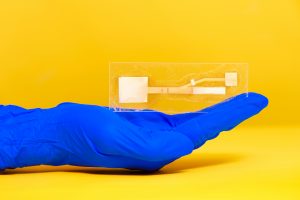
Dr. Linnes’ paper device. Image used courtesy of Erin Easterling, Purdue College of Engineering.

Despite great advancements in diagnostics technology over the past few decades, patient accessibility to these technologies remains one of the biggest challenges of the field today. Particularly in low-resource areas, even simple processes can end up taking weeks or months to return results from tests that are normally completed in days. But what if these tests could be simplified to smaller, at-home tests based on properties of microfluidics – something like a pregnancy test but for infectious diseases like HIV?

Jacqueline Linnes, Ph.D., and her team of researchers at Purdue University are working towards finding a way to do just that by creating paper-based devices that use microfluidics to help carry out the necessary diagnostic tests. Specifically, her lab designed such a paper-based system that can detect HIV nucleic acids within 90 minutes of receiving a drop of patient blood. The success of this design shows promise for producing devices for diseases whose diagnostics process involve similar pathways of pathogen detection, opening the door to more applications of at-home tests based in the properties of paper microfluidics.

Here at Penn, undergraduate bioengineering students enrolled in the two-semester laboratory course Bioengineering Modeling, Analysis, and Design (BE 309 & BE 310) have the chance to create their own models of paper microfluidics delivery systems based on given time constraints in a multi-step process. Though the students’ challenge only involves water as a substrate, Linnes’ research demonstrates the later implications of studying fluid flow through a medium as cheap and accessible as paper.

Watch the video below demonstrating Dr. Linnes’ device:

Funding for Cancer Research in Tumor Mimicry and Imaging

Two of the deadliest forms of cancer today are breast cancer and pancreatic cancer, with the latter having a five-year survival rate of only about 8%. Because cancer treatments are often adjusted according to a unique patient-to-patient basis, learning how to improve predictions of tumor behavior could help determine proper therapies sooner.

Chien-Chi Lin, Ph.D., an associate professor of biomedical engineering at Indiana University – Purdue University Indianapolis, recently received a grant from the National Institute of Health to advance his research in pancreatic cancer treatment. His project under the grant involves the development of bio-inspired, responsive, and viscoelastic (BRAVE) cell-laden hydrogels to help understand cell interactions in pancreatic ductal adenocarcinoma, which is the most common form of malignancy in the pancreas. These hydrogels mimic tumor tissue, as well as model tumor development over time, helping to eventually find better ways of treating pancreatic cancer.

In other news surrounding cancer-related research, a team of researchers led by Kenneth Tichauer, Ph.D., at the Illinois Institute of Technology won the university’s Nayar Prize for their development of the Agent-Dependent Early Photon Tomography (ADEPT) Cancer Imager, a machine designed to find early tumor development in the lymph nodes of breast cancer patients. Through the use of a special dyeing process that now dyes the entire lymph node, providing a sharper image that allows for a quicker discovery of smaller tumors.

Penn’s Women in Computer Science (WiCS) Hosts FemmeHacks

Penn President Amy Gutmann and Penn Engineering Dean Vijay Kumar stopped by FemmeHacks at the Pennovation Center Feb. 9. The annual event is a beginner-friendly collegiate hackathon for women-identifying people with an interest in computer programming, and featured a day of all-levels workshops Feb. 8. The event is sponsored by Penn’s Women in Computer Science student organization.

Though the event is not specifically tailored towards applications in bioengineering, skills relating to coding and software development are increasingly important for those interested in pursuing a career in medical device design. In fact, in the evaluation of new medical devices, the FDA often focuses more on software over hardware, as the former is associated with more security liabilities, due to its relative novelty.

Read the full story and see pictures on FemmeHacks on Penn Today.

People & Places

In December, the department of biomedical engineering at the University of Alabama at Birmingham received the Ruth L. Kirschstein National Research Service Awards Institutional Research Training Grant, which will support predoctoral students enrolled in the university’s biomedical engineering graduate programs. The department plans to use the grant for research in cardiovascular tissue engineering.

Case Western Reserve University and Cleveland Clinic announced the launch of an alliance last year with the goal of creating better synergy across the two renowned institutions, hoping to provide more opportunities for students with interest in medicine at all levels, from high school to postdoctoral education. Though researchers from both institutions frequently partner on projects, this new alliance will create a more structured platform for future collaborations.

We would like to commend Steven George, M.D./Ph.D., on his new position as the chair of the Department of Biomedical Engineering at the University of California at Davis. His research involves the development of “organ-on-a-chip” technologies using stem cells and microfluidics to mimic human organ functions of vascularized cardiac, tumor, and pancreatic tissues.

Finally, we want to congratulate Paul Yock, M.D., on his being chosen to receive the National Academy of Engineering’s 2019 Fritz J. and Dolores H. Russ Prize. The prize honors two of Dr. Yock’s inventions from his research in interventional cardiology, one of which is Rapid Exchange, which is a kind of stenting and balloon angioplasty system. Dr. Yock is the Martha Meier Weiland Professor in the School of Medicine and Professor of Bioengineering.


Week in BioE (May 10, 2018)

Advances in Cancer Detection

glioblastoma
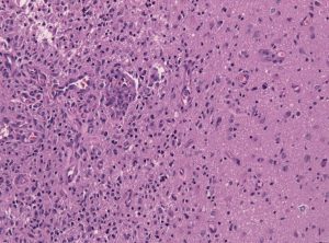
Tumor-brain-interface in a glioblastoma biopsy specimen.

Among the deadliest and most difficult to treat types of cancer is glioblastoma, an especially aggressive form of brain cancer. Widely available imaging techniques can diagnose the tumor, but often the diagnosis is too late to treat the cancer effectively. Although blood-based cancer biomarkers can provide for earlier detection of cancer, these markers face the difficult task of crossing the blood-brain barrier (BBB), which prevents all but the tiniest molecules from moving from the brain to the bloodstream.

A study recently published in Scientific Reports, coauthored by Hong Chen, PhD, Assistant Professor of Biomedical Engineering at Washington University in St. Louis (WUSTL), reports of successful deployment of a strategy consisting of focused ultrasound (FUS), enhanced green fluorescent protein (eGFP), and systemically injected microbubbles to see if the BBB could be opened temporarily to allow biomarkers to pass from the brain into the bloodstream. The authors used eGFP-activated mouse models of glioblastoma, injecting the microbubbles into the mice and then exposing the mice to varying acoustic pressures of FUS. They found that circulating blood levels of eGFP were several thousand times higher in the FUS-treated mice compared to non-treated mice, which would significantly facilitate the detection of the marker in blood tests.

The method has some way to go before it can be used in humans. For one thing, the pressures used in the Scientific Reports study would damage blood vessels, so it must be determined whether lower pressures would still provide detectable transmission of proteins across the BBB. In addition, the authors must exclude the possibility of FUS unexpectedly enhancing tumor growth.

In other body areas, with easier access from tissue to the bloodstream, engineers have developed a disease-screening pill that, when ingested and activated by infrared light, can indicate tumor locations on optical tomography. The scientists, led by Greg M. Thurber, PhD, Assistant Professor of Biomedical and Chemical Engineering at the University of Michigan, reported their findings in Molecular Pharmaceutics.

The authors of the study used negatively charged sulfate groups to facilitate absorption by the digestive system of molecular imaging agents. They tested a pill consisting of a combination of these agents and found that their model tumors were visible. The next steps will include optimizing the imaging agent dosage loaded into the pill to optimize visibility. The authors believe their approach could eventually replace uncomfortable procedures like mammograms and invasive diagnostic procedures.

Liquid Assembly Line to Produce Drug Microparticles

Pharmaceuticals owe their effects mostly to their chemical composition, but the packaging of these drugs into must be done precisely. Many drugs are encapsulated in solid microparticles, and engineering consistent size and drug loading in these particles is key. However, common drug manufacturing techniques, such as spray drying and ball milling, produce uneven results. 

University of Pennsylvania engineers developed a microfluidic system in which more than ten thousand of these devices run in parallel, all on a silicon-and-glass chip that can fit into a shirt pocket, to produce a paradigm shift in microparticle manufacturing. The team, led by David Issadore, Assistant Professor in the Department of Bioengineering, outlined the design of their system in the journal Nature Communications.

The Penn team first tested their system by making simple oil-in-water droplets, at a rate of more than 1 trillion droplets per hour. Using materials common to current drug manufacturing processes, they manufactured polycapralactone  microparticles at a rate of ‘only’ 328 billion particles per hour. Further testing backed by pharma company GlaxoSmithKline will follow.

Preventing Fungal Infections of Dental Prostheses

Dental prostheses are medical devices that many people require, particularly as they age. One of the chief complications with prostheses is fungal infections, with an alarming rate of two-thirds among people wearing dentures. These infections can cause a variety of problems, spreading to other parts of the digestive system and affecting nutrition and overall well-being. Fungal infections can be controlled in part by mouthwashes, microwave treatments, and light therapies, but none of them have high efficacy.

To address this issue, Praveen Arany, DDS, PhD, Assistant Professor, Department of Oral Biology and Biomedical Engineering at SUNY Buffalo, combined 3D printing technology and polycaprolactone microspheres containing amphotericin-B, an antifungal agent. Initial fabrication of the prostheses is described in an article in Materials Today Communications, along with successful in vitro testing with fungal biofilm. If further testing proves effective, these prostheses could be used in dental patients in whom the current treatments are either ineffective or contraindicated.

People and Places

West Virginia University has announced that it will launch Master’s and doctoral programs in Biomedical Engineering. The programs will begin enrolling students in the fall. The graduate tracks augment a Bachelor’s degree program begun in 2014.

Oncology/Engineering Review Published

oncology
Mike Mitchell, Ph.D.

Michael Mitchell, Ph.D., who will arrive in the Spring 2018 semester as assistant professor in the Department of Bioengineering, is the first author on a new review published in Nature Reviews Cancer on the topic of engineering and the physical sciences and their contributions to oncology. The review was authored with Rakesh K. Jain, Ph.D., who is Andrew Werk Cook Professor of Radiation Oncology (Tumor Biology) at Harvard Medical School, and Robert Langer, Sc.D., who is Institute Professor in Chemical Engineering at the David H. Koch Institute for Integrative Cancer Research at MIT. Dr. Mitchell is currently in his final semester as a postdoctoral fellow at the Koch Institute and is a member of Dr. Langer’s lab at MIT.

The review focuses on four key areas of development for oncology in recent years: the physical microenvironment of the tumor; technological advances in drug delivery; cellular and molecular imaging; and microfluidics and microfabrication. Asked about the review, Dr. Mitchell said, “We’ve seen exponential growth at the interface of engineering and physical sciences over the last decade, specifically through these advances. These novel tools and technologies have not only advanced our fundamental understanding of the basic biology of cancer but also have accelerated the discovery and translation of new cancer therapeutics.”

Week in BioE (October 13, 2017)

Seeing and Repairing Damaged Heart Vessels

angiography
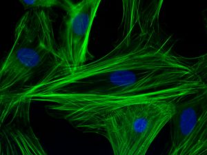
Immunofluorescent staining for F-actin filaments (green) and nuclei (blue) in neonatal cardiomyocyte

Two common diagnostic procedures in cardiology are intravascular ultrasound and cardiac angiography. These procedures are performed to quantify the amount of plaque affecting a patient’s blood vessels. This information is vital because it helps to determine how advanced heart degree is, as well as guiding treatment planning and even the course of bypass surgery. However, the current technologies used for these procedures have significant limitations. Although conventional angiography can help to quantify the plaque burden, it does not offer any information about how much of the diameter of a vessel is blocked. Intravascular ultrasound is very good at quantifying plaque burden, but it is poor at identifying smaller features of compromised blood vessels.

One solution suggested to these issues is the combination of these imaging technologies into a single multimodal technique. Scientists led by Laura Marcu, Ph.D., professor of biomedical engineering at the University of California, Davis, invented a method combining intravascular ultrasound with multispectral fluorescence lifetime imaging (FLIM). As published in Scientific Reports, the device resembles a typical cardiac catheter but contains an optical fiber within the catheter that emits fluorescent light to characterize the plaque components before treatment.

Dr. Marcu and her colleagues tested their new device in live pigs and in human coronary arteries obtained from cadavers. The fluorescence data acquired with the device were comparable to those acquired with traditional fluorescence angiography. Moreover, the device could acquire data without having to administer a contrast agent, which can be dangerous in some patients due to allergies or weakened kidneys. The authors are currently seeking FDA approval to test their combined catheter in humans.

In addition to treating vessels before a heart attack can occur, there is new work showing how to efficiently repair heart tissue after a heart attack. A team of scientists collaborating among Clemson University, the Medical University of South Carolina,  the University of South Carolina, and the University of Chicago has received a $1.5 million grant from the National Institutes of Health to examine a treatment that combines stem cells with nanowires. The principal investigator on the grant is Ying Mei, Ph.D., who is assistant professor of bioengineering at Clemson. Dr. Mei’s team mixes stem cells with nanowires so that they form spheroids that are larger than single cells and thus less likely to wash away. In addition, the investigators hope that the spheroids will mitigate the issue of the transplanted cells and the recipient’s heart beating at different rhythms.  If successful, the group’s treatment paradigm could be a major step forward in stem cell therapies and cardiology.

Look, Up in the Sky!

Drones became famous when deployed on battlefields for the first time a decade ago. Since then, they’ve been adopted as a technology for a variety of purposes. For example, Amazon introduced delivery drones almost a year ago, and it has plans to expand its drone fleet enormously in coming years. It was only a matter of time before engineers began to imagine medical applications for drones.

Engineers in Australia and Iraq recently investigated whether a drone could be used to monitor cardiorespiratory signals remotely. They reported their findings in BioMedical Engineering OnLine. The authors used imaging photoplethysmography (PPG), which employs a video camera to detect visual indications on the skin of heart activity. They also applied advanced digital processing technology due to the tendency of PPG to be affected by sound and movement in the area of detection. By testing the combined technologies in 15 healthy volunteers, the authors found that their data compared well with several traditional techniques for monitoring vital signs. Among the possible applications that the authors imagine for this technology is battlefield triage performed remotely using drones. In the meantime, they will seek to fine-tune the technology’s abilities.

Concussion Distressingly Common

A research letter published in a recent issue of JAMA reports that a study conducted in Canada found that one in five adolescents sustained a concussion on at least one occasion. Of the approximately 20% of the study respondents who had experienced concussions, one quarter had suffered more than one. The letter is particularly relevant to the United States because of the similar popularity in Canada of contact and semicontact sports such as ice hockey and football. In addition, the study included more than 13,000 teenagers, lending significantly reliability to the conclusions.

Ending the Time of Cholera

Although largely eradicated in the developed world, cholera remains a major public health issue in the Global South and other parts of the developing world. The disease is a bacterial infection that causes severe gastrointestinal distress. Because the disease is transmitted via water, effective public sanitation is a core requirement of an effective prevention campaign.

One technology being deployed in this fight is a smartphone microfluidics platform that can determine the presence of the pathogen that causes cholera in a sample and report the data almost immediately to public health authorities. This technology was produced by a company called PathVis, which was spun off at Purdue University based on science produced the laboratories of Tamara Kinzer-Ursem, Ph.D., and Jacqueline Linnes, Ph.D., both of whom are assistant professors in Purdue’s Weldon School of Biomedical Engineering. There are plans to test PathVis in Haiti and to expand it to detect other diseases in the future.

The Latest on CRISPR

CRISPR/Cas9 is the biggest bioengineering story to come along in some time — certainly the biggest in genetic engineering. But the mere fact that it’s here and already being used in animals and in human cell lines doesn’t mean that the story is over.  For instance, the Cas9 protein, which CRISPR deploys as part of its gene editing process, is currently developed most often using a viral vector. However, this system of delivery has certain drawbacks, not the least of which is a host immune system response when levels of the deployed viral vector reach the levels necessary for CRISPR to work.

A recent study published in Nature Biomedical Engineering reports on the successful use of gold nanoparticles to deliver Cas9. The new delivery system, called CRISPR-Gold, could obviate the need to use a viral vector as part of the CRISPR induction process. So far, the authors, led by University of California, Berkeley, bioengineers Irina Conboy, Ph.D., and Niren Murthy, Ph.D., have only used CRISPR-Gold in mice, but their successful results indicate that nonviral delivery with CRISPR is possible, so CRISPR could be used for more than previously thought.


Week in BioE (September 22, 2017)

Live Bone Cells Grown in Lab

osteogenesis
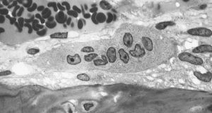
An osteoclast, one type of bone cell.

Bone injuries and bone loss can constitute major challenges for patients and the people who treat them. Beyond the need for bone grafts or artificial implants in cases such as severe fractures, cancers metastasizing to the bones can be disabling and disfiguring. Doctors are able to use autologous bone grafts, in which patients are their own bone donors and provides grafts from other bones in their bodies. However, the grafting process compromises the bone from the donor site. In addition, there are specific problems in cases of long bones, such as those in the arms and legs. With these bones, no site of the body can provide sufficient material without becoming severely compromised itself due to bone loss.

Stem cells have been intensively investigated as a source of bone grafts. With their ability to produce a variety of cell lines from the same source, these cells have the potential to be used in a variety of clinical situations. The mechanisms underlying the determination of the type of cell that an individual stem cell will become are known. However, the ability to produce living bone cells in the laboratory had remained elusive – until now.  In an article published online last week by Nature Biomedical Engineering, a group of scientists led in part by Professor Matthew Dalby, a cellular engineer with the Institute of Molecular, Cell and Systems Biology at the University of Glasgow, United Kingdom, reported its success.

Professor Dalby’s tissue engineering team used a nanoscale bioreactor to stimulate mesenchymal stem cells into osteogenesis (bone creation). The bioreactor applied vibrations on a microscopic scale of 1,000 hertz with 15 nanometers of vertical displacement. In their previous work, Professor Dalby and his colleagues could generate only one bone cell sample at a time. In the current paper, they showed the ability to generate multiple cells for three-dimensional tissue. In addition, they showed that the cells could be generated in environments with less rigidity than that in which osteogenesis normally occurs. This is an important advance because the body provides optimal conditions of stiffness for this process, but the lab does not. Should the techniques in the paper prove viable on a greater scale, they could revolutionize the field of bone grafting.

Microfluidics in the News

Since their introduction, organs on a chip (OOCs) have proliferated in the field of bioengineering. These chips use microfluidics technology to create a model of an organ system in the body. However, until now, OOCs have not been used to model the human placenta – the tissue that connects the embryonic sac to the uterine wall during pregnancy.

Responding to the lack of a OOC model of the placenta, two professors at Florida International University (FAU) have developed a placenta OOC. Sarah E. Du, Ph.D., assistant professor of ocean and mechanical engineering, and Andrew Oleinkov, Ph.D., associate professor of biomedical science, have collaborated to create this chip, which they to intend to use to determine the effects of malaria on the placental microenvironment. A $400,000 grant from the NIH will certainly help.

With malaria causing more than 200,000 perinatal deaths annually, beyond the burden we cited last week, there is an urgent need to determine the exact effects of this parasitic infection on the placenta. Without this knowledge, the development of technologies to mitigate or even prevent these effects will be much more difficult. In addition, because of the obvious ethical constraints on prospective testing in natural history studies, the placenta OOC offers an ideal model.

Elsewhere in the field of microfluidics, an NIH grant to scientists at the University of Illinois, Urbana-Champaign, has gone toward the development of a new test chip to detect sepsis, a condition in which the body’s reaction to infection results in inflammation of the blood vessels and which can cause lethal shock unless detected and treated promptly. The UIUC team developing this more rapid diagnostic technology is led by Rashid Bashir, Ph.D., professor of bioengineering and associate dean of UIUC’s Carle Illinois College of Medicine. Dr. Bashir was lead author on a paper published over the summer in Nature Communications.

Among the more remarkable aspects of the chip developed by Professor Bashir and his colleagues is that it can diagnose sepsis with a single drop of blood. Therefore, in addition to the device’s portability and size, which allows it to be used at the point of care, it is only necessary to use 10 microliters of blood to complete the test. Other available lab tests for sepsis can require as much as 300 times as much blood. Testing its device against the gold standard of flow cytometry, the UIUC team found that the findings obtained with its biochip were strongly correlated with those from flow cytometry. Unlike the new chip, flow cytometry cannot be performed outside the lab.

Since a large proportion of sepsis patients are treated in intensive care units, the ICU is a likely setting in which the biochip could be used, particularly because some ICUs might be in hospitals where the staff does not have 24-hour lab access. The ability to use this chip at the bedside immediately, rather than waiting until the next morning or longer, could make a key difference in detecting and treating sepsis.

Brains on the Internet

For years, Ray Kurzweil, the computer scientist turned author and inventor, has been discussing a future in which, he claims, the distinction between human and artificial intelligence will disappear. For example, Kurweil imagines brains being uploaded to computers. While what Kurzeil imagines has yet to materialize, scientists in South Africa have created the “Brainternet,” which streams brain waves onto the Internet in real time.

As a student project at the School of Electrical and Information Engineering of the University of Witerstand in Johannesburg led by Adam Pantanowitz, a lecturer in the school, the Brainternet was developed from pre-existing technology. The project starts with portable electroencephalography (EEG), which is worn by the subject and which transmits its signal by telemetry to a Raspberry Pi computer. Then, using open source software, the computer live streams the data to an application programming interface, which in turn allows the data to be published at a website accessible to others.

Beyond being an innovative use of these technologies, the Brainternet could be used in telemedicine applications. For instance, it could be helpful in situations where a specialist neurologist is not in the immediate geographic vicinity. Moreover, for research projects involving EEG measurement during tasks or under certain types of external stimulation, the Brainternet could allow for a much larger sample size to be enrolled, owing to its portability and use of the Internet.

People and Places

Dawn Elliott, Ph.D., chair of the Department of Biomedical Engineering at the University of Delaware, has been elected president of the Biomedical Engineering Society (BMES), for which she had served as treasurer. Dr. Elliott’s term as president will begin in October 2018 and last for two years. As president, she plans to take a closer look at education in the field to determine how bioengineering and biomedical engineering departments can graduate the most successful students. We wish her the best of luck and hearty congratulations.

Pancreatic Cancer Detection With Micropore Chip

Pancreatic cancer remains one of the deadliest types of cancer, with one- and five-year survival rates of only 20% and 7%, respectively, according to the American Cancer Society. The mortality is so high because the disease does not typically cause symptoms until it is too late. Therefore, earlier detection could be the key to better survival rates.

In a new paper published by Lab on a Chip, a research team from the lab of David Issadore, assistant professor of Bioengineering, reports on its development of a micropore chip, callled the circulating tumor cell fluorescence in situ hybridization (CaTCh FISH) chip, that could detect circulating tumor cells (CTCs) from mice and patients with pancreatic cancer, even at very low, previously undetectable levels.

pancreatic cancer
Jin A Ko

Jin A (Jina) Ko, who is a Ph.D. student in Bioengineering and first author on the paper, says that CTCs are a key mechanism underlying metastasis, which is another reason why pancreatic cancer has such a low survival rate. Not only can the chip that she helped design detect these cells, which circulate in the bloodstream, but more importantly, pancreatic tumors shed these cells even in their very early stages before any spread has occurred. Therefore, provided the test is performed early enough, the tumor can be detected and treated. Patients with family histories of pancreatic cancer or who have tested positive for certain gene mutations would likely benefit from this sort of test.

The study authors also tested the CaTCh FISH chip using blood samples from 14 patients with advanced pancreatic cancer and from healthy controls. They found that their micropore chip could detect several RNA markers of cancer in 10-mL samples — around 2 tsp. In addition, there were no false-negative results among the healthy controls, demonstrating a high level of reliability in that regard.

“We have developed a microchip platform that combines fast, magnetic micropore-based negative immunomagnetic selection with rapid on-chip in situ RNA profiling,” Jina said. “This integrated chip can isolate both rare circulating cells and cell clusters directly from whole blood and allow individual cells to be profiled for multiple RNA cancer biomarkers.”