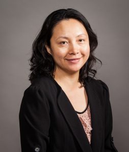
Jenny Jiang, the Peter & Geri Skirkanich Associate Professor of Innovation in the department of Bioengineering, has received a Lloyd J. Old STAR Program grant from the Cancer Research Institute (CRI), which is a major supporter of cancer immunotherapy research and clinical trials with the goal of curing all types of cancer.
The CRI Lloyd J. Old Scientists Taking Risks (STAR) Program “provides long-term funding to mid-career scientists, giving them the freedom and flexibility to pursue high-risk, high-reward research at the forefront of discovery and innovation in cancer immunotherapy.” This prestigious grant was give to six awardees this year, chosen from a pool of hundreds of applicants, and recognizes “future leaders in the field of cancer immunotherapy [who are expected to] carry out transformational research.”
The Old STAR Program Grant comes with $1.25 million in funding over 5 years to support the awardees’ cancer immunology research.
Jiang, who recently joined Penn Bioengineering, is a pioneer in developing tools in genomics, biophysics, immunology, and informatics and applying them to study systems immunology and immune engineering in human diseases. She was also inducted into the American Institute for Medical and Biological Engineering (AIMBE) College of Fellows in March 2021 for her outstanding contributions to the field of systems immunology and immunoengineering and devotion to the success of women in engineering. Jiang’s research focuses on systems immunology by developing technologies that enable high-throughput, high-content, single cell profiling of T cells in health and disease and she is recognized as one of the leading authorities in systems immunology and immunoengineering.
“The STAR Award from CRI allows my lab to answer some of the fundamental questions in T cell biology, such as is the T cell repertoire complete to cover all possible cancer antigens, as well as to improve the efficacy of T cell based cancer immunotherapies,” says Jiang.
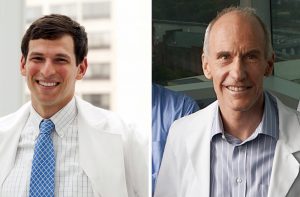
Penn Medicine researchers have developed a unifying definition of ‘cytokine storm’ to provide a framework to assess and treat patients whose immune systems have gone rogue.
Penn Medicine’s David Fajgenbaum (left) and Carl June (right). (Image: Penn Medicine News)
One of the most elusive aspects for clinicians treating COVID-19 is the body’s immune response to the virus. In the most severe cases of COVID-19, the immune system goes into overdrive, resulting in a fever, multiorgan system damage, and often death—a cytokine storm. But how to detect and treat a cytokine storm requires that clinicians can identify it as such.
Two Penn Medicine researchers have developed a unifying definition of “cytokine storm” to provide physicians with a framework to assess and treat severely-ill patients whose immune systems have gone rogue. Cytokine storms can be triggered by different pathogens, disorders, or treatments, from COVID-19 to Castleman disease to CAR T cell therapy.
In a paper published in the New England Journal of Medicine, David Fajgenbaum,an assistant professor of translational medicine & human genetics and director of the Center for Cytokine Storm Treatment & Laboratory (CSTL), and Carl June,a professor of pathology and laboratory medicine and director of the Center for Cellular Immunotherapies in the Abramson Cancer Center, and the Parker Institute for Cancer Immunotherapies define a cytokine storm as requiring elevated circulating cytokine levels, acute systemic inflammatory symptoms, and secondary organ dysfunction beyond what could be attributed to a normal response to a pathogen, if a pathogen is present.
“There has never been a defining central review of what a cytokine storm is and how to treat one, and now with COVID-19, that is a major issue,” says Fajgenbaum, a Castleman disease patient who has previously experienced five cytokine storms himself. “I’ve spent the last 10 years of my life as a cytokine storm patient and researcher, so I know the importance of having a comprehensive unified definition to find therapies that work across the various types of cytokine storms.”
There is widespread recognition that the immune response to a pathogen, but not the pathogen itself, can contribute to multiorgan dysfunction and other symptoms. Additionally, similar cytokine storm syndromes can occur with no obvious infection.
Title: “Engineering Next-Generation CAR-T Cells for Cancer Immunotherapy”
Abstract:
The adoptive transfer of T cells expressing chimeric antigen receptors (CARs) has demonstrated clinical efficacy in the treatment of advanced cancers, with anti-CD19 CAR-T cells achieving up to 90% complete remission among patients with relapsed B-cell malignancies. However, challenges such as antigen escape and immunosuppression limit the long-term efficacy of adoptive T-cell therapy. Here, I will discuss the development of next-generation T cells that can target multiple cancer antigens and resist immunosuppression, thereby increasing the robustness of therapeutic T cells against tumor defense mechanisms. Specifically, I will discuss the development of multi-input receptors and T cells that can interrogate intracellular antigens. I will also discuss the engineering of T cells that can effectively convert TGF-beta from a potent immunosuppressive cytokine into a T-cell stimulant. This presentation will highlight the potential of synthetic biology in generating novel mammalian cell systems with multifunctional outputs for therapeutic applications.
Bio:
Dr. Yvonne Chen is an Associate Professor of Microbiology, Immunology, and Molecular Genetics at the University of California, Los Angeles. She is also a faculty, by courtesy, in the Department of Chemical and Biomolecular Engineering. The Chen Laboratory focuses on applying synthetic biology and biomolecular engineering techniques to the development of novel mammalian-cell systems. The Chen Lab’s work on engineering next-generation T-cell therapies for cancer has been recognized by the NIH Director’s Early Independence Award, the NSF CAREER Award, the Hellman Fellowship, the ACGT Young Investigator Award in Cell and Gene Therapy for Cancer, the Mark Foundation Emerging Leader Award, and the Cancer Research Institute Lloyd J. Old STAR Award. Prior to joining UCLA in 2013, Yvonne was a Junior Fellow in the Harvard Society of Fellows. She received postdoctoral training at the Center for Childhood Cancer Research within the Seattle Children’s Research Institute, and in the Department of Systems Biology at Harvard Medical School. Yvonne received her B.S. in Chemical Engineering from Stanford University and her Ph.D. in Chemical Engineering from the California Institute of Technology.
Single cell sequencing aided researchers in identifying a previously undiscovered molecule in the brain.
Chimeric antigen receptor (CAR) T cell therapy has revolutionized treatment of leukemia, lymphoma, and multiple myeloma. But some people who have received this treatment experience neurotoxicity, or damage to the brain or nervous system.
New research from a team led by Avery Posey, an assistant professor of systems pharmacology and translational therapeutics in the Perelman School of Medicine, provides evidence that this side effect may owe to a molecule in the brain that scientists previously didn’t know was there.
The work, published in the journal Cell, revealed that the protein CD19 is present in brain cells that protect the blood-brain barrier. Prior to the finding, scientists believed CD19 was only expressed on B cells, and the protein served as a target for certain forms of CAR-T therapy. The discovery may chart a path forward for new strategies to effectively treat cancer while sparing the brain.
“The next question is,” says Posey, “can we identify a better target for eliminating B cell related malignancies other than CD19, or can we engineer around this brain cell expression of CD19 and build a CAR T cell that makes decisions based on the type of cell it encounters—for instance, CAR T cells that kill the B cells they encounter, but spare the CD19 positive brain cells?”
Avery Posey, PhD (Image: Penn Medicine Newsby Melissa Moody
Much of the world, including research at Penn Medicine, has focused its attention on how T cells–which play a central role in immune response—might shape the trajectory of COVID-19 infection, and how immunotherapy can shed light on treatment of the disease.
Already a leader in immunotherapy research and treatment, Penn Medicine pioneered the groundbreaking development of CAR T cell cancer therapy. Avery Posey, an assistant professor of systems pharmacology and translational therapeutics, trained as a postdoctoral fellow in the lab of Carl June, who pioneered CAR T cell immunotherapy to treat cancer. Now as a faculty member at Penn, Posey has maintained a focus on T cell therapeutics, mostly for the treatment of cancer.
“This research combines two of my biggest interests—the use of gene therapy to treat disease and the investigation of little known biology, such as the roles of glycans in cell behavior. The pursuit of new knowledge, the roads less traveled—those are my inspirations,” Posey says.
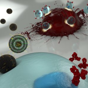
An artist’s illustration of nanoparticles transporting mRNA into a T cell (blue), allowing the latter to express surface receptors that recognize cancer cells (red). (Credit: Ryan Allen, Second Bay Studios)
New cancer immunotherapies involve extracting a patient’s T cells and genetically engineering them so they will recognize and attack tumors. This type of therapy is not without challenges, however. Engineering a patient’s T cells is laborious and expensive. And when successful, the alterations to the immune system immediately make patients very sick for a short period of time, with symptoms including fever, nausea and neurological effects.
Now, Penn researchers have demonstrated a new engineering technique that, because it is less toxic to the T cells, could enable a different mechanism for altering the way they recognize cancer, and could have fewer side effects for patients.
The technique involves ferrying messenger RNA (mRNA) across the T cell’s membrane via a lipid-based nanoparticle, rather than using a modified HIV virus to rewrite the cell’s DNA. Using the former approach would be preferable, as it only confers a temporary change to the patient’s immune system, but the current standard method for getting mRNA past the cell membrane can be too toxic to use on the limited number of T cells that can be extracted from a patient.
Michael Mitchell, Margaret Billingsley, and Carl June
The researchers demonstrated their technique in a study published in the journal Nano Letters. It was led by Michael Mitchell, Skirkanich Assistant Professor of Innovation of bioengineering in the School of Engineering and Applied Science, and Margaret Billingsley, a graduate student in his lab.
They collaborated with one of the pioneers of CAR T therapy: Carl June, the Richard W. Vague Professor in Immunotherapy and director of the Center for Cellular Immunotherapies in the Abramson Cancer Center and the director of the Parker Institute for Cancer Immunotherapy at the Perelman School of Medicine.
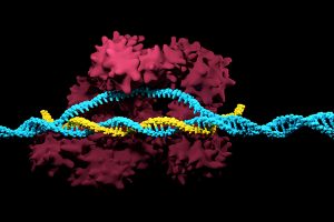
Positive results in first-in-U.S. trial of CRISPR-edited immune cells
3D render of the CRISPR-Cas9 genome editing system
Genetically editing a cancer patient’s immune cells using CRISPR/Cas9 technology, then infusing those cells back into the patient appears safe and feasible based on early data from the first-ever clinical trial to test the approach in humans in the United States. Researchers from the Abramson Cancer Center have infused three participants in the trial thus far—two with multiple myeloma and one with sarcoma—and have observed the edited T cells expand and bind to their tumor target with no serious side effects related to the investigational approach. Penn is conducting the ongoing study in cooperation with the Parker Institute for Cancer Immunotherapy and Tmunity Therapeutics.
“This trial is primarily concerned with three questions: Can we edit T cells in this specific way? Are the resulting T cells functional? And are these cells safe to infuse into a patient? This early data suggests that the answer to all three questions may be yes,” says the study’s principal investigator Edward A. Stadtmauer, section chief of Hematologic Malignancies at Penn. Stadtmauer will present the findings next month at the 61st American Society of Hematology Annual Meeting and Exposition.
Tulane researchers join NIH HEAL initiative for research into opioid crisis
A Tulane University professor and researcher of biomedical engineering will join fellow researchers from over 40 other institutions in the National Institute of Health’s Help to End Addiction Long-Term (HEAL) Initiative. Of the $945 million that make up the project, Michael J. Moore, Ph.D. will receive a share of $1.2 million to advance research in modeling human pain through computer chips, with the help of fellow Tulane researchers Jeffrey Tasker, Ph.D., and James Zadina, Ph.D., each with backgrounds in neuroscience.
Because of the opioid epidemic sweeping the nation, Moore notes that there’s a rapid search going on to develop non-addictive painkiller options. However, he also sees a gap in adequate models to test those new drugs before human clinical trials are allowed to take place. Here is where he hopes to step in and bring some innovation to the field, by integrating living human cells into a computer chip for modeling pain mechanisms. Through his research, Moore wants to better understand not only how some drugs can induce pain, but also how patients can grow tolerant to some drugs over time. If successful, Moore’s work will lead to a more rapid and less expensive screening option for experimental drug advancements.
New machine learning-assisted microscope yields improved diagnostics
Researchers at Duke University recently developed a microscope that uses machine learning to adapt its lighting angles, colors, and patterns for diagnostic tests as needed. Most microscopes have lighting tailored to human vision, with an equal distribution of light that’s optimized for human eyes. But by prioritizing the computer’s vision in this new microscope, researchers enable it to see aspects of samples that humans simply can’t, allowing for a more accurate and efficient diagnostic approach.
Led by Roarke W. Horstmeyer, Ph.D., the computer-assisted microscope will diffuse light through a bowl-shaped source, allowing for a much wider range of illumination angles than traditional microscopes. With the help of convolutional neural networks — a special kind of machine learning algorithm — Horstmeyer and his team were able to tailor the microscope to accurately diagnose malaria in red blood cell samples. Where human physicians typically perform similar diagnostics with a rate of 75 percent accuracy, this new microscope can do the same work with 90 percent accuracy, making the diagnostic process for many diseases much more efficient.
Case Western Reserve University researchers create first-ever holographic map of brain
A Case Western Reserve University team of researchers recently spearheaded a project in creating an interactive holographic mapping system of the human brain. The design, which is believed to be the first of its kind, involves the use of the Microsoft HoloLens mixed reality platform. Lead researcher Cameron McIntyre, Ph.D., sees this mapping system as a better way of creating holographic navigational routes for deep brain stimulation. Recent beta tests with the map by clinicians give McIntyre hope that the holographic representation will help them better understand some of the uncertainties behind targeted brain surgeries.
More than merely providing a useful tool, McIntyre’s project also brings together decades’ worth of neurological data that has not yet been seriously studied together in one system. The three-dimensional atlas, called “HoloDBS” by his lab, provides a way of finally seeing the way all of existing neuro-anatomical data relates to each other, allowing clinicians who use the tool to better understand the brain on both an analytical and visual basis.
Implantable cancer traps reduce biopsy incidence and improve diagnostic
Biopsies are one of the most common procedures used for cancer diagnostics, involving a painful and invasive surgery. Researchers at the University of Michigan are trying to change that. Lonnie Shea, Ph.D., a professor of biomedical engineering at the university, worked with his lab to develop implants with the ability to attract any cancer cells within the body. The implant can be inserted through a scaffold placed under the patient’s skin, making it a more ideal option than biopsy for inaccessible organs like lungs.
The lab’s latest work on the project, published in Cancer Research, details its ability to capture metastatic breast cancer cells in vivo. Instead of needing to take biopsies from areas deeper within the body, the implant allows for a much simpler surgical procedure, as biopsies can be taken from the implant itself. Beyond its initial diagnostic advantages, the implant also has the ability to attract immune cells with tumor cells. By studying both types of cells, the implant can give information about the current state of cancer in a patient’s body and about how it might progress. Finally, by attracting tumor and immune cells, the implant has the ability to draw them away from the area of concern, acting in some ways as a treatment for cancer itself.
People and Places
Cesar de la Fuente-Nunez, PhD
The Philadelphia Inquirer recently published an article detailing the research of Penn’s Presidential Assistant Professor in Psychiatry, Microbiology, and Bioengineering, Cesar de la Fuente, Ph.D. In response to a growing level of worldwide deaths due to antibiotic-resistant bacteria, de la Fuente and his lab use synthetic biology, computation, and artificial intelligence to test hundreds of millions of variations in bacteria-killing proteins in the same experiment. Through his research, de la Fuente opens the door to new ways of finding and testing future antibiotics that might be the only viable options in a world with an increasing level of drug-resistant bacteria
Emily Eastburn, a Ph.D. candidate in Bioengineering at Penn and a member of the Boerckel lab of the McKay Orthopaedic Research Laboratory, recently won the Ashton fellowship. The Ashton fellowship is an award for postdoctoral students in any field of engineering that are under the age of 25, third-generation American citizens, and residents of either Pennsylvania or New Jersey. A new member of the Boerckel lab, having joined earlier this fall, Eastburn will have the opportunity to conduct research throughout her Ph.D. program in the developmental mechanobiology and regeneration that the Boerckel lab focuses on.