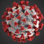
Hammer will offer a course on COVID-19 and the coronavirus pandemic during Penn’s Summer II session, which will be held online this year. The course will be co-taught with Miriam Wattenbarger, senior lecturer in CBE.
The course, “Biotechnology, Immunology, and COVID-19,” will culminate with a case study of the coronavirus pandemic including the types of drugs proposed and their mechanism of action, as well as the process of vaccine development.
“Obviously, the pandemic has been a life-altering event, causing an immense dislocation for everyone in our community, especially the students. Between me and Miriam, who has been trumpeting the importance of vaccines for some time in her graduate-level CBE courses, we have the expertise to inform students about this disease and how we might combat it,” says Hammer.
For more than ten years, Wattenbarger has run courses and labs focused on drug delivery and biotechnology, key elements of the vaccine development process.
“I invite both researchers and industry speakers to meet with my students,” Wattenbarger says, “so that they learn the crucial role engineers play in both vaccine development and manufacturing.”
Beyond studying the interactions between the immune system and viruses — including HIV, influenza, adenovirus and coronavirus — students will cover a variety of biotechnological techniques relevant to tracking and defending against them, including recombinant DNA technology, polymerase chain reaction, DNA sequencing, gene therapy, CRISPR-Cas9 editing, drug discovery, small molecule inhibitors, vaccines and the clinical trial process.
Students will also learn the mathematical principles used to quantify biomolecular interactions, as well as those found behind simple epidemiological models and methods for making and purifying drugs and vaccines.
“We all have to contribute in the ways that we can. Having taught biotechnology to freshmen for the past decade, this is something that I can do that can both inform and build community,” says Hammer. “Never has it been more important to have an informed and scientifically literate community that can fight this or any future pandemic.”
A message from Penn Bioengineering Professor and Chair Ravi Radhakrishnan:
In response to the unprecedented challenges presented by the global outbreak of the novel coronavirus SARS-CoV-2, Penn Bioengineering’s faculty, students, and staff are finding innovative ways of pivoting their research and academic projects to contribute to the fight against COVID-19. Though these projects are all works in progress, I think it is vitally important to keep those in our broader communities informed of the critical contributions our people are making. Whether adapting current research to focus on COVID-19, investing time, technology, and equipment to help health care infrastructure, or creating new outreach and educational programs for students, I am incredibly proud of the way Penn Bioengineering is making a difference. I invite you to read more about our ongoing projects below.
RESEARCH
Novel Chest X-Ray Contrast
David Cormode, Associate Professor of Radiology and Bioengineering
The Cormode and Noel labs are working to develop dark-field X-ray imaging, which may prove very helpful for COVID patients. It involves fabricating diffusers that incorporate gold nanoparticles to modify the X-ray beam. This method gives excellent images of lung structure. Chest X-ray is being used on the front lines for COVID patients, and this could potentially be an easy to implement modification of existing X-ray systems. The additional data give insight into the health state of the microstructures (alveoli) in the lung. This new contrast mechanics could be an early insight into the disease status of COVID-19 patients. For more on this research, see Cormode and Noel’s chapter in the forthcoming volume Spectral, Photon Counting Computed Tomography: Technology and Applications, edited by Katsuyuki Taguchi, Ira Blevis, and Krzysztof Iniewski (Routledge 2020).
Immunotherapy
Michael J. Mitchell, Skirkanich Assistant Professor of Innovation in Bioengineering
Mike Mitchell is working with Saar Gill (Penn Medicine) on engineering drug delivery technologies for COVID-19 mRNA vaccination. He is also developing inhalable drug delivery technologies to block COVID-19 internalization into the lungs. These new technologies are adaptations of prior research published Volume 20 of Nano Letters (“Ionizable Lipid Nanoparticle-Mediated mRNA Delivery for Human CAR T Cell Engineering” January 2020) and discussed in Volume 18 of Nature Reviews Drug Discovery (“Delivery Technologies for Cancer Immunotherapy” January 2019).
Respiratory Distress Therapy Modeling
Ravi Radhakrishnan, Professor, and Chair of Bioengineering and Professor of Chemical and Biomolecular Engineering
Computational Models for Targeting Acute Respiratory Distress Syndrome (ARDS). The severe forms of COVID-19 infections resulting in death proceeds by the propagation of the acute respiratory distress syndrome or ARDS. In ARDS, the lungs fill up with fluid preventing oxygenation and effective delivery of therapeutics through the inhalation route. To overcome this major limitation, delivery of antiinflammatory drugs through the vasculature (IV injection) is a better approach; however, the high injected dose required can lead to toxicity. A group of undergraduate and postdoctoral researchers in the Radhakrishnan Lab (Emma Glass, Christina Eng, Samaneh Farokhirad, and Sreeja Kandy) are developing a computational model that can design drug-filled nanoparticles and target them to the inflamed lung regions. The model combines different length-scales, (namely, pharmacodynamic factors at the organ scale, hydrodynamic and transport factors in the tissue scale, and nanoparticle-cell interaction at the subcellular scale), into one integrated framework. This targeted approach can significantly decrease the required dose for combating ARDS. This project is done in collaboration with Clinical Scientist Dr. Jacob Brenner, who is an attending ER Physician in Penn Medicine. This research is adapted from prior findings published in Volume 13, Issue 4 of Nanomedicine: Nanotechnology, Biology and Medicine: “Mechanisms that determine nanocarrier targeting to healthy versus inflamed lung regions” (May 2017).
Diagnostics
Sydney Shaffer, Assistant Professor of Bioengineering and Pathology and Laboratory Medicine
Arjun Raj, David Issadore, and Sydney Shaffer are working on developing an integrated, rapid point-of-care diagnostic for SARS-CoV-2 using single molecule RNA FISH. The platform currently in development uses sequence specific fluorescent probes that bind to the viral RNA when it is present. The fluorescent probes are detected using a iPhone compatible point-of-care reader device that determines whether the specimen is infected or uninfected. As the entire assay takes less than 10 minutes and can be performed with minimal equipment, we envision that this platform could ultimately be used for screening for active COVID19 at doctors’ offices and testing sites. Support for this project will come from a recently-announced IRM Collaborative Research Grant from the Institute of Regenerative Medicine with matching funding provided by the Departments of Bioengineering and Pathology and Laboratory Medicine in the Perelman School of Medicine (PSOM) (PI’s: Sydney Shaffer, Sara Cherry, Ophir Shalem, Arjun Raj). This research is adapted from findings published in the journal Lab on a Chip: “Multiplexed detection of viral infections using rapid in situ RNA analysis on a chip” (Issue 15, 2015). See also United States Provisional Patent Application Serial No. 14/900,494 (2014): “Methods for rapid ribonucleic acid fluorescence in situ hybridization” (Inventors: Raj A., Shaffer S.M., Issadore D.).
HEALTH CARE INFRASTRUCTURE
Penn Health-Tech Coronavirus COVID-19 Collaborations
Brian Litt, Professor of Bioengineering, Neurology, and Neurosurgery
In his role as one of the faculty directors for Penn Health-Tech, Professor Brian Litt is working closely with me to facilitate all the rapid response team initiatives, and in helping to garner support the center and remove obstacles. These projects include ramping up ventilator capacity and fabrication of ventilator parts, the creation of point-of-care ultrasounds and diagnostic testing, evaluating processes of PPE decontamination, and more. Visit the Penn Health-Tech coronavirus website to learn more, get involved with an existing team, or submit a new idea.
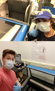
BE Educational Labs staff members Dana Abulez (BE ’19, Master’s BE ’20) and Matthew Zwimpfer (MSE ’18, Master’s MSE ’19) take shifts to laser-cut face shields.
The George H. Stephenson Foundation Educational Laboratory & Bio-MakerSpace staff have donated their PPE to Penn Medicine. Two staff members (Dana Abulez, BE ’19, Master’s BE ’20 and Matthew Zwimpfer, MSE ’18, Master’s MSE ’19) took shifts to laser-cut face shields in collaboration with Penn Health-Tech. Dana and Matthew are also working with Dr. Matthew Maltese on his low-cost ventilator project (details below).
Low-Cost Ventilator
Matthew Maltese, Adjunct Professor of Medical Devices and BE Graduate Group Member
Dr. Maltese is rapidly developing a low-cost ventilator that could be deployed in Penn Medicine for the expected surge, and any surge in subsequent waves. This design is currently under consideration by the FDA for Emergency Use Authorization (EUA). This example is one of several designs considered by Penn Medicine in dealing with the patient surge.
Face Shields
David F. Meaney, Solomon R. Pollack Professor of Bioengineering and Senior Associate Dean
Led by David Meaney, Kevin Turner, Peter Bruno and Mark Yim, the face shield team at Penn Health-Tech is working on developing thousands of rapidly producible shields to protect and prolong the usage of Personal Protective Equipment (PPE). Learn more about Penn Health-Tech’s initiatives and apply to get involved here.
Update 4/29/20: The Penn Engineering community has sprung into action over the course of the past few weeks in response to COVID-19. Dr. Meaney shared his perspective on those efforts and the ones that will come online as the pandemic continues to unfold. Read the full post on the Penn Engineering blog.
OUTREACH & EDUCATION
Student Community Building
Yale Cohen, Professor of Otorhinolaryngology, Department of Psychology, BE Graduate Group Member, and BE Graduate Chair
Yale Cohen, and Penn Bioengineering’s Graduate Chair, is working with Penn faculty and peer institutions across the country to identify intellectually engaging and/or community-building activities for Bioengineering students. While those ideas are in progress, he has also worked with BE Department Chair Ravi Radhakrishnan and Undergraduate Chair Andrew Tsourkas to set up a dedicated Penn Bioengineering slack channel open to all Penn Bioengineering Undergrads, Master’s and Doctoral Students, and Postdocs as well as faculty and staff. It has already become an enjoyable place for the Penn BE community to connect and share ideas, articles, and funny memes.
Undergraduate Course: Biotechnology, Immunology, Vaccines and COVID-19 (ENGR 35)
Daniel A. Hammer, Alfred G. and Meta A. Ennis Professor of Bioengineering and Chemical and Biomolecular Engineering
This Summer Session II, Professor Dan Hammer and CBE Senior Lecturer Miriam R. Wattenbarger will teach a brand-new course introducing Penn undergraduates to a basic understanding of biological systems, immunology, viruses, and vaccines. This course will start with the fundamentals of biotechnology, and no prior knowledge of biotechnology is necessary. Some chemistry is needed to understand how biological systems work. The course will cover basic concepts in biotechnology, including DNA, RNA, the Central Dogma, proteins, recombinant DNA technology, polymerase chain reaction, DNA sequencing, the functioning of the immune system, acquired vs. innate immunity, viruses (including HIV, influenza, adenovirus, and coronavirus), gene therapy, CRISPR-Cas9 editing, drug discovery, types of pharmaceuticals (including small molecule inhibitors and monoclonal antibodies), vaccines, clinical trials. Some quantitative principles will be used to quantifying the strength of binding, calculate the dynamics of enzymes, writing and solving simple epidemiological models, methods for making and purifying drugs and vaccines. The course will end with specific case study of coronavirus pandemic, types of drugs proposed and their mechanism of action, and vaccine development.
Update 4/29/20: Read the Penn Engineering blog post on this course published April 27, 2020.
Neuromatch Conference
Konrad Kording, Penn Integrates Knowledge University Professor of Bioengineering, Neuroscience, and Computer and Information Science
Dr. Kording facilitated Neuromatch 2020, a large virtual neurosciences conferences consisting of over 3,000 registrants. All of the conference talk videos are archived on the conference website and Dr. Kording has blogged about what he learned in the course of running a large conference entirely online. Based on the success of Neuromatch 1.0, the team are now working on planning Neuromatch 2.0, which will take place in May 2020. Dr. Kording is also working on facilitating the transition of neuroscience communication into the online space, including a weekly social (#neurodrinking) with both US and EU versions.
Neuromatch Academy
Konrad Kording, Penn Integrates Knowledge University Professor of Bioengineering, Neuroscience, and Computer and Information Science
Dr. Kording is working to launch the Neuromatch Academy, an open, online, 3-week intensive tutorial-based computational neuroscience training event (July 13-31, 2020). Participants from undergraduate to professors as well as industry are welcome. The Neuromatch Academy will introduce traditional and emerging computational neuroscience tools, their complementarity, and what they can tell us about the brain. A main focus is not just on using the techniques, but on understanding how they relate to biological questions. The school will be Python-based making use of Google Colab. The Academy will also include professional development / meta-science, model interpretation, and networking sessions. The goal is to give participants the computational background needed to do research in neuroscience. Interested participants can learn more and apply here.
Journal of Biomedical Engineering Call for Review Articles
Beth Winkelstein, Vice Provost for Education and Eduardo D. Glandt President’s Distinguished Professor of Bioengineering
The American Society of Medical Engineers’ (ASME) Journal of Biomechanical Engineering (JBME), of which Dr. Winkelstein is an Editor, has put out a call for review articles by trainees for a special issue of the journal. The call was made in March 2020 when many labs were ramping down, and trainees began refocusing on review articles and remote work. This call continues the JBME’s long history of supporting junior faculty and trainees and promoting their intellectual contributions during challenging times.
Update 4/29/20: CFP for the special 2021 issue here.
Are you a Penn Bioengineering community member involved in a coronavirus-related project? Let us know! Please reach out to ksas@seas.upenn.edu.
Cesar de la Fuente, Ph.D., a Presidential Assistant Professor in Psychiatry, Microbiology, and Bioengineering at Penn, recently published a literature review in Trends in Immunology entitled, “Emerging Frontiers in Microbiome Engineering.” The microbiome, in simple terms, consists of the genetic material of microorganisms in the gut, including bacteria, fungi, protozoa, viruses, and oral, vaginal, and skin microbiomes. Each human has a unique microbiome that depends both on predetermined factors like exposure to microorganisms within a mother’s birth canal or breastmilk in early life as well as environmental factors and diet in later life. The health of someone’s microbiome is extremely important, as an unhealthy microbiome with an imbalance of symbiotic and pathogenic microbes can make a person more susceptible to various diseases. The most common diseases or disorders associated with a problematic microbiome are rather far-reaching, including some of the most afflicting diseases of today like inflammatory bowel disease, diabetes, obesity, cardiovascular diseases, and neurological disorders.
In his recent literature review, de la Fuente provides an overview of microbiome engineering, and what the future might hold for the field. He defines microbiome engineering initially as a way of studying the “contribution of individual microbes and generating potential therapies against metabolic, inflammatory, and immunological diseases.” Currently, most treatments for issues with the microbiome are broad solutions like dietary adjustments to include more probiotics, antibiotics, or prebiotics, while more serious cases may require a fecal microbiota transplant. While these therapies may work for some patients, de la Fuente emphasizes the need for greater specificity in treatment targets and a need for precision in reprogramming existing microbial communities as an alternative to transplants.
De la Fuente highlights the current methods and tools in microbiome engineering such as the use of bacteriocins and bacteriophages to knock out specific bacteria within the microbiome. However, there are very few bacteriocins or bacteriophages commercially available on today’s market. Another common approach to microbiome engineering is in synthetic biology, or the use of “chassis” — a type of cell that maintains DNA constructs for different functions — to engineering interactions within the microbiome. De la Fuente continues his discussion of current methods by naming and describing several specific examples of these approaches, particularly in relation to synthetic biology options before moving on to examine future directions for these methods.
Before bringing up potential new frontiers for microbiome engineering, de la Fuente also outlines the way that microbiome engineering works in the first place, and dedicates sections of the review to the microbiome’s influence on its host’s immune system and how to engineer the microbiome to modulate that immune system. The main future methods for microbiome engineering that de la Fuente points out in his review include more precise regulation of gene expression through commensal organisms and the use of CRISPRi to find genes involved in bacterial maintenance. The conclusion of de la Fuente’s review brings up the notion of new personalized medicine or therapy for the microbiome that could come with further advances in the field. However, he also makes sure to bring up some still-outstanding questions about the human microbiome that require further research, most notably, what exactly makes a healthy human microbiome? Here’s hoping the research de la Fuente mentions can illuminate a path to the answer.
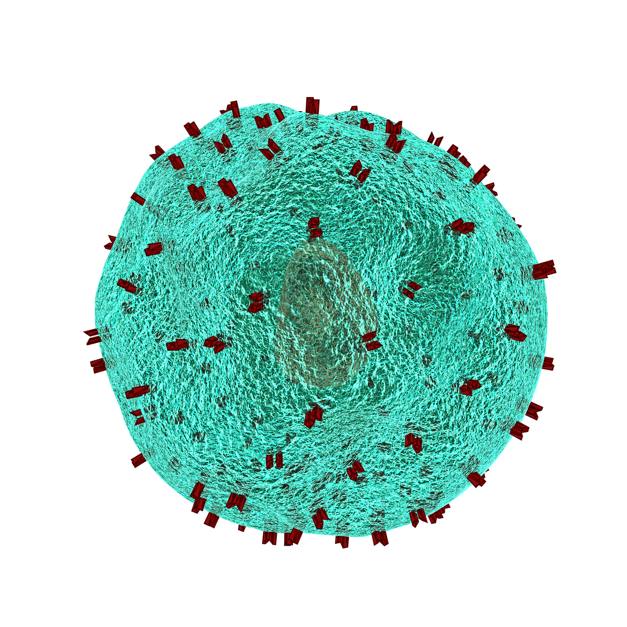
T lymphocytes in the immune system play a vital role in the body to recognize invasion by an outside element. When foreign bacteria enter the body, receptors on the T cell surface detect antigens associated with the bacteria and send a signal deploying phagocytes to attack and defeat the invading bacteria. While evolution and vaccination make the immune system very efficient, the inability of T cell receptors (TCRs) to detect cancer makes normal T cells relatively ineffective in resisting cancer. One of the ways to overcome this limitation of the immune system is to better understand how the TCRs respond to antigens. Analyses of the proteins involved in TSR responses are useful but limited by several factors, including the dizzying amount of data involved. Data analysis techniques have been helpful but have offered little information about the general reactions of TSRs, rather than how they react to specific antigens.
A possible solution to this obstacle is ImmunoMap, developed by scientists collaborating between Johns Hopkins University and Memorial Sloan Kettering Cancer Center. In a study recently published in Cancer Immunology Research, the authors, led by Jonathan P. Schneck, M.D., Ph.D., a professor of pathology at Johns Hopkins associated with the university’s Institute for Cell Engineering and Institute for Nanobiotechnology, describe their creation and deployment of ImmunoMap, a group of artificial intelligence algorithms that use machine learning to process large amounts of sequencing data and compare data from different antigens with each other.
The authors trained ImmunoMap initially using data from a mouse model of melanoma, in which the algorithm demonstrated significantly better performance than traditional methods. Subsequently, ImmunoMap was applied to patient response data from a melanoma clinical trial of the chemotherapy agent nivolumab. The algorithm discovered a new group of patients that would respond positively to nivolumab treatment — a finding missed by popular past methods. More testing of ImmunoMap is necessary, but the technology could make significant contributions to the monitoring of cancer patients receiving chemotherapy. In addition, it could to help to better predict response in patients before they begin specific chemotherapy regimes.
Wearables Improving Health
Among the most troubling health disparities related to global wealth inequality is the higher rate of mortality among children suffering from cancer. Fever is a common symptom of children undergoing cancer treatment, and this symptom may indicate more serious health issues that require the attention of a doctor. However, continuously monitoring skin temperature in children from low resource settings is difficult. Seeking to help remedy this problem, undergraduate engineering students at Harvard collaborated with the Dana-Farber/Boston Children’s Cancer & Blood Disorders Center’s Global Health Initiative to develop tools for earlier fever detection and treatment.
In a course taught by David Mooney, Ph.D., Robert P. Pinkas Family Professor of Bioengineering at Harvard, students developed a wearable device that sounds an alarm when the wearer needs medical help. The app can send patients’ recorded messages to their doctors, who can then review the temperature data and messages from the children before responding. Fashioned like a wristwatch, the extra-durable and waterproof device will next move into pilot testing among a larger patient population.
Meanwhile, at Northwestern, John A. Rogers, Ph.D., the Louis Simpson and Kimberly Querrey Professor of Materials Science and Engineering, Biomedical Engineering, and Neurological Surgery in Northwestern’s McCormick School of Engineering, has partnered with cosmetics giant L’Oréal to create the world’s smallest wearable. The device, which is smaller than an adult fingernail, measures UV sun exposure for the wearer and can tell when they should go back inside instead of risking overexposure. Unsurprisingly, it’s solar powered, and it was demonstrated a couple of weeks ago at a consumer electronics show in Las Vegas.
Growing Hydrogels Like Human Tissue
Scientists at Carnegie Mellon University and Nanyang Technological University in Singapore have collaborated in a process to create polyacrylamide gels that grow in a manner resembling natural tissue. K. Jimmy Hsia, Ph.D., Professor of Biomedical Engineering at Carnegie Mellon, is co-lead author of a new study in PNAS describing this new growth mode.
In the study, Dr. Hsia and his coauthors report that, in the same way that growth factors secreted by a living organism affect the generation of new tissue, oxygen can be modulated to control how hydrogels grow. Moreover, while growth is under way, the process could be continued to efficiently manage the mass transfer of nutrients from cell to cell. Finally, the authors detail the mechanical processes that help to shape the final product. With this new process, the ability to design and create materials for applications such as robotics and tissue engineering comes a step closer to resembling living tissue as closely as possible.
People and Places
Engineers at Virginia Tech have been awarded a $1.1 million grant from the Virginia Research Investment Committee to develop a device that uses low-energy electric fields for the treatment of brain tumors. Rafael Davalos, Ph.D., L. Preston Wade Professor of Biomedical Engineering and Mechanics, is the chief investigator on the grant.
The Department of Biomedical Engineering has announced the appointment of Kam W. Leong, Ph.D., as the Samuel Y. Sheng Professor of Biomedical Engineering. Dr. Leong earned his Ph.D. in chemical engineering from the University of Pennsylvania and taught at Duke and Johns Hopkins before arriving at Columbia in 2006. He was previously the James B. Duke Professor of Biomedical Engineering at Duke. Congratulations to Dr. Leong!
The human immune system deploys a variety of cells to counteract pathogens when they enter the body. B cells are a type of white blood cell specific to particular pathogens, and they form part of the adaptive immune system. As these cells develop, the cells with the strongest reactions to antigens are favored over others. This process is called clonal selection. Given the sheer number of pathogens out there, the number of different clonal lineages for B cells is estimated to be around 100 billion. A landscape like that can be difficult to navigate without a map.
Luckily, an atlas was recently published in Nature Biotechnology. It is the work of scientists collaborating between Penn’s own Perelman School of Medicine and faculty from the School of Biomedical Engineering, Science and Health Systems at our next-door neighbor, Drexel University. Using tissue samples from an organ donor network, the authors, led by Nina Luning Prak, MD, PhD, of Penn and Uri Heshberg, Ph.D., of Drexel, submitted the samples to a process called deep immune repertoire profiling to identify unique clones and clonal lineages. In total, they identified nearly a million lineages and mapped them to two networks: one in the gastointestinal tract and one that connects the blood, bone marrow, spleen, and lungs. This discovery suggests that the networks might be less complicated than initially thought. Also, it confirms a key role for the immune system in the gut.
Not only does this B cell atlas provide valuable information to the scientific community, but it also could serve as the basis for immune-based therapies for diseases. If we can identify these lineages and how clonal selection occurs, we could identify the most effective immunological cells and perhaps engineer them in the lab. At the very least, the extent to which scientists understand how B cells are formed and develop has received an enormous push with this research.
Understanding Muscle Movement
Natural movements of limbs require the coordinated activation of several muscle groups. Although the molecular composition of muscle is known, there remains a poor understanding of how these molecules coordinate their actions to confer power, strength, and endurance to muscle tissue. New fields of synthetic biology require this new knowledge to efficiently produce naturally inspired muscle substitutes.
Responding to this challenge, scientists at Carnegie Mellon University, including Philip R. LeDuc, Ph.D., William J. Brown Professor of Mechanical Engineering and Professor of Biomedical Engineering, have developed a computational system to better understand how mixtures of specific myosins affect muscle properties. Their method, published in PNAS, uses a computer model to show that mixtures of myosins will unexpectedly produce properties that are not the average of myosin molecular properties. Instead, the myosin mixtures coordinate and complement each other at the molecular level to create emergent behaviors, which lead to a robustness in how the muscle functions across a broad range. Dr. LeDuc and his colleagues then confirmed their model in lab experiments using muscle tissue from chickens. In the future, this new computational method could be used for other types of tissue, and it could prove useful in developing treatments for a variety of disorders.
Determining Brain Connectivity
How the brain forms and keeps memories is one of the greatest challenges in neuroscience. The hippocampus is a brain region considered critical for remembering sequences and events. The connections made by the hippocampus to other brain regions is considered critical for the hippocampus to integrate and remember experiences. However, this broad connectivity of the hippocampus to other brain areas raises a critical question: What connections are essential for rewiring the brain for new memories?
To offer an explanation for this question, a team of scientists in Hong Kong published a paper in PNAS in which they report on a study conducted in rats using resting-state function MRI. The study team, led by Ed X. Wu, Ph.D., of the University of Hong Kong, found that stimulation of a region deep in the hippocampus would propagate more broadly out into many areas of the cortex. The stimulation frequency affected how far this signal propagated from the hippocampus and pointed out the ability for frequency-based information signals to selectively connect the hippocampus to the rest of the brain. Altering the frequency of stimulation could affect visual function, indicating that targeted stimulation of the brain could have widespread functional effects throughout the brain.
Although human and rodent brains are obviously different, these findings from rats offer insights into how brain connectivity emerges in general. Similar studies in humans will be needed to corroborate these findings.
Seeing Inside a Tumor
Years of research have yielded the knowledge that the most effective treatments for cancer are often individualized. Knowing the genetic mutation involved in oncogenesis, for instance, can provide important information about the right drug to treat the tumor. Another important factor to know is the tumor’s chemical makeup, but far less is known about this factor due to the limitations of imaging.
However, a new study published in Nature Communications is offering some hope in this regard. In the study, scientists led by Xueding Wang, Ph.D., associate professor of biomedical engineering and radiology at the University of Michigan, used pH-sensing nanoprobes and multiwavelength photoacoustic imaging to determine tumor types in phantoms and animals. This new technology is based on the principle that cancerous cells frequently lower the pH levels in tissue, and designing probes with properties that are pH sensitive provides a method to find tumors with imaging methods and also treat these tumors.
With this technology, Dr. Wang and his colleagues were able to obtain three-dimensional images of pH levels inside of tumors. Importantly, it allowed them to noninvasively view the changes in a dye injected inside the tumor. Although a clinical application is years away, the information obtained using the Michigan team’s techniques could add significantly to our knowledge about tumorigenesis and tumor growth.
The Role of Bacteria in MS
The growing awareness of how bacteria interact with humans to affect health has led to the emergence of new scientific areas (e.g., human microbiome). Research findings from scientists collaborating between Caltech and UCSF suggest bacteria can play a role in the onset of multiple sclerosis. These investigators include Sarkis K. Mazmanian, Ph.D., Luis B. and Nelly Soux Professor of Microbiology and a faculty member in the Division of Biology and Biological Engineering at Caltech. Reporting their research results in PNAS, the researchers found several bacteria elevated in the MS microbiome. Study results showed that these bacteria regulated adaptive immune responses and helped to create a proinflammatory milieu. The identification of the bacteria interacting with immunity in MS patients could result in better diagnosis and treatment of this disabling disease.
People and Places
Faculty members at the University of California, Irvine, including biomedical engineer Zoran Nenadic, Ph.D., have received an $8 million grant to develop a brain-computer interface. The research using this grant aims to restore function in people with spinal cord injuries. Also, at the University of Texas, Austin, the lab of Amy Brock, Ph.D., an assistant professor in the Department of Biomedical Engineering, has received a three-year $180,000 R21 grant from the National Cancer Institute to develop a barcoding platform to isolate cancer cell lineages and to identify genetic targets for treatment.
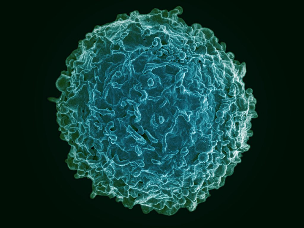
A healthy human T cell, one of the key immune system cells.
Organ transplantation is a lifesaving measure for people with diseases of the heart, lungs, liver, and kidneys that can no longer be treated medically or surgically. The United Network for Organ Sharing, a major advocacy group for transplant recipients, reports that a new person is added to a transplant list somewhere in America every 10 minutes. However, rejection of the donor organ by the recipient’s immune system remains a major hurdle for making every transplant procedure successful. Unfortunately, the drugs required to prevent rejection have serious side effects.
To address this problem, a research team at Cornell combined DNA sequencing and informatics algorithms to identify rejection earlier in the process, making earlier intervention more likely. The team, led by Iwijn De Vlaminck of the Department of Biomedical Engineering, report in PLOS Computational Biology that a computer algorithm they developed to detect donor-derived cell-free DNA, a type of DNA shed by dead cells, in the blood of the recipient could predict heart and lung allograft rejection with a 99% correlation with the current gold standard. The earlier that signs of rejection are detected, the more likely it is that an intervention can be performed to save the organ and, more importantly, the patient.
Meanwhile, at Yale, scientists have used nanoparticles to fight transplant rejection. Publishing their findings in Nature Communications, the study authors, led by Jordan S. Pober, Bayer Professor of Translational Medicine at Yale, and Mark Saltzman, Goizueta Foundation Professor of Chemical and Biomedical Engineering, used small-interfering RNA (siRNA) to “hide” donated tissue from the immune system of the recipient. Although the ability of siRNA to hide tissue in this manner has been known for some time, the effect did not last long in the body. The Yale team used poly(amine-co-ester) nanoparticles to deliver the siRNA that extended and extended its duration of effect, in addition to developing methods to deliver to siRNA to the tissue before transplantation. The technology has yet to be tested in humans, but provides an exciting new approach to help solve the transplant rejection challenge in medicine.
Africa in Focus
A group of engineering students at Wright State University, led by Thomas N. Hangartner, professor emeritus of biomedical engineering, medicine and physics, traveled to Malawi, a small nation in southern Africa, to build a digital X-ray system at Ludzi Community Hospital. Once on site, Hangartner and his student team trained the staff to use system on patients. The group hopes they have made a significant contribution to improving the standard of care in the country, which currently allocates only 9% of its annual budget to healthcare. While the project admitted has limited impact, it’s important to bear in mind that expanding public health on a global level is a game of inches. The developing world will rise to the standards of the developed world one village at a time, one hospital at a time.
Speaking of Africa, the recent Ebola outbreak in West Africa had global implications and prompted many international organizations to identify better methods to identify early signs of outbreak. Since diseases like Ebola can spread rapidly and aggressively, detecting the outbreak early can save thousands of lives. To this end, Tony Hu of Arizona State University’s School of Biological and Health Systems Engineering has partnered with the U.S. Army to develop a platform using porous silicone nanodisks that, coupled with a mass spectrometer, could be used to detect Ebola more quickly and less expensively. In particular, by determining the strain of the Ebola virus detected, treatment could be more specifically individualized for the patient. Dr. Hu presents the technology in a video available here.
Neurotech News
Karen Moxon, professor of biomedical and mechanical engineering at the University of California, Davis, recently showed that rats with spinal injuries recovered to a more significant extent when treated with a combination of serotonergic drugs and physical therapy. Dr. Moxon found that the treatment resulted in cortical reorganization to bypass the injury. Many consider combining two different drugs to treat a disease or injury; Moxon’s clever approach used a drug in combination with the activation of cortical circuits (electroceuticals), and approach that was not considered possible with some types of spinal cord injuries.
At Stanford, Karl Deisseroth, professor of bioengineering and of psychiatry and behavioral sciences, led a study team that recently reported in Science Translational Medicine that mice bred to have a type of autism could receive a genetic therapy that caused their brain cells to activate differently. Although the brains of the autistic mice were technically normal, the mice were unsocial and lacked curiosity. Treatment modulated expression of the CNTNAP2 gene, resulting in increased sociability and curiosity. Their findings could have tremendous implications for treating autism in humans.
Elsewhere in neurotech, Cornell announced its intention to create a neurotech research hub, using a $9 million grant from the National Science Foundation. Specializing in types of neurological imaging, the new NeuroNex Hub and Laboratory for Innovative Neurotechnology will augment the neurotech program founded at Cornell in 2015.
Academic Developments
Two important B(M)E department have developed new programs. In Montreal, McGill University has introduced a graduate certificate program in translational biomedical engineering (video here). Also at the annual meeting of the American Society for Engineering Education in Columbus, Ohio, an interdisciplinary group of scholars from Worcester Polytechnic Institute, including three professors of engineering, presented a paper entitled “The Theatre of Humanitarian Engineering.” The authors developed an experimental role-playing course in which the students developed a waste management solution for a city. According to the paper’s abstract, a core misunderstanding about engineering is the belief that it exists separately from social and political contexts. With the approach they detail, the authors believe they could address the largely unmet call for greater integration of engineering with the humanities and social sciences on the academic level.


 In response to the unprecedented challenges presented by the global outbreak of the novel coronavirus SARS-CoV-2,