Penn and CHOP Researchers Show Gene Editing Tools Can be Delivered to Perinatal Brain

Genetic diseases that involve the central nervous system (CNS) often impact children before birth, meaning that once a child is born, irreversible damage has already been done. Given that many of these conditions result from a mutation in a single gene, there has been growing interest in using gene editing tools to correct these mutations before birth.

However, identifying the appropriate vehicle to deliver these gene editing tools to the CNS and brain has been a challenge. Viral vectors used to deliver gene therapies have some potential drawbacks, including pre-existing viral immunity and vector-related adverse events, and other options like lipid nanoparticles (LNPs) have not been investigated extensively in the perinatal brain.

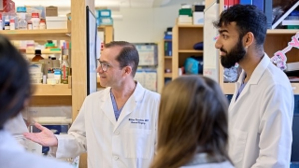
Now, researchers in the Center for Fetal Research at Children’s Hospital of Philadelphia (CHOP) and Penn Engineering have identified an ionizable LNP that can deliver mRNA base editing tools to the brain and have shown it can mitigate CNS disease in perinatal mouse models. The findings, published in ACS Nano, open the door to mRNA therapies that could be delivered pre- or postnatally to treat genetic CNS diseases.

The research team began by screening a library of ionizable LNPs – microscopic fat bubbles that have a positive charge at low pH but neutral charge at physiological conditions in the body. After identifying which LNPs were best able to penetrate the blood-brain barrier in fetal and newborn mice, they optimized their top-performing LNP to be able to deliver base editing tools. The LNPs were then used to deliver mRNA for an adenine base editor, which would correct a disease-causing mutation in the lysosomal storage disease, MPSI, by changing the errant adenine to guanine.

The researchers showed that their LNP was able to improve the symptoms of the lysosomal storage disease in the neonatal mouse brain, as well as deliver mRNA base editing tools to the brain of other animal models. They also showed the LNP was stable in human cerebrospinal fluid and could deliver mRNA base editing tools to patient-derived brain tissue.

“This proof-of-concept study – co-led by Rohan Palanki, an MD/PhD student in my lab, and Michael Mitchell’s lab at Penn Bioengineering – supports the safety and efficacy of LNPs for the delivery of mRNA-based therapies to the central nervous system,” said co-senior author William H. Peranteau, MD, an attending surgeon in the Division of General, Thoracic and Fetal Surgery at CHOP and the Adzick-McCausland Distinguished Chair in Fetal and Pediatric Surgery. “Taken together, these experiments provide the foundation for additional translational studies and demonstrate base editing facilitated by a nonviral delivery carrier in the NHP fetal brain and primary human brain tissue.”

This story was written by Dana Bate. It originally appeared on CHOP’s website.

Alexander Buffone Appointed Assistant Professor at New Jersey Institute of Technology

Alexander Buffone, Ph.D.

Penn Bioengineering is proud to congratulate Alexander Buffone, Ph.D. on his appointment as Assistant Professor in the Department of Biomedical Engineering at New Jersey Institute of Technology. His appointment began in the Spring of 2022.

Buffone got his Ph.D. in Chemical Engineering from SUNY Buffalo in Buffalo, NY in 2012, working with advisor Sriram Neelamegham, Professor of Chemical and Biological Engineering. Buffone completed previous postdoctoral studies at Roswell Park Comprehensive Cancer Center with Joseph T.Y. Lau, Distinguished Professor of Oncology in the department of Cellular and Molecular Biology. Upon coming to Penn in 2015, Buffone has worked in the Hammer Lab under advisor Daniel A. Hammer, Alfred G. and Meta A. Ennis Professor in Bioengineering and in Chemical and Biomolecular Engineering, first as a postdoc and later a research associate. Buffone also spent a year as a Visiting Scholar in the Center for Bioengineering and Tissue Regeneration, directed by Valerie M. Weaver, Professor at the University of California, San Francisco in 2019.

While at Penn, Buffone was a co-investigator on an R21 grant through the National Institutes of Health (NIH) which supported his time as a research associate. Buffone is excited to start his own laboratory where he plans to train a diverse set of trainees.

Buffone’s research area lies at the intersection of genetic engineering, immunology, and glycobiology and addresses how to specifically tailor the trafficking and response of immune cells to inflammation and various diseases. The work seeks to identify and subsequently modify critical cell surface and intracellular signaling molecules governing the recruitment of various blood cell types to distal sites. The ultimate goal of his research is to tailor and personalize the innate and adaptive immune response to specific diseases on demand.

“None of this would have been possible without the unwavering support of all of my mentors, past and present, and most especially Dan Hammer,” Buffone says. “His support in helping me transition into an independent scientist and his understanding of my outside responsibilities as a dad with two young children is truly the reason why I am standing here today. It’s a testament to Dan as both a person and a mentor.”

Bioengineering Graduate Students Take the Annual BETA Day Online

By GABE Outreach Chairs and Ph.D. students David Gonzalez-Martinez and David Mai

BETA Day Biomaterials workshop

Every spring, the Graduate Association of Bioengineers (GABE) at Penn partners up with iPraxis, an educational non-profit organization based in Philadelphia, to organize BETA Day, an event that brings together Bioengineering graduate students and local Philadelphia grade school students to introduce them to the field of bioengineering, the life of graduate students, and hands-on scientific demonstrations. Due to COVID-19 restrictions, we adapted the traditional in-person BETA Day into a virtual event on Zoom. This year, we assembled kits containing the necessary materials for our chosen demonstrations and worked with iPraxis to coordinate their delivery to partner schools and their students. This enabled students to perform their demonstrations in a hands-on manner from their own homes; over 40 students were able to participate in extracting their own DNA and making biomaterials with safe household materials.

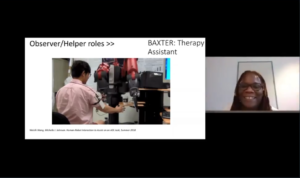
Michelle Johnson presents on her work in robotics

The day began with a fantastic lecture by Michelle Johnson, Associate Professor in Bioengineering and Physical Medicine and Rehabilitation, who introduced students to the field of rehabilitation robotics and shared her experience as a scientist. Students then learned about DNA and biomaterials through lectures mediated by the graduate students Dayo Adetu and Puneeth Guruprasad. After each lecture, students broke into breakout rooms with graduate student facilitators where they were able to get some hands-on scientific experience as they extracted DNA from their cheek cells and fabricated alginate hydrogels. Michael Sobrepera, a graduate student in Dr. Johnson’s lab, concluded the event by giving a lecture on the process of robotics development and discussed where the field is heading and some important considerations for the field.

Dayo Adetu, Bioengineering Master’s student and GABE President, teaches the students about Genetic Engineering

While yet another online event may seem unexciting, throughout the lectures students remained exceptionally engaged and raised fantastic questions ranging from the accessibility of low income communities to novel robotic therapeutic technologies to the bioethical questions robotic engineers will face as technologies advance. The impact of BETA day was evident as the high school students began to discuss the possible majors they would like to pursue for their bachelor’s degrees. Events like BETA Day give a glimpse into possible STEM fields and careers students can pursue.

Week in BioE (April 2, 2018)

Beer Gets a Hand From Bioengineering

beerBeer is among the oldest beverages known to humankind. While we don’t know what the beer that the ancient Egyptians drank tasted like, there’s little question that the trend toward craft brewing over the past generation has resulted in a proliferation of brews with a range of colors and flavors. Beers with a strong flavor of hops are currently popular, resulting in high demand for the plant that bears them. Like many plants, however, the hop plant requires significant water and energy to produce, adding to the burden of global climate change.

Bioengineers from the University of California, Berkeley, might have found a solution to this problem. In an article recently published in Nature Communications, the Berkeley scientists, led by Jay D. Keasling, Ph.D., from the Department of Bioengineering, described how they engineered brewer’s yeast cells using DNA from mint, basil, and yeast to produce the terpenoid that produces the taste of hops. Taste testers found the beer made from the engineered yeast to be hoppier in flavor than two commercial beers.

The authors of the study acknowledge that a true hop flavor is likely subtler than the register of tastes they used to engineer their brewers yeast. That said, they believe their research could serve as a basis for the genetic engineering of plant products for a range of uses. In addition, the amount of saved money could be enormous — currently, the cost of growing an acre of hop plants is nearly $7,000, which is approximately 10 times that of corn.

Wearables Monitor Digestion

We may be getting a much closer look at certain aspects of digestion thanks to two wearable devices developed by engineers on two sides of the country. On the West Coast, bioengineers at the University of California, San Diego, led by Todd P. Coleman, PhD, professor in UCSD’s Department of Bioengineering,  developed a device to monitor the electrical activity of the stomach over a 24-hour period. Unlike other technologies, this approach doesn’t require the user to drink a barium solution or ingest a tiny camera. The device, which they describe in an article in Scientific Reports, consists of a series of wearable sensors based on EKG technology and an event-logging app for computers or mobile devices. In testing, the new device performed comparably to gastric manometry, a ‘gold standard’ in clinical care that requires insertion of a nasogastric tube. The authors believe their device could have use in gastric motility disorders.

Across the country at Tufts, Fiorenzo Omenetto, PhD, Frank C. Doble Professor in the Department of Biomedical Engineering, led a team of scientists in developing a tooth-mounted sensor that can monitor food intake. The device, which measures 4 square millimeters, is described in an article in Advanced Materials. The sensor could replace the much bulkier mouthguards used for such research in the past.

Painless Lupus Testing

Despite being among the more common autoimmune disorders, lupus is difficult to diagnose. Blood tests often do not provide conclusive results. Ultimately, a kidney biopsy is often necessary to confirm the diagnosis, an invasive procedure that can be problematic with sick patients.

Now, a research team at the University of Houston has developed a saliva test that might eliminate the need for kidney biopsy. Chandra Mohan, MD, PhD, Hugh Roy and Lillie Cranz Cullen Endowed Professor of Biomedical Engineering at UH, received an NIH grant to develop a new technology which identifies proteins in the saliva as biomarkers of lupus. An important aspect of the assay is that it can be used to monitor the disease, as well as diagnose it, so the efficacy of medication to treat the disease can be followed without having to obtain multiple kidney biopsies.

Fluid Shear Stress Affects Ovarian Cells

Like most cancers, survival rates for ovarian cancer have gone up in past decades. As with most cancers, early detection remains a key step to extending survival, but a large proportion of ovarian cancers are not diagnosed until they have metastasized to other organs. Therefore, understanding the process by which normal ovarian cells become cancerous and the mechanism underlying their metastasis could improve our ability to predict these cancers and perhaps detect them earlier.

In response to this issue, researchers at Virginia Tech have uncovered part of the mechanism by which ovarian cancer cells metastasize. Led in part by  Rafael V. Davalos, PhD, L. Preston Wade Professor of Biomedical Engineering and Mechanics, the researchers report in PLOS One that fluid-induced shear stress plays a key role. Ovarian cancer causes the accumulation of fluid in the abdomen, called ascites. Using a mouse model, the study authors found that benign ovarian cells actually became malignant, and malignant cells were more likely to metastasize under this stress. This knowledge could go a long way toward developing more effective treatments for ovarian cancer.

People and Places

Two universities have announced they will begin offering degree programs in biomedical sciences. Among three programs added by Loma Linda University in California is one with an emphasis on neuroscience, systems biology, and bioengineering. Enrollment begins in the fall. In Huntington, W.V., Marshall University has added a bachelor’s degree program in BME, also beginning in the fall.

The University of North Texas, near Dallas, broke ground on a new BME building. The college anticipates the building will open for the Fall 2019 semester.

Finally, we are very proud to report that Elsie Effah Kaufmann, PhD, who earned bachelor’s, master’s, and doctoral degrees from Penn Bioengineering, was honored last week at the 44th annual meeting of the National Society of Black Engineers in Pittsburgh, where she received the 2018 Golden Torch Award for International Academic Leadership. Congratulations!
Week in BioE (February 27, 2018)

Pain Relief Using Spearmint Aromatherapy

spearmintPain is the body’s way of telling you there’s something wrong. For most of us, the pain goes away after the body fixes itself. However, more than 10% of Americans suffer from chronic pain after the healing period. Many chronic pain patients need drugs to reduce their symptoms.  Given the pervasive use of opioid drugs to treat chronic pain, opioid addiction is common among chronic pain patients.

However, a remarkably clever and elegant cellular engineering technology may provide a new approach for treating chronic pain. Martin Fussenegger, Ph.D., a professor in the Department of Biosystems Science and Bioengineering at the Swiss Federal Institute of Technology, is the lead author of a new study published in Nature Biomedical Engineering combining cellular and genetic engineering to alleviate pain using cells as factories to produce spearmint. The strategy employed by the authors used engineered human cells to express huwentoxin IV, a blocker of sodium channels regulating pain signals in neurons, upon exposure to carvone, a terpenoid found in spearmint.

Testing their concept in a mouse model of pain, the authors found that mice exposed to spearmint both orally and via aromatherapy showed fewer signs of pain. Looking forward, Dr. Fussenegger and his colleagues believe that their technology, called AromaCell, should be tested next in human cell lines to alleviate concerns about immunological responses to the cells when implanted into patients.

Press Button to Bleed

Another recent article in Nature Biomedical Engineering details the work of the Boston-area biotech firm Seventh Sense Biosystems on their push-button blood collection device, called TAP. As we have discussed here before, currently used blood-drawing procedures are often uncomfortable to patients because of the sharp needle prick used to collect blood. TAP was designed to collect 100 microliters of whole blood using a device the size of a stethoscope bell in a “virtually painless” manner.

The scientists from Seventh Sense designed the patch using microneedle technology. With this approach, they designed TAP with multiple microneedles deployed at high velocity to collect blood from capillaries — the tiniest vessels that connect veins and arteries and that lie closest to the surface of the skin — rather than from a vein tied off with a tourniquet. Testing the device in 144 volunteers, the study authors found that the device was as accurate as current methods for obtaining blood to measure hemoglobin (important for diabetics) and was significantly less painful.

Seventh Sense predicts this disposable device will cost only $5 per use, but this is still almost double the materials cost for standard blood draws. However, the company believes that the pain-free nature of and time saved with TAP will offset the higher cost of the device.

Advances in Global Health

The positively epidemic nature of human papilloma virus (HPV), affecting nearly one quarter of all Americans, has drawn particular attention over the last decade or so. The clear association between HPV and cervical cancer (as well as head and neck cancers) has led to the development and deployment of vaccines (controversial due to the sexually transmitted nature of HPV) and to increased calls for more regular and accurate screening. In developing nations, implementing either effective vaccination or early screening programs remains an uphill struggle.

Responding to the need for more accessible screening technologies, Jessica Ramella-Roman, Ph.D., Associate Professor of Biomedical Engineering at Florida International University (FIU), and Purnima Madhivanan, Ph.D., an epidemiology professor at FIU, traveled to Mysore, India, to install a device developed by Dr. Ramella-Roman at the Public Health Research Institute of India. The device is a hand-held imaging tool that uses a technology called Mueller matrix imaging to provide high-resolution digital images of the cervix in about 5 seconds. The resolution of the images eliminates the need to use dyes or stains to detect malignant cells. The testing of the device is currently ongoing.

Elsewhere in global health, researchers at Google have teamed with medical faculty from Stanford to produce a machine learning algorithm that could examine the human retina and determine whether the person in question is at risk for cardiovascular disease. They report their findings in Nature Biomedical Engineering.

The technology is not ready for actual patients yet — the study authors concede that the algorithm does not outperform the currently available technologies. However, if improved with additional research and testing, the algorithm could be deployed virtually anywhere, including in patients’ homes.

People and Places

Yale University has launched a new Center for Biomedical Data Science, dedicated to collecting, studying, and managing big data. The interim directors are Mark Gerstein, Ph.D., Albert L Williams Professor of Biomedical Informatics, Molecular Biophysics, and Biochemistry, and Hongyu Zhao, Ph.D., Ira V. Hiscock Professor of Biostatistics and Professor of Genetics and Professor of Statistics and Data Science.

The University of Virginia has announced a partnership with Smithfield Bioscience, a subsidiary of Smithfield Foods, Inc. The goal of the partnership is to advance a variety of tissue engineering applications using tissue samples from pigs. George J. Christ, Ph.D., Professor of Biomedical Engineering and Orthopaedic Surgery, heads UVA’s $3 million Center for Advanced Biomanufacturing, which is involved in the partnership.

Finally, we offer our congratulations to Guoqiang Yu, Ph.D., Professor of Biomedical Engineering at the University of Kentucky and a former research faculty member in physics here at Penn, for being awarded a two-year $420,000 R21 research grant from the NIH’s Eunice Kennedy Shriver National Institute of Child Health and Human Development. Dr. Yu will use the money to develop a device to measure cerebral hemodynamics in neonatal ICU patients.

Week in BioE (February 12, 2018)

Engineering Recovery?

opioidsThe introduction of morphine in the 19th century to alleviate pain revolutionized medicine in a way few innovations do, but it brought with it a grave unintended consequence: addiction. In today’s society, opioid addiction is creating the biggest health crisis of the last half century. Affecting nearly 1 in 100 people, opioid addiction occurs more than type 1 diabetes, multiple sclerosis, or a number of other diseases. The addiction crisis also appears in global affairs and impacts our national security: heroin production in Afghanistan over the last 40 years has been critical to funding military actions by insurgent groups against both the US and, in the past, the Soviet Union.
However, bioengineers at Stanford have begun to tackle the issue of production and might have begun to tackle the issue of addiction. In the lab of Christine Smolke, Ph.D., Professor of Bioengineering at Stanford, they have been genetically engineering yeast to produce opioids. They described the process in a 2015 article from Science. Now, in a recent interview in Fast Company, Dr. Smolke discusses the possibility of using the yeast producing method she pioneered to produce opioids without addiction potential. These alternative drugs are very expensive to produce, and Dr. Smolke’s process could provide safer, less addictive compounds to people in need.

Breaking the Barrier

Among the challenges faced by bioengineers working on therapies for brain disease is the blood-brain barrier (BBB), a tightly regulated boundary between the circulatory system and the brain that prevents all but the tiniest molecules from getting into the brain. The poor permeability of the BBB to many molecules means that one needs to use higher drug dosages to reach the brain, which is one of the reasons why most psychiatric medications have a broad array of side effects.
One way of circumventing this issue is to deliver the drugs directly to the brain, rather than using oral or intravenous delivery methods that need to cross the BB. Here, the challenge is one of size — unless a needle used to administer such a drug is very small, it will invariably damage brain tissue, which can have devastating consequences. Answering this call has been Robert Langer, Ph.D., David H. Koch Institute Professor at MIT, whose lab has successfully microfabricated delivery cannulas as small as 30 microns, about one-third the diameter of human hair. As they report in Science Translational Medicine, the new cannula can target brain areas as small as 1 cubic millimeter.
Dr. Langer and his colleagues used the new cannula to create an implantable device, called the miniaturized neural drug delivery system (MiNDS), that they subsequently tested in rats and rhesus monkeys. They found that the device could modulate neuronal activity in both animals. In addition, MiNDS could also record and transmit information from the treatment site to enable feedback control. Going forward, the study authors envision the use of non-metallic materials to fashion cannulas and hydrogel coatings to facilitate MR imaging and increase biocompatibility.

Unlocking the Mystery of IPF

Idiopathic pulmonary fibrosis (IPF) is a lung disease that causes permanent scarring of the lung tissue. The disease affects around five million people worldwide, mainly people 50 or older, and the five-year mortality rate is very high. Although risk factors, such as cigarette smoking, have been identified, as the word “idiopathic” implies, the cause is unknown, making it difficult to create effective therapies other than ones that merely slow the progression of the disease.
However, thanks to a new discovery, we might be closer to effective treatments. In an article in the Journal of Clinical Investigation Insight, a team of scientists from Yale University report that the tissue lesions that constitute IPF are made up of roughly one-fifth pericytes — a type of contractile cell that plays an important role in the proper function of capillaries, including those in the lungs.
The study authors, led by Anjelica Gonzalez, Ph.D., Donna L. Dubinsky Associate Professor of Biomedical Engineering at Yale, found that IPF caused  pericytes to take on the properties of myofibroblasts, a cell type that is important to the wound-healing process. They found further that treatment of these myofibroblast-like pericytes with nintedanib, a drug approved for IPF treatment, reversed this effect. Armed with this knowledge, we come a step closer to designing and producing more effective therapies for IPF, as well as for diseases with similar effects.

People and Places

Washington University in St. Louis has announced it will launch a Ph.D. program in imaging science, to enroll its first cohort this fall. The program will be headed by Mark Anastasio, Ph.D., Professor of Biomedical Engineering and a 1993 recipient of an MSE from Penn.  WashU’s program is only the second such program in the country, following the program at the Rochester Institute of Technology.
Closer to home, Johns Hopkins is the recipient of a $50 million donation from the United Arab Emirates. The money will be used to create the Sheikh Khalifa Stroke Institute, which will unite faculty members from biomedical engineering, neurology, and rehabilitation medicine to advance research into stroke.

Week in BioE (December 15, 2017)

A New Model of the Small Intestine

small intestine
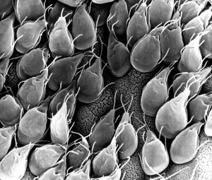
Small intestinal mucosa infested with Giardia lamblia parasites

Diseases of the small intestine, including Crohn’s disease and microbial infections, impose a huge burden on health. However, finding treatments for these diseases is challenged by the lack of optimal models for studying  disease. Animal models are only so close to human disease states, and laboratory models using cell lines do not completely mimic the environment inside the gut.

However, these limitations might be overcome soon thanks to the research of scientists at Tufts University.  In an article recently published in PLOS ONE, a team led by David L. Kaplan, Ph.D., of the Tufts Department of Biomedical Engineering, describes how they used donor stem cells and a compartmentalized biomimetic scaffold to model and generate small intestine cells that could differentiate into the broad variety of cell types common to that organ.

The study team tested the response of its cell model to E. coli, a common pathogen. At the genetic level, the model matched the reaction of the human small intestine when exposed to this bacterium. The success of the model could translate into its use in the near future to better understand the digestive system’s response to infection, as well as to test individualized treatments for inflammatory bowel diseases such as Crohn’s.

Saving Battle-wounded Eyes

The increase in combat survival rates has led to a higher incidence of veterans with permanent vision loss due to catastrophic damage to the eye. Globe injuries will recover of some vision, if caught in time. However, combat care for eye injuries often occurs hundreds or thousands of miles away from emergency rooms with attending ophthalmologists. With this unavoidable delay in treatment, people with globe injuries suffer blindness and often enucleation.

However, battle medics might soon have something in their arsenals to prevent such blinding injuries immediately in the combat theater. As reported recently in Science Translational Medicine, engineers at the University of Southern California (USC) and ophthalmologists from USC’s Roski Eye Institute have collaborated in creating a new material for temporary sealing of globe injuries. The study authors, led by John J. Whalen, III, Ph.D., used a gel called poly(N-isopropylacrylamide) (PNIPAM), already under investigation for treating retinal injuries. PNIPAM is a thermoresponsive sealant, meaning it is a liquid at cooler temperature but an adhesive gel at warmer temperature. These interesting properties mean PNIPAM can be applied as a liquid and then solidifies quickly on the eye. The authors manipulated PNIPAM chemically to make it more stable at body temperature. As envisioned, the gel, when used with globe injuries, could be applied by medics and then removed with cold water just before the eye is treated.

The study team has tested the gel in rabbits, where it showed statistically significant improvement in wound sealing and no negative effects on the eyes or overall health of the rabbits. The authors believe the material will be ready for human testing in 2019.

Predicting Seizures in Epilepsy

Epilepsy is a central nervous system disorder characterized by seizure activity that can range in severity from mild to debilitating. Many patients with epilepsy experience adequate control of seizures with medications; however, about a third of epileptic patients have intractable cases requiring surgery or other invasive procedures.

In what could be a breakthrough in the treatment of refractive epilepsy, scientists from Australia in collaboration with IBM Research-Australia have used big data from epilepsy patients to develop a computer model that can predict when seizures will occur. So far, the technology predicts 69% of seizures in patients. While it’s still short of a range of accuracy making it feasible for use in patients outside of experimental settings, the acquisition of ever-increasing amounts of data will render the model more accurately.

The Art of Genetic Engineering

Among the techniques used in genetic engineering is protein folding, which is one of the naturally occurring processes that DNA undergoes as it takes on three dimensions. Among the major developments in genetic engineering was the discovery of the ability to fold DNA strands artificially, in a process called DNA origami.

Now, as suggested by the name “origami,” some people have begun using the process in quasi-artistic fashion. In an article recently published in Nature, bioengineers at CalTech led by Lulu Qian, Ph.D., assistant professor of bioengineering, showed they were able to produce a variety of shapes and designs using DNA origami, including a nanoscale replica of Leonardo da Vinci’s Mona Lisa.

DNA now also has another unique artistic application — tattoos, although people’s opinions of whether tattooing constitutes art might vary. Edith Mathiowitz, Ph.D., of Brown University’s Center for Biomedical Engineering, is among the patenters of Everence, a technology that takes DNA provided by a customer and incorporates it into tattoo ink. Potential tattooees can now have the DNA of loved ones incorporated into their bodies permanently, if they should so wish.

People and Places

The University of Washington has launched its new Institute for Nano-engineered Systems, cutting the ribbon on the building on December 4. The center will house facilities dedicated to scalable nanomanufacturing and integrated photonics, among others. Meanwhile, at the University of Chicago, Rama Ranganathan, M.D., Ph.D., a professor in the Department of Biochemistry and Molecular Biology and the Institute for Molecular Engineering, will lead that college’s new Center for Physics of Evolving Systems. Congratulations!

Week in BioE (December 8, 2017)

Brain Implant Advance to Cure Dementia

Alzheimer's
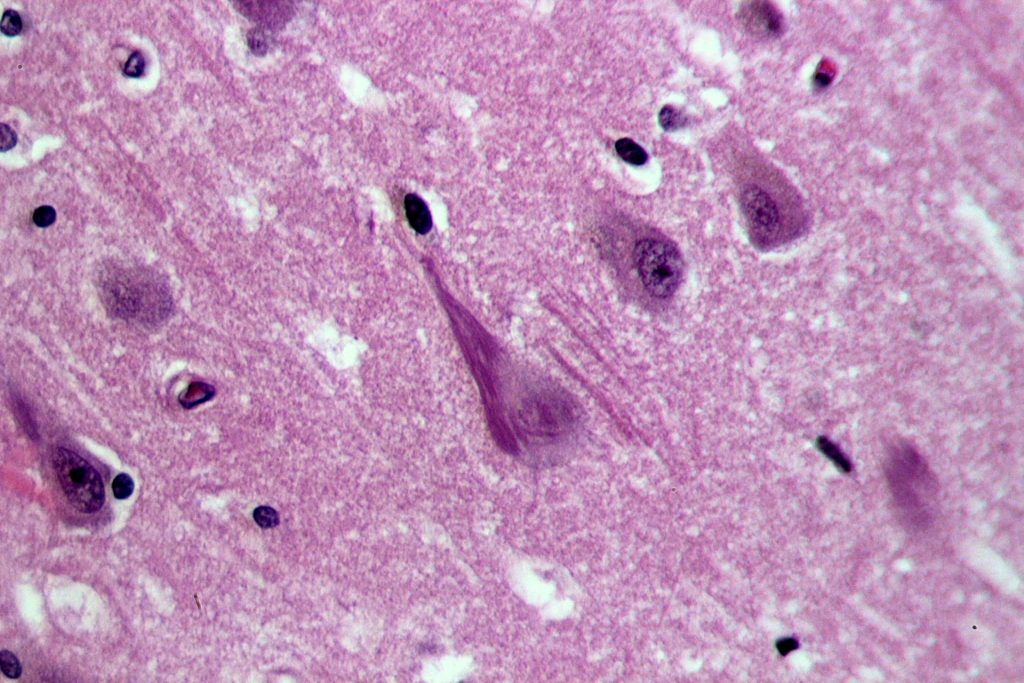
Neurofibrillary tangles in the hippocampus of a patient with Alzheimer’s disease.

The dramatic increase in life expectancy over the past couple of generations has one unfavorable consequence: an increase in the incidence of age-related dementias that include Alzheimer’s disease. Drugs like donepezil, which inhibits hydrolization of acetylcholine and thus increases its presence at the neural synapses, is one treatment that can slow the progression of these diseases, but there is currently no cure. 

An alternative technology that directly stimulates the brain with an implantable chip holds promise to reverse the effects of Alzheimer’s. At the annual meeting of the Society for Neuroscience, held last month in Washington, D.C., Dong Song, Ph.D., Research Associate Professor of Biomedical Engineering at USC’s Viterbi School of Engineering, gave a lecture on his lab’s device, which uses an array of implantable electrodes to improve human memory.

Dr. Song tested his device in epilepsy patients, who often receive implants designed to control their seizures in intractable cases. Twenty such patients volunteered to receive Dr. Song’s implant, and data from these patients showed that short-term memory increased by 15% and working memory by 25%. While additional testing is needed on more patients, it might not be long before implants like Dr. Song’s become the standard of care in treatment dementias.

Genetic Variation in the Human Microbiome

The human body is host to a veritable universe of microbes that play important roles in the organ systems and other bodily processes. E. coli, for example, is present in the large intestine and it participates in the breaking down of food for energy. Like all other forms of life, these microbes evolve. creating variations in genetic information and, ultimately, new bacteria species. Within any given species of bacteria, the number of differences in the genome sequences can vary broadly; with E. coli, some areas of the genome can vary radically between strains and cannot be explained by DNA copying errors.
   
To determine why the genome of E. coli subject to such variation, scientists at the University of Illinois, Urbana-Champaign (UIUC), led by Sergei Maslov, Ph.D., professor of bioengineering and physics at UIUC, investigated the issue by developing computational models using Multi Locus Sequence Typing (MLST). In their findings, published in Genetics, they concluded that the variation can be ascribed to the process of recombination, by which different sequences from different sources are combined into the same chromosome. When such events are frequent, they result in a sort of genetic stability in which variation in genetic information increases without speciation.

The study provides an important contribution to basic science in helping to better explain how different strains of bacteria develop, including virulent and drug-resistant strains. In addition, it sheds further light on the mechanisms underlying evolution.

A Step Closer to Water-efficient Agriculture

Drought and famine are closely related phenomena. Some plants are more resistant to drought than others, but few of these plants are fit for human consumption. Determining how plants resist drought could provide a key to engineering crops to become drought-resistant.
Investigating this topic, scientists at the Oak Ridge National Laboratory of the U.S. Department of Energy sought to understand better the process of crassulacean acid metabolism (CAM), by which drought-resistant plants keep their stomata, or pores, closed during sunlight hours to retain water and open them at night. The team reports in Nature Communications that they compared the genomes of three drought-resistant plants — orchid, pineapple, and Kalanchoë fedtschenkoi, a species of plant native to Madagascar.  Among the authors’ discoveries was a variation in a gene encoding phosphoenolpyruvate carboxylase, an enzyme that plays a role in CAM.

With this increased knowledge of the evolutionary development of drought resistance, we come a step closer to being able to expedite the evolution of plants that are typically not resistant to drought to developing the CAM mechanism and developing this resistance.

Computer Model Can Mimic Heart Attack

Heart disease remains the leading cause of death in developed countries. A major obstacle in reducing the deaths due to cardiac arrest is the inability to determine the precise mechanics unfolding in the heart when it stops suddenly. Abnormal heart rhythms (arrythmias) are a major cause of death, but the reasons how arrhythmias occur at the cellular level is poorly understood.

In a recent study published in PLOS Computational Biology,  Raimond L. Winslow, Ph.D. who is Raj and Neera Singh Professor in the Department of Biomedical Engineering at Johns Hopkins University, and his colleagues developed a computer model of calcium dynamics in cardiac cells. The model predicted a new mechanism for arrythmia that would occur when cardiac cells expelled calcium, creating an electrical charge outside the cell that could evoke an arrhythmia.

The authors believe that their research will facilitate the development of drugs to prevent cardiac arrhythmias and treatments for sudden cardiac arrest. In addition, the work shows that it could be easier to predict the statistical relationship between arrhythmias and cardiac arrest on the basis of far less data.

People and Places

Stevens Institute of Technology in Hoboken, N.J., has announced plans to divide its Department of Biomedical Engineering, Chemistry and Biological Sciences (BCB) into two new departments: the Department of Biomedical Engineering and the Department of Chemistry and Chemical Biology. Hongjun Wang, Ph.D., associate professor in the BCB department, will be the new chair of BME. Congratulations Hongjun!

Week in BioE (August 3, 2017)

There’s news in bioengineering every week, to be sure, but the big story this past week is one that’s sure to continue appearing in headlines for days, weeks, and months — if not years — to come. This story is CRISPR-Cas9, or CRISPR for short, the gene-editing technology that many geneticists are viewing as the wave of the future in terms of the diagnosis and treatment of genetic disorders.

Standing for clustered regularly interspaced short palindromic repeats, CRISPR offers the ability to cut a cell’s genome at a predetermined location and remove and replace genes at this location. As a result, if the location is one at which the genes code for a particular disease, these genes can be edited out and replaced with healthy ones. Obviously, the implCRISPRications for this technology are enormous.

This week, it was reported that, for the first time, CRISPR was successfully used by scientists to edit the genomes of human embryos. As detailed in a paper published in Nature, these scientists edited the genomes of 50 single-cell embryos, which were subsequently allowed to undergo division until the three-day mark, at which point the multiple cells in the embryos were assessed to see whether the edits had been replicated in the new cells.  In 72% of them, they had been.

In this particular case, the gene edited out was one for a type of congenital heart defect, and the embryos were created from the eggs of healthy women and the sperm of men carrying the gene for the defect. However, the experiments prove that the technology could now be applied in other disorders.

Needless to say, the coverage of this science story has been enormous, so here is a collection of links to coverage on the topic. Enjoy!