Week in BioE (May 10, 2018)

Advances in Cancer Detection
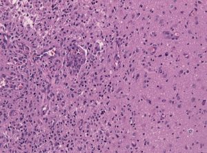
glioblastoma
Tumor-brain-interface in a glioblastoma biopsy specimen.

Among the deadliest and most difficult to treat types of cancer is glioblastoma, an especially aggressive form of brain cancer. Widely available imaging techniques can diagnose the tumor, but often the diagnosis is too late to treat the cancer effectively. Although blood-based cancer biomarkers can provide for earlier detection of cancer, these markers face the difficult task of crossing the blood-brain barrier (BBB), which prevents all but the tiniest molecules from moving from the brain to the bloodstream.

A study recently published in Scientific Reports, coauthored by Hong Chen, PhD, Assistant Professor of Biomedical Engineering at Washington University in St. Louis (WUSTL), reports of successful deployment of a strategy consisting of focused ultrasound (FUS), enhanced green fluorescent protein (eGFP), and systemically injected microbubbles to see if the BBB could be opened temporarily to allow biomarkers to pass from the brain into the bloodstream. The authors used eGFP-activated mouse models of glioblastoma, injecting the microbubbles into the mice and then exposing the mice to varying acoustic pressures of FUS. They found that circulating blood levels of eGFP were several thousand times higher in the FUS-treated mice compared to non-treated mice, which would significantly facilitate the detection of the marker in blood tests.

The method has some way to go before it can be used in humans. For one thing, the pressures used in the Scientific Reports study would damage blood vessels, so it must be determined whether lower pressures would still provide detectable transmission of proteins across the BBB. In addition, the authors must exclude the possibility of FUS unexpectedly enhancing tumor growth.

In other body areas, with easier access from tissue to the bloodstream, engineers have developed a disease-screening pill that, when ingested and activated by infrared light, can indicate tumor locations on optical tomography. The scientists, led by Greg M. Thurber, PhD, Assistant Professor of Biomedical and Chemical Engineering at the University of Michigan, reported their findings in Molecular Pharmaceutics.

The authors of the study used negatively charged sulfate groups to facilitate absorption by the digestive system of molecular imaging agents. They tested a pill consisting of a combination of these agents and found that their model tumors were visible. The next steps will include optimizing the imaging agent dosage loaded into the pill to optimize visibility. The authors believe their approach could eventually replace uncomfortable procedures like mammograms and invasive diagnostic procedures.

Liquid Assembly Line to Produce Drug Microparticles

Pharmaceuticals owe their effects mostly to their chemical composition, but the packaging of these drugs into must be done precisely. Many drugs are encapsulated in solid microparticles, and engineering consistent size and drug loading in these particles is key. However, common drug manufacturing techniques, such as spray drying and ball milling, produce uneven results. 

University of Pennsylvania engineers developed a microfluidic system in which more than ten thousand of these devices run in parallel, all on a silicon-and-glass chip that can fit into a shirt pocket, to produce a paradigm shift in microparticle manufacturing. The team, led by David Issadore, Assistant Professor in the Department of Bioengineering, outlined the design of their system in the journal Nature Communications.

The Penn team first tested their system by making simple oil-in-water droplets, at a rate of more than 1 trillion droplets per hour. Using materials common to current drug manufacturing processes, they manufactured polycapralactone  microparticles at a rate of ‘only’ 328 billion particles per hour. Further testing backed by pharma company GlaxoSmithKline will follow.

Preventing Fungal Infections of Dental Prostheses

Dental prostheses are medical devices that many people require, particularly as they age. One of the chief complications with prostheses is fungal infections, with an alarming rate of two-thirds among people wearing dentures. These infections can cause a variety of problems, spreading to other parts of the digestive system and affecting nutrition and overall well-being. Fungal infections can be controlled in part by mouthwashes, microwave treatments, and light therapies, but none of them have high efficacy.

To address this issue, Praveen Arany, DDS, PhD, Assistant Professor, Department of Oral Biology and Biomedical Engineering at SUNY Buffalo, combined 3D printing technology and polycaprolactone microspheres containing amphotericin-B, an antifungal agent. Initial fabrication of the prostheses is described in an article in Materials Today Communications, along with successful in vitro testing with fungal biofilm. If further testing proves effective, these prostheses could be used in dental patients in whom the current treatments are either ineffective or contraindicated.

People and Places

West Virginia University has announced that it will launch Master’s and doctoral programs in Biomedical Engineering. The programs will begin enrolling students in the fall. The graduate tracks augment a Bachelor’s degree program begun in 2014.


Pancreatic Cancer Detection With Micropore Chip

Pancreatic cancer remains one of the deadliest types of cancer, with one- and five-year survival rates of only 20% and 7%, respectively, according to the American Cancer Society. The mortality is so high because the disease does not typically cause symptoms until it is too late. Therefore, earlier detection could be the key to better survival rates.

In a new paper published by Lab on a Chip, a research team from the lab of David Issadore, assistant professor of Bioengineering, reports on its development of a micropore chip, callled the circulating tumor cell fluorescence in situ hybridization (CaTCh FISH) chip, that could detect circulating tumor cells (CTCs) from mice and patients with pancreatic cancer, even at very low, previously undetectable levels.

pancreatic cancer
Jin A Ko

Jin A (Jina) Ko, who is a Ph.D. student in Bioengineering and first author on the paper, says that CTCs are a key mechanism underlying metastasis, which is another reason why pancreatic cancer has such a low survival rate. Not only can the chip that she helped design detect these cells, which circulate in the bloodstream, but more importantly, pancreatic tumors shed these cells even in their very early stages before any spread has occurred. Therefore, provided the test is performed early enough, the tumor can be detected and treated. Patients with family histories of pancreatic cancer or who have tested positive for certain gene mutations would likely benefit from this sort of test.

The study authors also tested the CaTCh FISH chip using blood samples from 14 patients with advanced pancreatic cancer and from healthy controls. They found that their micropore chip could detect several RNA markers of cancer in 10-mL samples — around 2 tsp. In addition, there were no false-negative results among the healthy controls, demonstrating a high level of reliability in that regard.

“We have developed a microchip platform that combines fast, magnetic micropore-based negative immunomagnetic selection with rapid on-chip in situ RNA profiling,” Jina said. “This integrated chip can isolate both rare circulating cells and cell clusters directly from whole blood and allow individual cells to be profiled for multiple RNA cancer biomarkers.”

Secondary Projects From Ghana: Group 4

While brainstorming and writing a proposal for a device to detect pediatric tuberculosis has been extremely valuable, we recognize the challenge of developing our devices as undergraduate/graduate students. This acknowledgement led us to try to identify a healthcare problem in Ghana and to come up with a solution that undergraduates could potentially pursue. The process began after we arrived in Ghana, with each student independently identifying a problem and brainstorming a solution. Next, we played an entrepreneurial game, in which each student gave a pitch for an idea, and everyone gave hypothetical money to his or her favorite idea. The ideas with the most hypothetical monetary investments would move on to the next round. After two rounds of pitches, we narrowed our list down to two ideas: Big Data and the Multi-Cot. Splitting up our group between the two ideas, we then prepared a presentation to give to Kumasi Center for Collaborative Research in Tropical Medicine (KCCR) researchers. Yesterday and today, we present the summaries of our ideas.

Ghana group 4-1
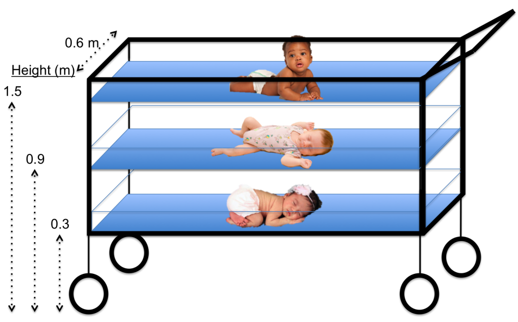
The Multi-Cot aims to tackle the issue of limited space in large regional hospitals within Ghana and other similar situations of overcrowding.

Kate Panzer (gave first-round pitch) ’18, Katharine Cocherl ’20, Kaila Helm ’20, Hope McMahon ’18

Throughout our time in Ghana, we had the opportunity to visit many hospitals and smaller health clinics. While visiting Komfo Anokye Teaching Hospital (KATH) in Kumasi, Ghana, we noticed that there was a poster on a pediatrician’s wall for the “One Baby One Cot” initiative. We soon learned that there is very limited space per patient at the large regional hospitals — certainly not enough space for each individual baby to occupy his or her own cot. For example, in some hospitals, there can be up to eight babies in one cot! This can be problematic when trying to prevent the spread of infection but also difficult for mothers who have little to no space to watch over their newborns when they stay at the hospital to breastfeed.

There are several implications of having multiple babies in a single cot that we would like to address. First, the risk of hospital-acquired infections greatly increases because of the close contact of the babies. This close contact also makes it difficult for nurses and caretakers to monitor each baby. In addition, many babies may need to be transported to other hospitals because of a lack of bed space, moving the patients and their caretakers farther from home.

Ghana 4-2
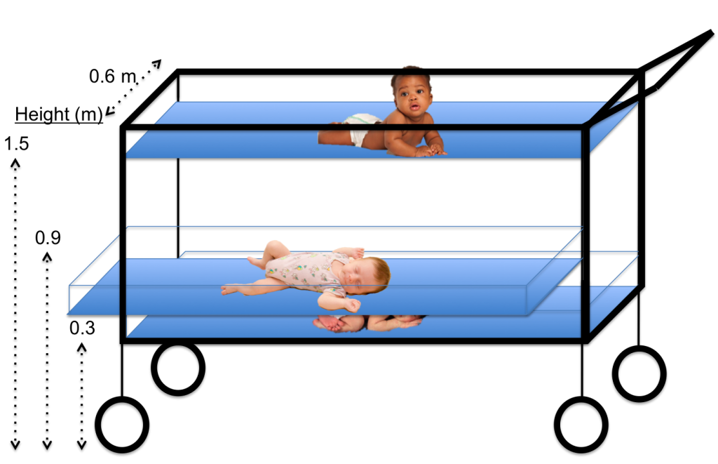
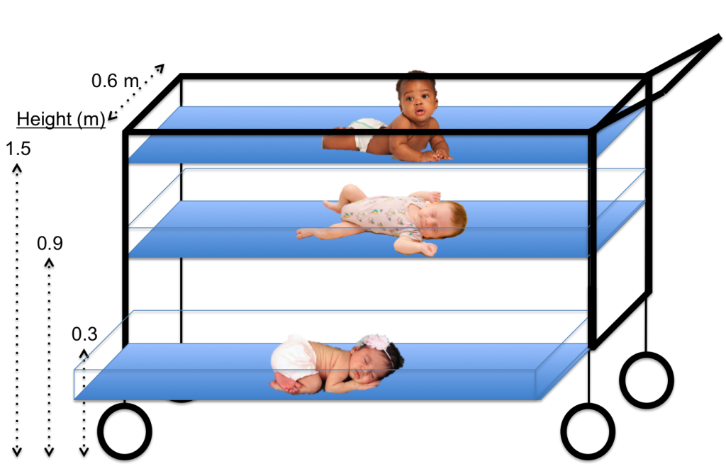
The horizontal sliding mechanism of the Multi-Cot allows each newborn to be safely removed from the structure, regardless of the cot level.

Ghana group 4-3

After learning about this problem, we began thinking of ways to decrease the complications associated with having multiple newborns in one cot. During the brainstorming session, the key element that led to our solution was actually how we view the problem. We started to see the issue as a lack of horizontal space – meaning the inability to add more cots horizontally without physically expanding the newborn ward. If expanding the horizontal space is not possible, then why not try to make better use of the vertical space that is already available? This concept of vertical space led us to the idea of the Multi-Cot, which involves three smaller newborn cots stacked vertically, with space between each cot to provide proper airflow. With clear plastic sides and an open top, each baby would be easily seen from every direction. Finally, to ensure safety when removing newborns from the lower levels, we added a sliding mechanism to our design to allow the lower cots to slide horizontally and eliminate any vertical obstructions when picking up the baby.

As we anticipate developing the Multi-Cot, we must consider multiple factors. Our main consideration is safety, which includes the Multi-Cot’s stability, the visibility of every child, and the ability to be sanitized. Other factors to be considered include the cost, as well as the ease of physical construction and dismantling; however, we would address these details later in the design process.

Secondary Projects From Ghana: Group 3

While brainstorming and writing a proposal for a device to detect pediatric tuberculosis has been extremely valuable, we recognize the challenge of developing our devices as undergraduate/graduate students. This acknowledgement led us to try to identify a healthcare problem in Ghana and to come up with a solution that undergraduates could potentially pursue. The process began after we arrived in Ghana, with each student independently identifying a problem and brainstorming a solution. Next, we played an entrepreneurial game, in which each student gave a pitch for an idea, and everyone gave hypothetical money to his or her favorite idea. The ideas with the most hypothetical monetary investments would move on to the next round. After two rounds of pitches, we narrowed our list down to two ideas: Big Data and the Multi-Cot. Splitting up our group between the two ideas, we then prepared a presentation to give to Kumasi Center for Collaborative Research in Tropical Medicine (KCCR) researchers. Today and Friday we present the summaries of our ideas.

Ghana secondary 3

Big Data: Deciphering Acoustic Trends in Tuberculosis, Pneumonia and Healthy Coughs

David Pontoriero (gave first-round pitch) ’18, Kathleen Givan ’20, Jason Grosz ’19, Danielle Tsougarakis ’20, Ethan Zhao ’19

Our goal was to think of a project that a team of undergraduates at Penn could complete in one year to produce something of value to KCCR in the scope of Ghanaian healthcare. We turned our attention toward big data science and the difficulties in tuberculosis diagnosis. One of the difficulties identified was the lack of diagnostic tools in more remote arms of the healthcare system. This lack leads to unnecessary and numerous referrals to larger care centers, inconveniencing the patient and placing a burden on the efficiency of the healthcare system.

Specifically, the only standard-of-care diagnostic ubiquitous throughout all clinics was patient-reported symptoms — the most notable of which is prolonged coughing. Moreover, this symptom can often be confused with asthma or pneumonia. However, asthma involves bronchial constriction, and TB and pneumonia have different sputum distribution profiles. We theorized that this difference would correlate with differentiated sound profiles for patient coughs or baseline breathing and, subsequently, measurable biomarkers. The idea proposed was that, if blind data could be collected from KCCR with sound recordings of patients coughing and breathing, along with their demographics and final diagnoses, then analyses could be run to produce an algorithm capable of differentiating between each cough or breath. This algorithm could then be extended to a phone app that could be used to more empirically diagnose patients in any setting and increase overall healthcare efficiency.

Primary Projects From Ghana: Group 2

Throughout the Spring 2017 semester, our professor, Dr. David Issadore, taught us (a class of eight undergraduates students and one graduate student) about microfluidics and point-of-care diagnostics. The next phase of the course was to come up with a new diagnostic for pediatric tuberculosis. At the end of the semester, our final assignments included submitting an NIH Research Project Grant (R01) proposal and giving a 20-minute presentation for our devices. These assignments greatly prepared us for our trip to Ghana, as we were able to ask questions and get feedback on our proposed devices by speaking to healthcare professionals at Ghanaian hospitals, clinics, and research facilities. The semester course was mainly focused on the technical design of our devices, which enabled us to hone in on the practical and real-world implementation of the devices while in Ghana. This week, the BE Blog will publish our summaries.

The LAMinator: Urine Diagnostic for Pediatric Tuberculosis

Danielle Tsougarakis ’20, Ethan Zhao ’19, Jason Grosz ’19, Kate Panzer ’18

Current devices that detect Mycobacterium tuberculosis include chest X-ray, smear microscopy, and GeneXpert. Although the combination of these techniques can lead to a proper diagnosis for adults, there are three main limitations of their use: (1) necessary infrastructure; (2) required sputum samples; and (3) time. First, many clinics in rural Ghana do not currently have the infrastructure or electricity sources to support these machines. Second, both smear microscopy and the GeneXpert rely on analyzing sputum samples (bacteria-containing phlegm), but children have difficulty providing sufficient samples. Finally, since sputum samples are best taken in the morning, these techniques often require patients to go home and return the next day to provide a sample.

ghana group 2-1
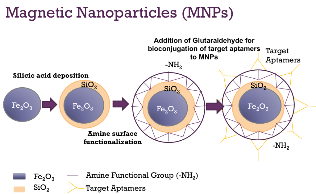
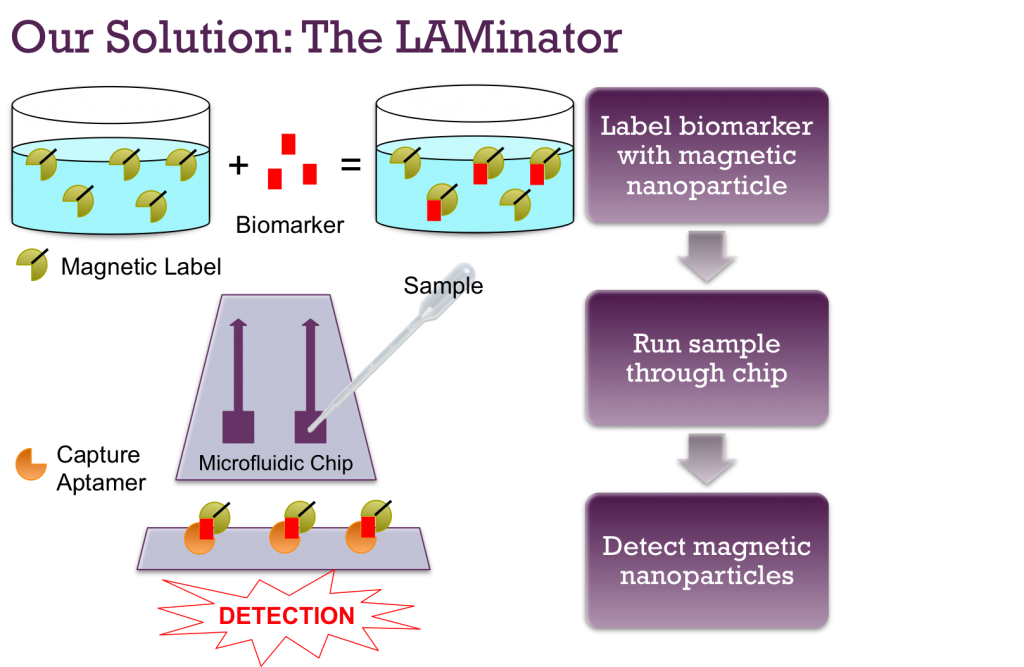
Since all biological molecules are inherently non-magnetic, these magnetic nanoparticles can be attached to ManLAM using aptamers to allow for detection by the spin-valve sensor.

To address these limitations in our own design, we proposed a diagnostic device that does not require electricity, relies on a urine sample instead of a sputum sample, and is anticipated to take one hour to obtain a diagnosis. By incorporating these three characteristics, we propose a device that can be used to more easily diagnose children during their first initial visit at any healthcare facility in Ghana.

ghana group 2-2
This overview of our device shows how the biomarker will be magnetically labeled, pushed through microfluidic channels, captured on the surface, and detected by the spin-valve sensor.

After doing a literature search of publications on pediatric tuberculosis, we learned that M. tuberculosis sheds a glycolipid called lipoarabinomannan (ManLAM) that is excreted in the urine. Therefore, ManLAM is the biomarker we hope to detect. Next, after learning that biology is inherently nonmagnetic, we figured that we could detect ManLAM specifically and sensitively if we could label it magnetically. Our proposed design does this labeling by adding magnetic nanoparticles (MNPs) to the ManLAM. This magnetic labeling involves aptamers, which are synthetic oligonucleotides that can be created to bind to a specific target. By combining the MNPs with aptamers that bind only to ManLAM, we can ultimately give the urine biomarker a magnetic property.

ghana group 2-3
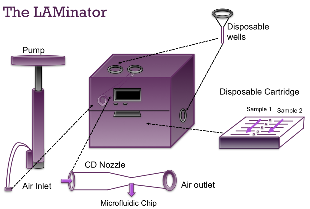
The LAMinator has a reusable box component to house the electronics as well as a disposable cartridge to hold the microfluidic chip and disposable wells to avoid sample contamination.

Therefore, the first step of our device is treating the urine sample with the custom aptamer-bound MNPs. The electronic components of our diagnostic device consist of specialized sensors, called spin-valve sensors, that can detect the presence of magnetic particles. Small fluid channels containing the urine sample traverse the surface of these sensors. If ManLAM is present in the urine as it passes by the spin-valve sensors, the surface-bound aptamers bind to the magnetically labeled ManLAM and capture them on the surface. The presence of these magnetic particles activates the spin-valve sensors and produces a change in voltage that can be detected by computer-like microprocessors. If ManLAM is not in the sample, then nothing will bind to the capture aptamers and no TB will be detected.

ghana group 2-4
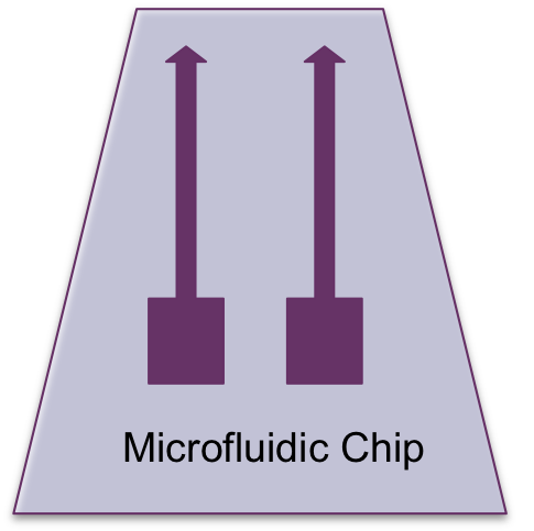
The microfluidic chip design has two channels to allow for two urine samples to be analyzed at the same time.

We would like to thank Penn Engineering and everyone who has helped to make this program possible. As you can see from our blog posts, our time in the classroom and the month in Ghana have been an unforgettable academic and cultural experience. The APOC program has been an amazing opportunity to get out of our comfort zones and to see the potential of engineering solutions in the world around us.

Primary Projects From Ghana: Group 1

Throughout the Spring 2017 semester, our professor, Dr. David Issadore, taught us (a class of eight undergraduates students and one graduate student) about microfluidics and point-of-care diagnostics. The next phase of the course was to come up with a new diagnostic for pediatric tuberculosis. At the end of the semester, our final assignments included submitting an NIH Research Project Grant (R01) proposal and giving a 20-minute presentation for our devices. These assignments greatly prepared us for our trip to Ghana, as we were able to ask questions and get feedback on our proposed devices by speaking to healthcare professionals at Ghanaian hospitals, clinics, and research facilities. The semester course was mainly focused on the technical design of our devices, which enabled us to hone in on the practical and real-world implementation of the devices while in Ghana. This week, the BE Blog will publish our summaries.

Fecal Diagnostics for Pediatric Tuberculosis

Katharine Cocherl ’20, Kathleen Givan ’20, Kaila Helm ’20, Hope McMahon ’18, David Pontoriero ’18

In order to address the numerous diagnostic problems specific to pediatric tuberculosis in low-resource settings, we have designed a device that uses a fecal sample rather than the current method of sputum samples. Because many children cannot produce sputum samples with the required quality and quantity of sputum, we decided to use stool samples. This noninvasive substitute will ideally allow us to collect all the bacteria swallowed by the patient. The bacterium that causes the disease, Mycobacterium tuberculosis (MTB), is very hardy and has been found to appear in fecal matter. However, this method may be difficult because there are many other substances in fecal matter that need to be removed. By filtering out these impurities, the presence of the bacteria can be detected.

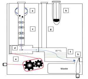
The device we designed is essentially a disposable cartridge that separates  virulent TB bacteria from all other fecal material. This collection can be performed with no power and minimal technician input and can be obtained in any desired volume. The total operation time is predicted to be 90 minutes.

ghana group 1
The figure shows an overview of the steps (a-h) for use of the fecal diagnostic for pediatric TB (click to enlarge).

The first aim of our project is to identify a target protein on the surface of the bacteria so that the bacteria can be isolated from the solution. Next, the MTB will be enriched from fecal samples with a single-use filtration device so that a final sample can be provided in a similar form as a sputum sample. This final sample can then be used for smear microscopy, in which technicians look for the presence of the bacteria under a microscope, or for use with the GeneXpert. The GeneXpert is an automated diagnostic test that can identify MTB DNA and resistance to the most potent TB drug, rifampicin. These devices have been distributed to labs and hospitals across Ghana, but they are not yet widely used for general diagnostics.

Because the number of GeneXperts available and the infrastructure supporting them are increasing, we are hopeful that, in the near future, our diagnostic will be able to be used in conjunction with this technology. Upon integration with the GeneXpert system, our device would be able to increase sample specificity for the underserved demographic of pediatric TB patients. In addition, as technology becomes available in smaller, more local clinics, we foresee lower travel burdens for families and lower operational costs for healthcare facilities.

We are beyond grateful for the opportunity to engage with Ghana’s medical system. Before traveling to Ghana, we created a proposal for our fecal diagnostic for pediatric TB. After learning more about the current medical system and infrastructure in place, we were able to revise our ideas in a meaningful way. It is our hope that one day a project of this magnitude can come to fruition.

Ghana Trip to Study Tuberculosis: Day 29

Ghana 29.1
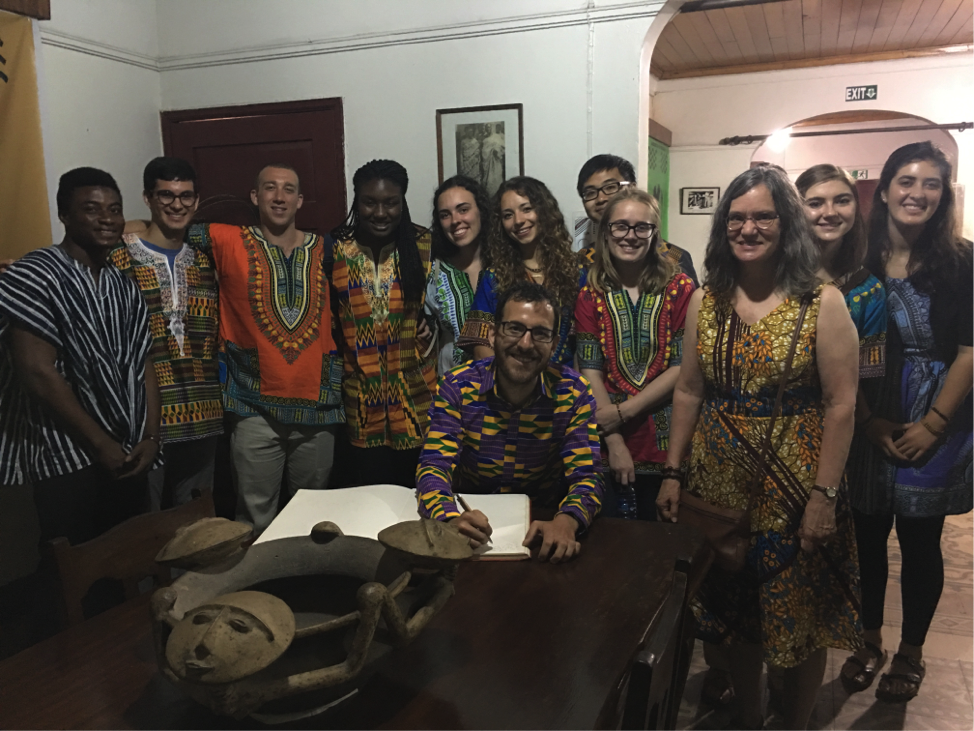
One of our favorite memories was visiting King Otumfuo Nana Osei Tutu II of the Asante region (left to right: Salim, Jason Grosz, David Pontoriero, Kaila Helm, Hope McMahon, Dr. David Issadore, Danielle Tsougarakis, Ethan Zhao, Kathleen Givan, Dr. Miriam Wattenbarger, Katharine Cocherl, Kate Panzer).

David Issadore, a faculty member in the Department of Bioengineering at the University of Pennsylvania teaches an engineering course ENGR566 – Appropriate Point of Care Diagnostics. As part of this course, he and Miriam Wattenberger from CBE, have taken nine Penn students, most of them majoring in Bioengineering, to Kumasi, Ghana, to study the diagnosis of pediatric tuberculosis. While in Ghana, these students are blogging daily on their experiences.

As we woke up early to prepare for the nine-hour flight ahead of us, we all acknowledged that time really does fly. Arriving at the Accra airport, we had to say goodbye to our Ghanaian friends Salim, Uncle Ebo, and Nana Yaa. The month has come and gone. It feels like the trip went quickly, but we have learned so much and gained many valuable experiences along the way. From our hospital and clinic visits, to our interactions with an herbalist and a fetish priestess, we were exposed to many healthcare settings found in Ghana. We had the opportunity to present our pediatric tuberculosis diagnostic ideas to a room filled with researchers and clinicians, getting invaluable feedback from multiple experts. Along with our academic pursuits, we also got to explore the Ghanaian culture and learn about customs, traditions, food, and much more. We met many friendly people along the way. These aspects are the memories that we will remember for years to come. As we move beyond this course, we are excited to continue pursuing our interests in biomedical diagnostics and problem solving that can be applied globally. We would like to thank everyone who helped make this unforgettable experience possible.

Ghana Trip to Study Tuberculosis: Day 28

Ghana 28.2
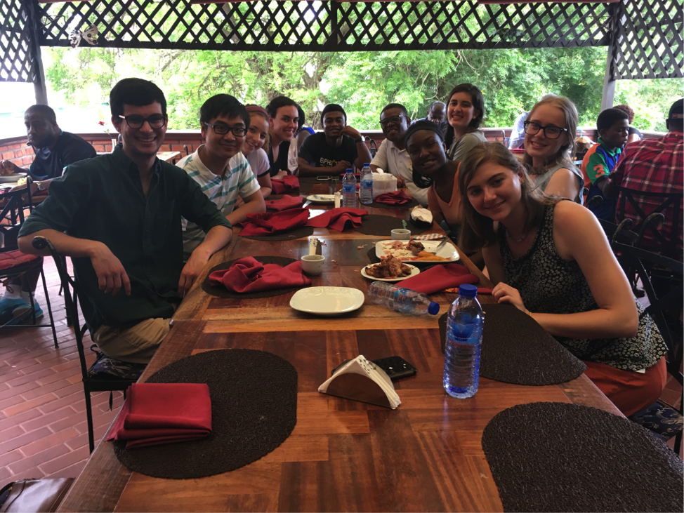
Students enjoy their last dinner in Ghana at Buka, a Ghanaian and Nigerian restaurant in Accra (left to right: Jason Grosz, Ethan Zhao, Danielle Tsougarakis, Hope McMahon, Salim, Uncle Ebo, Kaila Helm, Kate Panzer, Katharine Cocherl, Kathleen Givan).

David Issadore, a faculty member in the Department of Bioengineering at the University of Pennsylvania teaches an engineering course ENGR566 – Appropriate Point of Care Diagnostics. As part of this course, he and Miriam Wattenberger from CBE, have taken nine Penn students, most of them majoring in Bioengineering, to Kumasi, Ghana, to study the diagnosis of pediatric tuberculosis. While in Ghana, these students are blogging daily on their experiences.

Today marked our last full day in Ghana. In the morning, we set off rather early to start our day in Accra. But first, we had to drop one of our students, Dave, at the airport so he could make his way to Rwanda to visit a college friend. As we traveled to the airport, we had the opportunity to get a better picture of what life is like in Ghana’s capital. It was nice to go back to Accra and see how different it was from Kumasi. It is a much larger city, with various government buildings, people walking about, and large advertising signs every few yards.

Ghana 28.1
Kwame Nkrumah stands with past Vice Provost Roy Nichols in front of the Benjamin Franklin statue on College Green.

Our first stop was the Kwame Nkrumah Memorial Park. Kwame Nkrumah was the first president of Ghana when the country gained independence in 1957. Interestingly, he went to Penn to earn a Master of Arts in philosophy and a Master of Science in education. The mausoleum in Accra contains his and his wife’s bodies. It is surrounded by various water fountains, which are a symbol of life to provide a sense of immortality for Nkrumah. Many Ghanaians want to continue the work that Nkrumah did not get to finish by helping Ghana to continue developing as an independent country. In addition, there is a museum that contains many of his clothes and pictures of him as he met with various world leaders. We even saw a picture of him on Penn’s campus, shaking the hand of then Vice Provost Roy Nichols.

After the tour, we met Dr. Ellis from KCCR for lunch at a nice open-air restaurant, called Buka. Many of us stuck to our favorites of chicken and fried plantains, but some ventured out to try guinea fowl and snails. After lunch, we walked around the area to some nearby vendors, where we were able to shop for last minute gifts. We soon realized how much more expensive Accra was, compared to Kumasi.

We headed back to the hotel to relax a bit before dinner. For our last night in Ghana, we went out to a restaurant that had a live jazz band. We had our last taste of Ghanaian cuisine and had fun dancing to highlife music. Highlife is a genre of music that we only recently learned is popular in both Ghana and Nigeria. To end our last night in Ghana, we headed back to the hotel. After spending some time to prepare, we huddled in the hotel’s lobby for our talent show, and as night turned into morning, we reluctantly headed to our rooms to finish packing for our early departure.

Ghana 28.3
The APOC program began and ended in Accra, the capital of Ghana.

Ghana Trip to Study Tuberculosis: Day 27

by Ethan Zhao, Bioengineering ’19; and Jason Grosz, Bioengineering ’19

Ghana 27.1
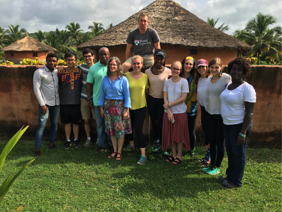
The APOC team poses in front of the rooms at Coconut Grove, resembling huts on the outside and furnished with beds and a bathroom on the inside (left to right: Salim, Ethan Zhao, Jason Grosz, Dr. Ocek Eke, Dr. Miriam Wattenbarger, Hope McMahon, David Pontoriero, Kaila Helm, Kathleen Givan, Kate Panzer, Danielle Tsougarakis, Katharine Cocherl, Nana Yaa).

David Issadore, a faculty member in the Department of Bioengineering at the University of Pennsylvania teaches an engineering course ENGR566 – Appropriate Point of Care Diagnostics. As part of this course, he and Miriam Wattenberger from CBE, have taken nine Penn students, most of them majoring in Bioengineering, to Kumasi, Ghana, to study the diagnosis of pediatric tuberculosis. While in Ghana, these students are blogging daily on their experiences.

Today was the second day that we spent in the coastal city of Cape Coast. Many of us woke up earlier than usual to walk along the beach and explore the resort. While walking along the beach, we noticed large rowboats in the distance that were anchored to the shore by ropes. We originally thought that they were fishing boats, but it turned out that they were digging up sand from the ocean floor to restore sand erosion on the beach.

Ghana 27.2
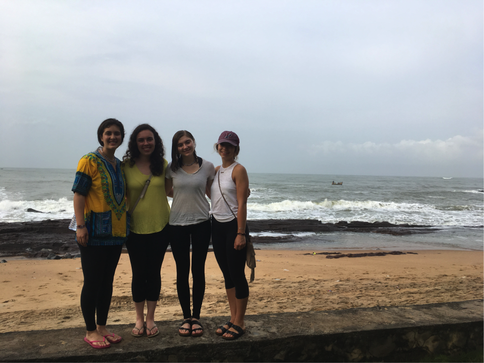
Students (left to right) Kate Panzer, Hope McMahon, Katharine Cocherl, and Danielle Tsougarakis stand along the beachfront of Coconut Grove in Cape Coast, with the Atlantic Ocean in the background.

After breakfast, we traveled outside of Cape Coast to Kakum National Park, which is a dense tropical rainforest on the coast that is home to many wildlife species, including monkeys, leopards, elephants, and antelope. It is also the home of one of Africa’s largest canopy walkways, consisting of rope suspension bridges more than one hundred feet above the forest floor. The views from the bridges were amazing, as we could see for miles across the tops of the rainforest trees. While we were on the bridges, it started drizzling, which was refreshing given the heat. After leaving Kakum National Park, we drove back to Accra, the capital of Ghana, where we will stay for the remainder of our trip.

Ghana 27.3
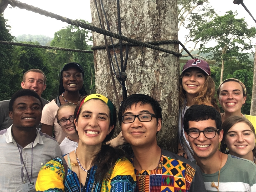
The APOC students stand on a platform among the treetops of Kakum National Park, 100 feet above the ground (left to right: Salim, David Pontoriero, Kathleen Givan, Kate Panzer, Ethan Zhao, Danielle Tsougarakis, Jason Grosz, Hope McMahon, Katharine Cocherl)

Ghana Trip to Study Tuberculosis: Day 26

by Dave Pontoriero, Biotechnology MS ’18

Ghana 26.1
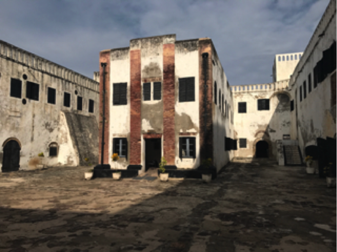
A Portuguese church found in the center of the Elmina Slave Castle.

David Issadore, a faculty member in the Department of Bioengineering at the University of Pennsylvania teaches an engineering course ENGR566 – Appropriate Point of Care Diagnostics. As part of this course, he and Miriam Wattenberger from CBE, have taken nine Penn students, most of them majoring in Bioengineering, to Kumasi, Ghana, to study the diagnosis of pediatric tuberculosis. While in Ghana, these students are blogging daily on their experiences.

Today, we said goodbye to our Kumasi friends and left the Ashanti region for the final leg of our trip. After our bonus night in the new student hostel (dormitory), we boarded the KNUST bus for a six-hour road trip to Cape Coast. The drive was pleasant, and the scenery became more coastal as we continued. Most people slept through it, but once the ocean became visible, everyone woke up in excitement because we knew we were getting close to Elmina, a beach town just west of Cape Coast.

As we drove through the town, we noticed that it resembled many of the beach towns back home. Our driver, Uncle Ebo, then parked in front of an enormous white castle. It was located on the edge of a peninsula, with a narrow beach to its left and crashing waves to its right. It had cannons situated all along its upper levels and a bustling group of locals hanging out in front of its entrance. It was the Elmina Slave Castle, also known as St. George’s Castle, and the team started to prepare for the tour.

Ghana 26.2
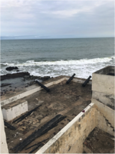
Cannons are found on the perimeter of the Elmina Slave Castle, which point out toward the Atlantic Ocean.

As we entered the castle, the mood became somber. A tour guide provided us with a background of the building, which was a Dutch fort used over the years to facilitate the sale and transport of people from Ghana and the surrounding countries during the slave trade. The first portion of the tour followed the path of a slave during their internment, beginning with the female quarters, then the courtyards used for public punishment, the male quarters, punishment cells, and lastly the final exit where people were loaded onto the ships for their journey across the Atlantic. It was a grim tour to take, and the guide shared some incredibly harrowing stories throughout. The second portion of the tour focused more on other aspects of the fort.

Once we loaded back onto the bus, the team reflected on the experience we had at Elmina Castle as we drove away. After a half-hour drive, we soon arrived to our new rooms at a local beachfront resort called Coconut Grove. Its beautiful facility included a private beach, an ocean-facing restaurant/bar, beachfront swings, a golf course, horse stables, and a crocodile pond (with ~10 real crocodiles!). We went out to a local restaurant with live music, danced a bit, then headed home to enjoy the amenities during one of our last nights together as a team.

Ghana 26.3
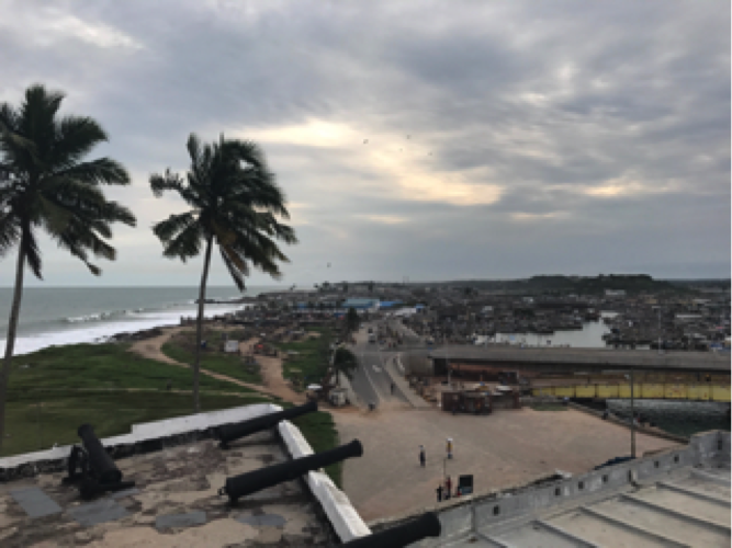
The breathtaking view of Elmina, a beach town on the coast of Ghana near Cape Coast.