Week in BioE (July 31, 2018)

New Data Analysis Methods

Like many other fields, biomedical research is experiencing a data explosion. Some estimates suggest that the amount of data generated from the health sciences is now doubling every eighteen months, and experts expect it to double every seventy-three days by 2020.  One challenge that researchers face is how to meaningfully analyze this data tsunami.

The challenge of interpreting data occurs at all scales, and a recent collaboration shows how new approaches can allow us to handle the volumes of data emerging at the level of individual cells to infer more about how biology “works” at this level.  Wharton Statistics Department researchers Mo Huang and Jingshu Wang (PhD Student and Postdoctoral Researcher, respectively) collaborated with Arjun Raj’s lab in Bioengineering and published their findings in recent issues of Nature Methods and Proceedings of the National Academy of Sciences.  One study focused on a de-noising technique called SAVER to provide more precise data from single cell experiments and significantly improves the ability to detect trends in a dataset, similar to how increasing sample size helps improve the ability to determine differences between experimental groups.  The second method, termed DESCEND, creates more accurate characterization of gene expression that occur in individual cells. Together these two methods will improve data collection for biologists and medical professionals working  to diagnose, monitor, and treat diseased cells.

Dr. Raj’s team contributed data to the cause and acted as consultants on the biological aspects of this research. Further collaboration involved Mingyao Li, PhD, Professor of Biostatistics at the Perelman School of Medicine, and Nancy Zhang, PD, Professor Statistics at the Wharton School. “We are so happy to have had the chance to work with Nancy and Mingyao on analyzing single cell data,” said Dr. Raj. “The things they were able to do with our data are pretty amazing and important for the field.”

Advancements in Biomaterials

This blog features many new biomaterials techniques and substances, and there are several exciting new developments to report this week. First, the journal of Nature Biomedical Engineering published a study announcing a new therapy to treat or even eliminate lung infections, such as those acquired while in hospital or as the result of cystic fibrosis, which are highly common and dangerous. Researchers identified and designed viruses to target and kill the bacteria which causes these infections, but the difficulty of administering them to patients has proven prohibitive. This new therapy, developed by researchers at the Georgia Institute of Technology, is administered as a dry powder directly to the lungs and bypasses many of the delivery problems appearing in past treatments. Further research on the safety of this method is required before clinical trials can begin.

A team at Harvard University published another recent study in Nature Biomedical Engineering announcing their creation of a tissue-engineered scale model of the left human heart ventricle. This model is made from degradable fibers that simulate the natural fibers of heart tissue. Lead investigator Professor Kevin Kit Parker, PhD, and his team eventually hope to build specific models culled from patient stem cells to replicate the features of that patient’s heart, complete with the patient’s unique DNA and any heart defects or diseases. This replica would allow researchers and clinicians to study and test various treatments before applying them to a specific patient.

Lastly, researchers at the Tufts University School of Engineering published in the Proceedings of the National Academy of Sciences on their creation of flexible magnetic composites that respond to light. This material is capable of macroscale motion and is extremely flexible, allowing its adaptation into a variety of substances such as sponges, film, and hydrogels. Author and graduate student Meg Li explained that this material differs from similar substances in its complexity; for example, in the ability for engineers to dictate specific movements, such as toward or away from the light source. Co-author Fiorenzo Omenetto, PhD, suggests that with further research, these movements could be controlled at even more specific and detailed levels.

CFPS: Getting Closer to “On Demand” Medicine

A recent and growing trend in medicine is the move towards personalized or “on demand” medicine, allowing for treatment customized to specific patients’ needs and situations. One leading method is Cell-Free Protein Synthesis (CFPS), a way of engineering cellular biology without using actual cells. CFPS is used to make substances such as medicine, vaccines, and chemicals in a sustainable and portable way. One instance if the rapid manufacture of insulin to treat diabetic patients. Given that many clinics most in need of such substances are found in remote and under-served locations far away from well-equipped hospitals and urban infrastructure, the ability to safely and quickly create and transport these vital substances to patients is vitally important.

The biggest limiting factor to CFPS is difficulty of replicating Glycosylation, a complex modification that most proteins undergo. Glycosylation is important for proteins to exert their biological function, and is very difficult to synthetically duplicate. Previously, achieving successful Glycosylation was a key barrier in CFPS. Fortunately, Matthew DeLisa, PhD, the Williams L. Lewis Professor of Engineering at Cornell University and Michael Jewett, PD, Associate Professor of Chemical and Biological Engineering at Northwestern University, have created a “single-pot” glycoprotein biosynthesis that allows them to make these critical molecules very quickly. The full study was recently published in Nature Communications. With this new method, medicine is one step closer to being fully “on demand.”

People and Places

The Institute of Electrical and Electronics Engineers (IEEE) interviewed our own Penn faculty member Danielle Bassett, PhD, the Edwardo D. Glandt Faculty Fellow and Associate Professor in Bioengineering, for their website. Dr. Bassett, who shares a joint appointment with Electrical Systems Engineering (ESE) at Penn, has published groundbreaking research in Network Neuroscience, Complex Systems, and more. In the video interview (below), Dr. Bassett discusses current research trends in neuroscience and their applications in medicine.

Finally, a new partnership between Case Western Reserve University and Cleveland Clinic seeks to promote education and research in biomedical engineering in the Cleveland area. Cleveland Clinic Lerner Research Institute‘s Chair of Biomedical Engineering, Geoff Vince, PhD, sees this as an opportunity to capitalize on the renown of both institutions, building on the region’s already stellar reputation in the field of BME. Dozens of researchers from both institutions will have the opportunity to collaborate in a variety of disciplines and projects. In addition to professional academics and medical doctors, the leaders of this new partnership hope to create an atmosphere that can benefit all levels of education, all the way down to high school students.

Erdős-Rényi Prize for Danielle Bassett

    Erdos-Renyi PrizeDanielle Bassett, PhD

Danielle S. Bassett, PhD, Eduardo D. Glandt Faculty Fellow and Associate Professor of Bioengineering at the University of Pennsylvania, has been named the 2018 recipient of the Erdős-Rényi Prize in Network Science by the Network Science Society (NetSci). NetSci has recognized Dr. Bassett for “fundamental contributions to our understanding of the network architecture of the human brain, its evolution over learning and development, and its alteration in neurological disease.” Dr. Bassett will receive the award and deliver a lecture on June 14 at the International Conference on Network Science in Paris. She is the seventh scientist and fourth American to receive the prize.

“Receiving the Erdos prize is a clear recognition from her colleagues that Dani is a true pioneer with many significant accomplishments to date and even more ahead of her,” says Bioengineering Chair Dave Meaney. “She is an amazing role model for all of us.”

The Erdős-Rényi Prize is awarded annually to a scientist younger than 40 years old for his/her achievements in the field of network science. It is named for the Hungarian mathematicians Paul Erdős, whose surname provided a measurement for research collaboration by academic mathematicians, and Alfréd Rényi, whose work focused on probability and graph theory. In network science, an Erdős-Rényi model is a model for generating random graphs. Dr. Bassett’s research applies the principles of network science in neuroscience, with the intention of understanding the brain better by modeling the networks and circuits of our most complex organ.

“I am thrilled and honored to receive this prestigious award,” Dr. Bassett says. “Network science is a true passion for me, and it is heartwarming to see my work, and that of my fantastic collaborators and brilliant students, acknowledged in this way.”

Week in BioE (March 14, 2018)

Nanotube Yarn Used for Neurological Applications

nanotubes
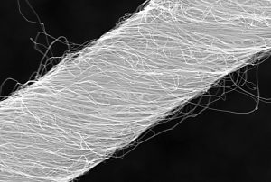
Microscopic image of carbon nanotube yarn.

Tapping into the autonomous nervous system – the control center for things like heartbeat and breathing – is a relatively new part of neurostimulation technologies to both record and direct organ function. Implants designed for stimulating peripheral nerves often fail because the protective tissue surrounding nerve bundles (the perineurium) is difficult to penetrate, and the body’s immune response often builds a scar around the implanted device.

Now, a team of scientists from Case Western Reserve University (CWRUL) has used carbon nanotubes to overcome these obstacles, reporting their findings in Scientific Reports. The authors, led by Dominique M. Durand, Ph.D., Director of the Neural Engineering Center and El Lindseth Professor of Biomedical Engineering, Neurosciences, Physiology and Biophysics at CWRU, fabricated yarn made of carbon nanotubes that was 10 to 20 µm in diameter. The yarn was then used to create electrodes, which were implanted into rats to monitor activity of the glossopharyngeal and vagus nerves.

The authors found that they could use the implants to monitor nerve activity under conditions of hypoxia and stomach distention. They report that the success of their experiments likely derives from the similarity of the nanotube yarn to the actual neural tissue surrounding the implant. The implants are a long way from being tried in humans, but the large number of functions controlled by just these two nerves indicates that such implants could find use in an enormous number of diseases.

Better Screening of Nanoparticle Delivery

As we discussed last week, the development of gene-based therapies is hindered by the sheer size of the human genome. The immense volume of information involved can quickly become difficult to manage, so one way in which scientists “keep track” of genetic information during the process of introducing new genetic material into an organism is DNA barcoding. This process attaches a small piece of DNA to the gene being studied; if and when the gene causes cells to replicate, these cells will bear the barcode, thus allowing the observer to be certain of the gene identities the whole time.

Seeking to determine whether DNA barcoding of lipid nanoparticles for injection into living models would outperform in vitro testing, a team of investigators at Georgia Tech and Emory University conducted a comparison of the two techniques, reporting their findings in Nano Letters. The authors, led by James Dahlman, Ph.D., Assistant Professor of Biomedical Engineering at GT/Emory, found that in vitro testing did not predict in vivo delivery. Further, they were able to track several dozen barcodes delivered by nanoparticles to eight different cell lines.

The authors believe that their technique, which they call JOint Rapid DNA Analysis of Nanoparticles (JORDAN), is superior to in vitro screening of nanoparticles to predict successful transplantation. They are offering JORDAN online as open source software so other scientists can use the technology to more accurately screen nanomaterials.

Synthetic Biologists Create Gene Circuits

Among the many types of molecules that regulate genetic expression in the body are microRNAs, non-coding strands of RNA that are responsible for gene silencing and other forms of gene expression regulation. The ability to harness and control the functions of microRNAs could have important implications for disease prevention and treatment.

In a recent article in Systems Biology and Applications, researchers at the University of Texas, Dallas, report on their engineering of a microRNA-based genetic circuit and its deployment in living cells. They created the circuit using strands of RNA from a variety of organisms, including viruses and jellyfish. The authors, led by Leonidas Bleris, Ph.D., Associate Professor of Bioengineering at UT Dallas, used the circuits to better understand how microRNAs change gene expression under different conditions.

More importantly, the authors found that their circuit had the ability to outproduce types of gene expression, which decreased as the number of gene replications increased.  The authors believe that their discoveries could have applications in a number of genetic disorders.

Discouraging Smoking at the Level of the Brain

Cigarette smoking is the single greatest contributor to negative health outcomes in the population.  Nicotine addiction often appears during the teenage years, and aggressive advertising has been used for the last couple of decades to encourage people to quit smoking and younger people not to start. Despite the widespread use of advertising to change human behavior, remarkably little is known on how the brain responds to advertising messages.

Danielle S. Bassett, Ph.D., Eduardo D. Glandt Faculty Fellow and Associate Professor of Bioengineering at Penn, recently collaborated with faculty from Penn’s Annenberg School of Communication to determine the neuroscience underlying this outcome. The collaborators showed graphic warning labels to a cohort of smokers while they were subjected to functional magnetic resonance imaging, which images brain activity during specific tasks. They found that smokers whose brains showed greater coherence between regions in the valuation network were more likely to quit smoking. Determining why these brain regions acted as they did could yield even more effective smoking-cessation messaging.

Purdue Startup Working to Expand MRI

Engineers at Purdue, including Zhongming Liu, Ph.D., Assistant Professor of Biomedical Engineering and Electrical and Computer Engineering, have cofounded at startup company, called MR-Link, to develop and produce a coin-sized device that can be inserted into MRI machines, allowing them to perform multiple scans simultaneously.

The device could be useful in reducing the amount of electromagnetic force to which patients are exposed during an MRI scan. In addition, Dr. Liu and his colleagues believe the device will cost perhaps less than a tenth of what similar devices currently cost. Given the widespread use of MRI, the device could ultimately impact how a number of diseases and disorders are diagnosed and tracked.

A Call to Understand Brain Network Mechanisms of Mental Disorders

The sheer complexity of the human brain means that, despite the tremendous advances made in neuroscience, there is still much we don’t know about what goes on inside our heads and how it goes awry in mental disorders. Even with the most advanced techniques, much of what we’ve learned about the brain is descriptive — telling that something is different between health and unhealthy function — but not why that something is different or how we could change it.

mental disorders
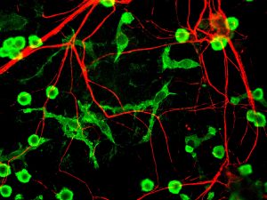
Rat microglia and neurons stained for different proteins

Among the approaches that have provided important insights into these questions is network science, which seeks to understand the brain as a complex system of multiple interacting components. Now, in a review published recently in Neuron, Danielle Bassett, Ph.D., Eduardo D. Glandt Faculty Fellow and Associate Professor of Bioengineering, and Richard Betzel, Ph.D., a postdoc in Dr. Bassett’s lab, have collaborated with scientists from the University of Heidelberg in Germany. The review covers a broad range of discoveries and innovations, moving from earlier, two-dimensional approaches to understanding the brain, such as graph theory, to newer approaches including multilayer networks, generative network models, and network control theory.

“Stating what is different in brain networks of individuals with disorders of mental health is not the same as identifying why” says Bassett. “Here we propose that emerging tools from network science can be used to identify true mechanisms of mental health disorders, and bridge molecular and genetic mechanisms through brain physiology, thus informing interventions in the form of pharmacological manipulations and brain stimulation.”

Brain Network Control Emerges over Childhood and Adolescence

network control


The developing human brain contains a cacophony of electrical and chemical signals from which emerge the powerful adult capacities for decision-making, strategizing, and critical thinking. These signals support the trafficking of information across brain regions, in patterns that share many similarities with traffic patterns in railway and airline transportation systems. Yet while air traffic is guided by airport control towers, and railway routes are guided by signal control rooms, it remains a mystery how the information traffic in the brain is guided and how that guidance changes as kids grow.

In part, this mystery has been complicated by the fact that, unlike transportation systems, the brain is not hooked up to external controllers. Control must happen internally. The problem becomes even more complicated when we think about the sheer number of routes that must exist in the brain to support the full range of human cognitive capabilities. Thus, the controllers would need to produce a large set of control signals or use different control strategies. Where internal controllers might be, how they produce large variations in routing, and whether those controllers and their function change with age are important open questions.

A recent paper published in Nature Communications – a product of collaboration among the Departments of Bioengineering and Electrical & Systems Engineering at the University of Pennsylvania and the Department of Psychiatry of Penn’s Perelman School of Medicine – offers some interesting answers. In their article, Danielle Bassett, Ph.D., Eduardo D. Glandt Faculty Fellow and Associate Professor in the Penn BE Department, Theodore D. Satterthwaite, M.D., Assistant Professor in the Penn Psychiatry Department, postdoctoral fellow Evelyn Tang, and their colleagues suggest that control in the human brain works in a similar way to control in man-made robotic and other mechanical systems. Specifically, controllers exist inside each human brain, each region of the brain can perform multiple types of control, and this control grows as children grow.

As part of this study, the authors applied network control theory — an emerging area of systems engineering – to explain how the pattern of connections (or network) between brain areas directly informs the brain’s control functions. For example, hubs of the brain’s information trafficking system (like Grand Central Station in New York City) show quite different capacities for and sensitivities to control than non-hubs (like Newton Station, Kansas). Applying these ideas to a large set of brain imaging data from 882 youths in the Philadelphia area between the ages of 8 and 22 years old, the authors found that the brain’s predicted capacity for control increases over development. Older youths have a greater predicted capacity to push their brains into nearby mental states, as well as into distant mental states, indicating a greater potential for diversity of mental operations than in younger youths.

The investigators then asked whether the principles of network control could explain the specific manner in which connections in the brain change as youths age. They used tools from evolutionary game theory – traditionally used to study Darwinian competition and evolving populations in biology – to ‘evolve’ brain networks in silico from their 8-year old state to their 22-year-old state. The results demonstrated that the optimization of network control is a principle that explains the observed changes in brain connectivity as youths develop over childhood and adolescence. “One of the observations that I think is particularly striking about this study,” Bassett says, “is that the principles of network controllability are sufficient to explain the observed evolution in development, suggesting that we have identified a quintessential rule of developmental rewiring.”

This research informs many possible future directions in scientific research. “Showing that network control properties evolve during adolescence also suggests that abnormalities of this developmental process could be related to cognitive deficits that are present in many neuropsychiatric disorders,” says Satterthwaite. The discovery that the brain optimizes certain network control functions over time could have important implications for better understanding of neuroplasticity, skill acquisition, and developmental psychopathology.

Lagrange Goes to Dani Bassett

Lagrange
Danielle Bassett, Ph.D.

Danielle S. Bassett, Eduardo D. Glandt Faculty Fellow and Associate Professor in the University of Pennsylvania’s Department of Bioengineering, is the recipient of the 2017 Lagrange-CRT Foundation Prize. The prize, given by the Institute for Scientific Interchange Foundation in Turin, Italy, was created to encourage and honor researchers working in the field of complex systems.

Complex systems feature many interconnected parts whose individual behavior influences the outcomes of the whole. Examples include social media networks, ecological webs, stock markets, and in Bassett’s case, the brain. Her research maps and analyzes the networks of neurons that enable all manners of cognitive abilities, as well as how those networks evolve during development or malfunction in disease.

The prize comes with an award of €50,000, or roughly $60,000. It will be formally presented to Bassett at a ceremony in Turin next week. Bassett is the first woman to be the sole recipient of the prize since its inception in 2008. Lada Adamic won it alongside Xavier Gabaix in 2012.

Read more at the SEAS blog on Medium.

Mind Control and an Ethical Appeal

mind control brain
A “wiring diagram of the human brain,” produced using diffusion MRI scans of the brain.

A group of four scholars from the University of Pennsylvania, including Bioengineering professor Danielle Bassett, have issued a call in the journal Nature Human Behaviour for greater safeguards for patients as treatments in the field of neuroscience evolve and come ever closer to resembling “mind control.”

“While we don’t believe,” Bassett said, “that the science-fiction idea of mind control, totally overriding a person’s autonomy, will ever be possible, new brain-focused therapies are becoming more specific, targeted and effective at manipulating individuals’ mental states. As these techniques and technologies mature, we need systems in place to make sure they are applied such that they maximize beneficial effects and minimize unwanted side effects.”

Read more at the Penn News Web Site.

Bassett on Improvements in Executive Function

executive function bassett
Danielle Bassett, Ph.D.

Danielle Bassett, Eduardo D. Glandt Faculty Fellow and Associate Professor in the departments of Bioengineering and Electrical and Systems Engineering, recently collaborated with colleagues from the Perelman School of Medicine on a study that looks at how brain networks change as children develop into adolescence. Bassett’s previous work on applying network science principles to neuroscience has suggested that the organization of these networks helps lead to “cognitive control” and that they reorganize as children age, improving executive function.

In a new paper published in Current Biology, Bassett and her colleagues delve deeper into the network changes that lead to this improvement.

“The work,” Bassett says, “significantly extends our understanding of the role of modular network organization in development, and its importance for executive function.”

Center for Curiosity Partners with Bioengineering

by Perry Zurn and Dani Bassett

Do not stop to think about the reasons for what you are doing, about why you are questioning. The important thing is not to stop questioning. Curiosity has its own reasons for existence. One cannot help but be in awe when he contemplates the mysteries of eternity, of life, of the marvelous structure of reality. It is enough if one tries merely to comprehend a little of this mystery each day. Never lose a holy curiosity.

–Albert Einstein1

This haunting passage prompts a series of difficult questions. Should we ever worry about where our curiosity goes? Is it true that curiosity is an end in itself? Or, are its justifications so obvious to us as to go unquestioned? Have we lost our sense of mystery? What makes curiosity holy? Einstein himself did not study curiosity, nor could he revolutionize the field of curiosity studies, which is just coming into its own today. But he does capture the compulsion of curiosity and its tantalizing promise.

Curious 1
Kushal Sacheti, Founder and Director of the Center for Curiosity

The Center for Curiosity was established in New York in 2014 by Kushal Sacheti, a diamond merchant who was formerly an engineer. Its mission is to advance both the academic study of curiosity and the public practice of curiosity. A year after its founding, the first of its satellite centers was established at the University of Pennsylvania, in the School for Social Policy and Practice, under the leadership of Dean John Jackson, Jr. It is here that Mr. Sacheti’s dream of uniting engineering and curiosity came alive.

Given her work on the network neuroscience of human learning, Dr. Danielle Bassett, Associate Professor of Bioengineering, was one of the first faculty spotlighted in Penn’s Center for Curiosity seminar series. Her talk, “Flexible Brain Network Dynamics During Learning,” so perfectly represented the Center’s mission that she was quickly appointed to its advisory board. Shortly thereafter, Dr. Bassett invited the Center’s two postdoctoral fellows, Dr. Arjun Shankar and Dr. Perry Zurn, to lead curiosity workshops at the 2016 Penn Network Visualization program. This program provides young artists the opportunity to understand and creatively reimagine network science. Dr. Zurn’s seminar on structural models of curiosity, coupled with Dr. Shankar’s workshop on the affective elements of curiosity, inspired program fellows to explore curiosity not only in network science, but also in their own artistic praxis.

Curious 3a
Dr. Arjun Shankar, Center for Curiosity, Postdoctoral Fellow
Curious 3b
Dr. Perry Zurn, Center for Curiosity, Postdoctoral Fellow
Curious 4
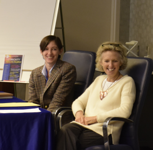
Dr. Danielle Bassett (left) and Dr. Susan Engel (right) at the Curiosity Across the Disciplines Symposium, December 9, 2016


Behind Dr. Bassett’s Network Visualization program is a passion for thinking between the arts and sciences and a conviction that they are richer enterprises together. An even broader commitment to interdisciplinarity energizes Penn’s Center for Curiosity. Last December, Drs. Zurn and Shankar organized the Curiosity Across the Disciplines symposium. This day-long event explored the concept of curiosity across major academic disciplines (history, medicine, ecology, neuroscience, psychology, education, anthropology, comparative literature, ethnic studies, political philosophy, and film). As presenters (including Dr. Bassett) reflected on their fields’ contributions to curiosity studies, as well as the role of curiosity in their own scholarship, a deeper, shared conversation emerged about how curiosity can help us to collectively navigate the scientific, educational, and political challenges of our times.

The collaboration between Penn’s Center for Curiosity and the Department of Bioengineering has really only begun. This fall, Drs. Zurn and Bassett are co-organizing a symposium on The Network Neuroscience of Curiosity. Speakers will include Dr. Danielle Bassett, Dr. David Danks (Carnegie Mellon University), Dr. Jacqueline Gottlieb (Columbia University), and Dr. Celeste Kidd (University of Rochester). And, as a long-term project, they have started a conversation about reinvigorating the Bioengineering curriculum with an emphasis on student curiosity and creativity. Sharing Penn’s commitment to community outreach, moreover, the Center for Curiosity and Department of Bioengineering are also in conversation with Westtown School about building an art- and science-centered curiosity initiative there.

If indeed one cannot help but be curious about life and its mysterious design, that journey is perhaps best undertaken together—Einstein’s fabled solipsism notwithstanding. This exciting new partnership at Penn is yet another step in that direction.

1 Albert Einstein, Statement to William Miller, as quoted in LIFE magazine (2 May 1955); reprinted in Joseph S. Willis, Finding Faith in the Face of Doubt: A Guide for Contemporary Seekers (Quest Books, 2001), 58; and William Hermanns, Einstein and the Poet: In Search of the Cosmic Man (1983; Brandon Books, 2013), 138.

Allen Foundation Awards Major Grant to Study Concussions

Faculty members in the Department of Bioengineering at the University of Pennsylvania are among the recipients of a major $9.25 million grant from the Paul G. Allen Family Foundation to study the mechanism underlying concussion and to investigate possible interventions.

allen foundation meaneyallen foundation smith


David Meaney, PhD, Solomon R. Pollack Professor and Chair of the Bioengineering Department (above left), is one of two principal investigators, with Douglas H. Smith, MD,  professor of neurosurgery at Penn’s Perelman School of Medicine (above right). In addition, Danielle S. Bassett, PhD, Eduardo D. Glandt Faculty Fellow and Associate Professor (below left), Dongeun (Dan) Huh, PhD, Wilf Family Term Assistant Professor (below center), and David Issadore, PhD, assistant professor (below right), all of BE Department, are co-investigators. The Allen Foundation grant also involves investigators from Columbia University (Barclay Morrison, Ph.D.), Duke University (Cameron Bass, Ph.D.), and Children’s Hospital of Philadelphia (Akiva Cohen, Ph.D.).

allen foundation bassettallen foundation huhallen foundation issadore

Selected from a large national pool of applicants, the Allen Foundation grant will bring together new technology platforms developed by Drs. Huh and Issadore to study how concussions occur at the microtissue scale and release markers of rewiring  during recovery. Network theory models from Dr. Bassett’s group will provide an entirely new view on how concussion recovery occurs at all scales in the brain. The overall impact of the project will be to move away from the widely held perspective that all concussions should be treated identically and towards a view that concussions can follow several recovery pathways, some of which must be monitored closely in the days to weeks following injury.