Bioengineering Round-Up (October 2019)

by Sophie Burkholder

Innovations in Advancing a Cure for Diabetes

The blue circle is the global symbol for diabetes. Wikimedia Commons.

Diabetes is one of the more common diseases among Americans today, with the American Diabetes Association estimating that approximately 9.5 percent of the population battles the condition today. Though symptoms and causes may vary across types and patients, diabetes generally results from the body’s inability to produce enough insulin to keep blood sugar levels in check. A new experimental treatment from the lab of Sha Jin, Ph.D., a biomedical engineering professor at Binghamton University, aims to use about $1.2 million in recent federal grants to develop a method for pancreatic islet cell transplantation, as those are the cells responsible for producing insulin.

But the catch to this new approach is that relying on healthy donors of these islet cells won’t easily meet the vast need for them in diabetic patients. Sha Jin wants to use her grants to consider the molecular mechanisms that can lead pluripotent stem cells to become islet-like organoids. Because pluripotent stem cells have the capability to evolve into nearly any kind of cell in the human body, the key to Jin’s research is learning how to control their mechanisms and signaling pathways so that they only become islet cells. Jin also wants to improve the eventual culture of these islet cells into three-dimensional scaffolds by finding ways of circulating appropriate levels of oxygen to all parts of the scaffold, particularly those at the center, which are notoriously difficult to accommodate. If successful in her tissue engineering efforts, Jin will not only be able to help diabetic patients, but also open the door to new methods of evolving pluripotent stem cells into mini-organ models for clinical testing of other diseases as well.

A Treatment to Help Others See Better

Permanently crossed eyes, a medical condition called strabismus, affects almost 18 million people in the United States, and is particularly common among children. For a person with strabismus, the eyes don’t line up to look at the same place at the same time, which can cause blurriness, double vision, and eye strain, among other symptoms. Associate professor of bioengineering at George Mason University, Qi Wei, Ph.D., hopes to use almost $2 million in recent funding from the National Institute of Health to treat and diagnose strabismus with a data-driven computer model of the condition. Currently, the most common method of treating strabismus is through surgery on one of the extraocular muscles that contribute to it, but Wei wants her model to eventually offer a noninvasive approach. Using data from patient MRIs, current surgical procedures, and the outcomes of those procedures, Wei hopes to advance and innovate knowledge on treating strabismus.

A Newly Analyzed Brain Mechanism Could be the Key to Stopping Seizures

Among neurological disorders, epilepsy is one of the most common. An umbrella term for a lot of different seizure-inducing conditions, many versions of epilepsy can be treated pharmaceutically. Some, however, are resistant to the drugs used for treatment, and require surgical intervention. Bin He, Ph. D., the Head of the Department of Biomedical Engineering at Carnegie Mellon University, recently published a paper in collaboration with researchers at Mayo Clinic that describes the way that seizures originating at a single point in the brain can be regulated by what he calls “push-pull” dynamics within the brain. This means that the propagation of a seizure through the brain relies on the impact of surrounding tissue. The “pull” he refers to is of the surrounding tissue towards the seizure onset zone, while the “push” is what propagates from the seizure onset zone. Thus, the strength of the “pull” largely dictates whether or not a seizure will spread. He and his lab looked at different speeds of brain rhythms to perform analysis of functional networks for each rhythm band. They found that this “push-pull” mechanism dictated the propagation of seizures in the brain, and suggest future pathways of treatment options for epilepsy focused on this mechanism.

Hyperspectral Imaging Might Provide New Ways of Finding Cancer

A new method of imaging called hyperspectral imaging could help improve the prediction of cancerous cells in tissue specimens. A recent study from a University of Texas Dallas team of researchers led by professor of bioengineering Baowei Fei, Ph.D., found that a combination of hyperspectral imaging and artificial intelligence led to an 80% to 90% level of accuracy in identifying the presence of cancer cells in a sample of 293 tissue specimens from 102 patients. With a $1.6 million grant from the Cancer Prevention and Research Institute of Texas, Fei wants to develop a smart surgical microscope that will help surgeons better detect cancer during surgery.

Fei’s use of hyperspectral imaging allows him to see the unique cellular reflections and absorptions of light across the electromagnetic spectrum, giving each cell its own specific marker and mode of identification. When paired with artificial intelligence algorithms, the microscope Fei has in mind can be trained to specifically recognize cancerous cells based on their hyperspectral imaging patterns. If successful, Fei’s innovations will speed the process of diagnosis, and potentially improve cancer treatments.

People and Places

A group of Penn engineering seniors won the Pioneer Award at the Rothberg Catalyzer Makerthon led be Penn Health-Tech that took place from October 19-20, 2019. SchistoSpot is a senior design project created by students Vishal Tien (BE ‘20), Justin Swirbul (CIS ‘20), Alec Bayliff (BE ‘20), and Bram Bruno (CIS ‘20) in which the group will design a low-cost microscopy dianostic tool that uses computer vision capabilities to automate the diagnosis of schistosomiasis, which is a common parasitic disease. Read about all the winners here.

Virginia Tech University will launch a new Cancer Research Initiative with the hope of creating an intellectual community across engineers, veterinarians, biomedical researchers, and other relevant scientists. The initiative will focus not only on building better connections throughout departments at the university, but also in working with local hospitals like the Carilion Clinic and the Children’s National Hospital in Washington, D.C. Through these new connections, people from all different areas of science and engineering and come together to share ideas.

Associate Professor of Penn Bioengineering Dani Bassett, Ph.D., recently sat down with the Penn Integrates Knowledge University Professor Duncan Watts, Ph.D., for an interview published in Penn Engineering. Bassett discusses the origins of network science, her research in small-world brain networks, academic teamwork, and the pedagogy of science and engineering. You can read the full interview here.

An all-female group of researchers from Northern Illinois University developed a device for use by occupational therapists that can capture three-dimensional images of a patient’s hand, helping to more accurately measure the hand or wrist’s range of motion. The group presented the abstract for their design at this year’s meeting of the Biomedical Engineering Society here in Philadelphia, where Penn students and researchers presented as well.

Penn Researchers’ Model Optimizes Brain Stimulation Therapies, Improving Memory in Tests

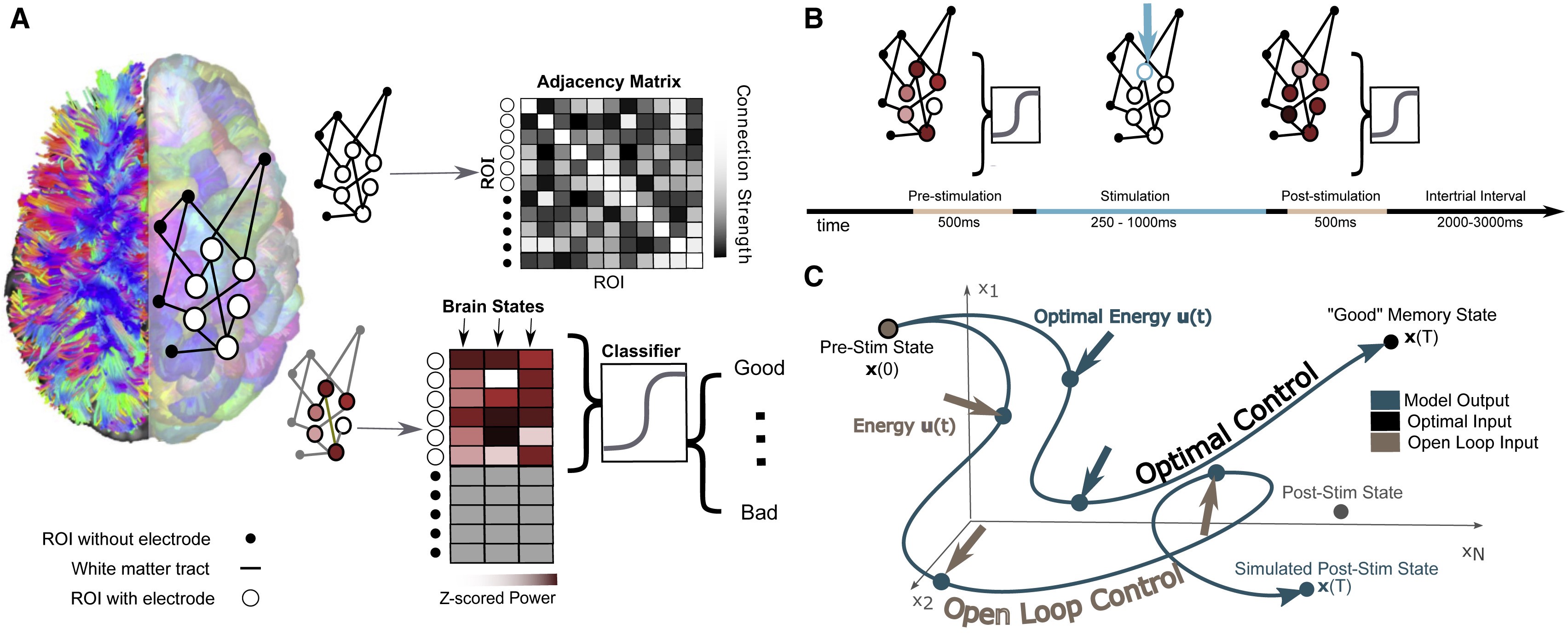
The researchers’ model involves mapping the connections between different regions of an individual’s brain while they performed a basic memory task, then using that data to predict how electrical stimulation in one region would affect activity throughout the network. Individuals’ improved performance on the same memory task after stimulation suggests the model could eventually be generalized toward a variety of stimulation therapies.

Brain stimulation, where targeted electrical impulses are directly applied to a patient’s brain, is already an effective therapy for depression, epilepsy, Parkinson’s and other neurological disorders, but many more applications are on the horizon. Clinicians and researchers believe the technique could be used to restore or improve memory and motor function after an injury, for example, but progress is hampered by how difficult it is to predict how the entire brain will respond to stimulation at a given region.

In an effort to better personalize and optimize this type of therapy, researchers from the University of Pennsylvania’s School of Engineering and Applied Science and Perelman School of Medicine, as well as Thomas Jefferson University Hospital and the University of California, Riverside, have developed a way to model how a given patient’s brain activity will change in response to targeted stimulation.

To test the accuracy of their model, they recruited a group of study participants who were undergoing an unrelated treatment for severe epilepsy, and thus had a series of electrodes already implanted in their brains. Using each individual’s brain activity data as inputs for their model, the researchers made predictions about how to best stimulate that participant’s brain to improve their performance on a basic memory test.

The participants’ brain activity before and after stimulation suggest the researchers’ models have meaningful predictive power and offer a first step towards a more generalizable approach to specific stimulation therapies.

Danielle Bassett and Jennifer Stiso.

The study, published in the journal Cell Reports, was led by Danielle Bassett, J. Peter Skirkanich Professor in Penn Engineering’s Department of Bioengineering, and Jennifer Stiso, a neuroscience graduate student in Penn Medicine and a member of Bassett’s Complex Systems Lab.

Read the full post on the Penn Engineering Medium blog. Media contact Evan Lerner.

Congratulations to Danielle Bassett and Arjun Raj on Their Promotions to Full Professors

We would like to congratulate Penn Bioengineering faculty members Arjun Raj, Ph.D., and Danielle Bassett, Ph.D., on their promotions from associate to full professors.

Arjun Raj, Ph.D.

The Raj lab studies how biological processes work at the level of individual cells. Their work combines quantitative tools from genomics, imaging, biology, math, and computer science to develop models for how individual cells function, and in particular, how these individual cells can behave differently from each other. One major interest is in cancer, in which the lab is studying how individual cells can drive resistance to anti-cancer drugs. He and his lab also have a regularly updated blog discussing general topics related to scientific academia.

Danielle Bassett, Ph.D.

The Bassett lab takes an in-depth look at the use of network science and complex systems theory to study computational neuroscience in projects that involve the architecture of knowledge networks, the controllability of brain networks, and the dynamic networks in neuroscience. This fall, she will teach an elective course in network neuroscience open to graduate and undergraduate students that covers the use of network science in understanding overall brain circuitry. Bassett was recently profiled in Science Magazine.

Penn Researchers Detect Brain Differences between Fast and Slow Learners

By Lauren Salig

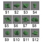
These 12 object-number value pairs were taught to the participants, who had to properly learn the associations to succeed in value judgement tests. The researchers investigated the differences in their brain activity patterns to see why some were faster learners than others.

Why do some people naturally excel at learning instruments, languages or technology while others take longer to pick up new knowledge? Learning requires the brain to encode information, changing its neural “wiring” and creating networks between brain regions.

In a new study, researchers at the University of Pennsylvania’s School of Engineering and Applied Science and the Max Planck Institute for Dynamics and Self-Organization in Germany looked at how brain activation patterns might affect how long it takes for new information to really stick in the brain.

Earlier research has suggested that part of what might slow down learners is over-thinking. A 2015 study led by Danielle Bassett, Eduardo D. Glandt Faculty Fellow and associate professor in the Department of Bioengineering, showed a correlation between slow learning and cognitive control: the brain’s ability to regulate itself by activating the necessary networks and inhibiting unnecessary activity. In that study, when people unnecessarily engaged parts of the brain linked to cognitive control, they were more likely to take longer to learn a simple task.

But beyond what might make an individual learn more slowly, the researchers want to know what sort of geometric patterns of brain activity make for better learning.

Evelyn Tang and Danielle Bassett

Their new study was led by Bassett and Evelyn Tang, who was an Africk Family Postdoctoral Fellow in Bassett’s Complex Systems Lab before starting at the Max Planck Institute this fall. Sharon Thompson-Schill, Christopher H. Browne Distinguished Professor and chair of Psychology, also contributed to the study.

The study was published in the journal Nature Neuroscience.

Read the full story at the Penn Engineering Medium Blog.

BE’s Danielle Bassett Profiled in Science Magazine

Danielle Bassett, PhD

Danielle Bassett, Eduardo D. Glandt Faculty Fellow and Associate Professor in theDepartment of Bioengineering, grew up in central Pennsylvania where she and her 10 siblings were homeschooled. Back then, Bassett had aspirations to become a professional pianist, a dream shattered by stress fractures in her arm at age 16.

Now, Bassett is a renowned physicist and MacArthur fellow who has pushed the field of network science, which studies connections and interactions between parts of a whole, to new realms. Bassett’s research focuses on brain function, including work on how brains of people with schizophrenia are organized, how brain communication changes with learning, and how the brain is able to switch between tasks.

Kelly Servick of Science sat down with Bassett to talk through her incredible journey from child pianist to leading network scientist:

““By 17, discouraged by her parents from attending college and disheartened at her loss of skill while away from the keys, she expected that responsibilities as a housewife and mother would soon eclipse any hopes of a career. ‘I wasn’t happy with that plan,’ she says.

Instead, Bassett catapulted herself into a life of research in a largely uncharted scientific field now known as network neuroscience. A Ph.D. physicist and a MacArthur fellow by age 32, she has pioneered the use of concepts from physics and math to describe the dynamic connections in the human brain. ‘She’s now the doyenne of network science,’ says theoretical neuroscientist Karl Friston of University College London. ‘She came from a formal physics background but was … confronted with some of the deepest questions in neuroscience.’”

Continue reading about Bassett’s career path and evolving research interests at Science.

Reposted from the Penn Engineering Medium blog. Media contact Evan Lerner.

Week in BioE: April 19, 2019

by Sophie Burkholder

New Vascularized Patches Could Help Patient Recovery from Heart Attacks

Heart attacks are the result of a stoppage of blood flow to the heart – an interruption to normal function that can result in severe tissue damage, or even tissue death. This loss of healthy tissue function is one of the biggest challenges in treating patients that undergo heart attacks, as the damaged tissue increases their risk of having future attacks. One of the main solutions to this issue right now is the creation of cardiac tissue scaffolds using stem cells to create a platform for new and healthy tissue to grow in vivo. A group of biomedical engineers at Michigan Technological University hopes to expand on this basis by focusing not just on cellular alignment in the scaffold but on that of microvessels too. Led by Feng Zhao, Ph.D., Associate Professor of Biomedical Engineering, the team hopes that this new attention on microvessel organization will improve the vasculature of the scaffolds, and thus improve the success of the scaffolds in vivo, allowing for a better recovery from heart attacks.

Some Stem Cells May Be More Fit Than Others

Stem cells are one of the hottest research areas in the field of bioengineering today. Widely known as the cells in the human embryo that have the ability to eventually transform into specific cells for the brain, lung, and every other organ, stem cells are also of recent interest because researchers found ways to reverse this process, transforming organ-specific cells back to the pluripotent stem cell level. This achievement however, is mostly applicable to individual stem cells, and doesn’t fully encapsulate the way this process might work on a larger population level. So Peter Zandstra, Ph. D., a bioengineering faculty member at the University of British Columbia, decided to research just that.

Using mouse embryonic fibroblasts (MEFs), Zandstra and his lab attempted to track the cells throughout their reprogramming, to more clearly trace each back to its respective parent population. Surprisingly, they found that after only one week of reprogramming, nearly 80% of the original cell population had been removed, meaning that most of the parent generation was not “fit” enough to undergo the process of reprogramming, indicating that perhaps some stem cells will have a better chance of survival in this process than others. This research may suggest that not all cells have the capacity to undergo reprogramming, as many researchers originally thought.

A New Microdevice Will Help Model Bronchial Spasms

The difficulty in breathing associated with asthma is the result of bronchial spasms, which are a kind of muscle contraction in the airways. But little was known about just how these spasms occurred in patients, so Andre Levchenko, Ph.D., Professor of Biomedical Engineering at Johns Hopkins, and his lab created a microdevice to model them. Calling the device a “bronchi on a chip,” Levchenko and his team used a microphysiological model to look at some of the biochemical and mechanical signals associated with these kinds of muscle contractions. They found that the contractions operate in a positive feedback system, so that those caused by disturbance from allergens will subsequently cause even more contractions to occur. But surprisingly, they also found that a second contraction, if triggered at the right time during the initial contraction, could actually stop the process and allow the muscles to relax. Because asthma is a notoriously difficult disease to translate from animal to human models, this new device opens the door to understanding different mechanisms of asthma before taking research to clinical trials.

New CHOP Research Center to Focus Research on Pediatric Airway Disorders

A new bioengineering lab at the Children’s Hospital of Philadelphia called the Center for Pediatric Airway Disorders will specialize in a variety of airway procedures for pediatric patients such as tracheal reconstruction and recurrent laryngeal nerve reinnervation. This new lab will be one of the first to give a unique focus to the application of bioengineering to pediatric laryngology. The interdisciplinary center brings together students and researchers from all different fields, including materials science and microbiology, to find new ways of repairing tissue and regenerating organs related to respiratory disorders. Specific areas of research will involve the modeling of children’s vocal cords, understanding the mechanisms of fibrosis, and improving surgical procedures.

Deeper Understanding of Sickle Cell Anemia Could Lead to New Treatments

Though sickle cell anemia is a common and well-known disease, a new study of its causes at the nanoscale level might reveal previously unknown information about the assembly of hemoglobin fibers. Using microscopes with the ability to visualize these molecules at such a small level, researchers at the University of Minnesota found that the beginning organizations that lead to sickle cell anemia are much less ordered than originally thought. Led by Associate Professor of Biomedical Engineering David Wood, Ph.D., the team of researchers used this higher level of microscopy to find that hemoglobin self-assembly process, which was originally thought to be 96% efficient, is actually only 4% efficient. Wood hopes that this new knowledge will help allow for the development of new and better treatments for patients with sickle cell anemia, as there are currently only two FDA-approved ones on the market.

People & Places

Penn Today asked five Penn researchers about the women in STEM who have been a source of inspiration and encouragement throughout their own careers. Their responses include active researchers who have paved the way for better inclusion in STEM and famous female scientists from the past who broke boundaries as they made strides with their research.

Dr. Danielle Bassett, the Eduardo D. Glandt Faculty Fellow and associate professor of bioengineering and electrical and systems engineering in the School of Engineering and Applied Science, has two heroes: “Ingrid Daubechies for her work on wavelets, or “little waves,” which are beautiful mathematical objects that can be used to extract hidden structure in complex data. “Also, Maryam Mirzakhani for inspiring a child to believe that mathematics is simply painting. Would that we all could see the world just that bit differently.”

Read the full story on Penn Today.

Joel Boerckel, Ph.D, Assistant Professor of Orthopaedic Surgery and Bioengineering

This week, we want to congratulate Joel Boerckel, Ph.D., Assistant Professor of Orthopaedic Surgery and Bioengineering, and his lab on receiving a second R01 Grant from the National Institute of Arthritis and and Musculoskeletal Skin Diseases for their work on defining the roles of YAP and TAZ in embryonic bone morphogenesis and mechanoregulation of fracture repair. Dr. Boerckel is a member of the McKay Orthopaedic Research Laboratory.

We would also like to congratulate Christopher Yip, Ph. D., on being appointed as the new dean of the University of Toronto’s Faculty of Applied Science and Engineering. A professor in both the Department of Chemical Engineering and Applied Chemistry the Institute of Biomaterials and Biomedical Engineering, Dr. Yip’s research involves the use of molecular imaging to understand the self-assembly of proteins.

Week in BioE (February 13, 2019)

by Sophie Burkholder

Bioengineers Tackle Heart Disease

Heart disease is currently the leading cause of death in the United States, resulting in about 630,000 deaths every year according to the Center for Disease Control. One of the most common side effects of heart disease is damage to blood vessels and cardiac tissue, which can ultimately lead to conditions like high blood pressure, arrhythmia, and even cardiac arrest. In serious cases of irreversible heart damage, often the only option for patients is a full heart transplant, and efforts to engineer vascularized cardiac tissue grafts have proved challenging in research so far.

But researchers Ying Zheng, Ph.D., and Charles Murry, M.D., Ph.D., both of whom have joint appointments in Bioengineering at the University of Washington, have found success in using human microvascular grafts to create working blood vessels in vitro to treat infarcted rat hearts. The new heart muscle, developed from human embryonic stem cell-derived endothelial cells in petri dishes, was grown with a focus on not only being able to easily integrate it in vivo, but also in creating a patch of vasculature that closely mirrored that of the heart. In concentrating more on the mechanical aspects of the blood vessel network, Zheng and Murry were able to better restore normal blood flow to the damaged rat hearts after integration of the grafts. The study appears in a recent edition of Nature Communications.

Another team of bioengineers, led by Michael Sacks, Ph.D. at the University of Texas at Austin, recently invented a software-based method for repairing mitral valves in the heart. Their work, published in the International Journal for Numerical Methods in Biomedical Engineering, uses computational modeling techniques to create a noninvasive way of simulating repairs to the mitral valve, which will allow for a better prediction of surgical procedures and postoperative side effects on a more patient-specific basis. This ability to know which treatment plan may be best-suited for a given patient is important especially for valve repair, as heart valves are notoriously difficult to model or image due to the complexity of their functions. But through the use of advanced technology in 3D echocardiography, Sacks and his team say that their new model is accurate enough to rely on in clinical settings.

Virtual Reality Assists in the Evaluation of Surgery

Any form of surgery is always a high risk procedure, as it is subject to a wide variety of sources of human error and irregularity, even with the best surgeons. Certainly, there should be a system in place to not only continually assess the knowledge of surgeons throughout their careers, but also to evaluate their practices and techniques during operation. Such an evaluation, however, would put patients at risk during the assessment of the surgeon.

But now a team of researchers from Rensselaer Polytechnic Institute has developed a way of simulating colorectal surgical procedures using virtual reality technology. Suvranu De, Sc.D. — the J. Erik Jonsson ‘22 Distinguished Professor of Engineering and Head of the Department of Mechanical, Aerospace and Nuclear Engineering with joint appointments in Biomedical Engineering and Information Technology and Web Science —leads the project which incorporates both visual and tactile feedback for users to employ as a tool for both training and evaluating colorectal surgeons. While virtual reality simulators have been used for similar applications related to procedures like the colonoscopy, they have yet to be fully developed for open surgical procedures, because of the difficulties in creating a fully engaged and immersive environment. Nonetheless, De and his team hope that their work will lead to the creation of the first “Virtual Intelligent Preceptor,” which will allow for more advanced technological innovations in aspects of surgical education that have so far been difficult to standardize. Support for the project comes from the National Institute of Biomedical Imaging and Bioengineering (NBIB).

Penn BE’s Dr. Bassett on Understanding Knowledge Networks in the Brain

Dr. Danielle Bassett, Ph.D., Eduardo D. Glandt Faculty Fellow and Associate Professor of Bioengineering

As a network neuroscientist, Danielle Bassett, Ph.D., Eduardo D. Glandt Faculty Fellow and Associate Professor in the Department Bioengineering, brings together insights from a variety of fields to understand how the brain’s connections form and change: mathematics, physics, electrical engineering and developmental biology, to name a few. Bassett’s recent work on the learning process also draws from linguistics, educational theory and other domains even further afield.

The intersection and interaction of knowledge from multiple sources doesn’t just describe Bassett’s methodology; it’s at the heart of her research itself. At the Society for Industrial and Applied MathematicsAnnual Meeting last year, Bassett provided an address on how the structure of knowledge networks can influence what our brains can do when it comes to learning new things.

Read the full story on Penn Engineering’s Medium blog.

People & Places:

Tammy Dorsey, a graduate student at Wichita State University, created a non-invasive in utero tool to help read the oxygen levels of unborn babies as part of her senior design project. Dorsey says the inspiration for the project came from complications during the birth of her middle child, who despite having a normal heart rate throughout the entire pregnancy, was born blue. The device Dorsey created uses measurements of the baby’s pH to read fetal oxygen levels. She hopes that the design will help doctors better detect when a fetus is in distress during pregnancy and childbirth.

The field of bioengineering is constantly growing, and new programs are always in development. Boise State University has announced the launch of a new doctoral program in bioengineering that will begin in the fall of 2019. Developed through the collaboration of the university’s College of Health Sciences, College of Engineering, Graduate College, and College of Arts and Sciences, this new opportunity to do research in the field of bioengineering will have three study tracks available in biomechanics, mechanobiology, and human performance.

The new biomedical engineering department at the University of Massachusetts Amherst has announced the department’s first faculty appointments. The founding department head will be Professor Tammy L. Haut Donahue, Ph.D., whose research focus is on the biomechanics of the musculoskeletal system. Another professor joining the department’s new faculty is Seth W. Donahue, Ph.D., who has also done research in the field of biomechanics, and specifically how it pertains to tissue regeneration.

Since we last posted, there have also been several significant academic appointments in the field of Bioengineering. This week, we would like to congratulate Bruce Tromberg, Ph.D., on his appointment as the director of the National Institute of Biomedical Imaging and Bioengineering (NIBIB). Dr. Tromberg is currently a Professor with appointments in Biomedical Engineering and Surgery at the University of California at Irvine, where he leads research in bioimaging and biophotonics. He has also served on the External Advisory Board of NIH P41 Center for Magnetic Resonance and Optical Imaging here at Penn since 2009, and has also given several lectures here on his work in bioimaging.

Secondly, we congratulate the University of Toronto’s Professor Warren Chan, Ph.D., who was recently named as a Tier 1 Canada Research Chair in Nanobioengineering. Professor Chan, who is also the director of the Institute of Biomaterials and Biomedical Engineering at the University of Toronto, conducts research in the field of nanotechnology for applications in the treatment and diagnosis of cancer and viral diseases.

And finally, we also want to congratulate Frank Pintar, Ph.D., on his appointment as the Founding Chair of the Marquette University and Medical College of Wisconsin. Dr. Pintar’s research in bioengineering involves the study of the biomechanics involved with brain and spinal cord injury, with a focus on motor vehicle crash trauma.

Week in BioE (July 31, 2018)

New Data Analysis Methods

Like many other fields, biomedical research is experiencing a data explosion. Some estimates suggest that the amount of data generated from the health sciences is now doubling every eighteen months, and experts expect it to double every seventy-three days by 2020.  One challenge that researchers face is how to meaningfully analyze this data tsunami.

The challenge of interpreting data occurs at all scales, and a recent collaboration shows how new approaches can allow us to handle the volumes of data emerging at the level of individual cells to infer more about how biology “works” at this level.  Wharton Statistics Department researchers Mo Huang and Jingshu Wang (PhD Student and Postdoctoral Researcher, respectively) collaborated with Arjun Raj’s lab in Bioengineering and published their findings in recent issues of Nature Methods and Proceedings of the National Academy of Sciences.  One study focused on a de-noising technique called SAVER to provide more precise data from single cell experiments and significantly improves the ability to detect trends in a dataset, similar to how increasing sample size helps improve the ability to determine differences between experimental groups.  The second method, termed DESCEND, creates more accurate characterization of gene expression that occur in individual cells. Together these two methods will improve data collection for biologists and medical professionals working  to diagnose, monitor, and treat diseased cells.

Dr. Raj’s team contributed data to the cause and acted as consultants on the biological aspects of this research. Further collaboration involved Mingyao Li, PhD, Professor of Biostatistics at the Perelman School of Medicine, and Nancy Zhang, PD, Professor Statistics at the Wharton School. “We are so happy to have had the chance to work with Nancy and Mingyao on analyzing single cell data,” said Dr. Raj. “The things they were able to do with our data are pretty amazing and important for the field.”

Advancements in Biomaterials

This blog features many new biomaterials techniques and substances, and there are several exciting new developments to report this week. First, the journal of Nature Biomedical Engineering published a study announcing a new therapy to treat or even eliminate lung infections, such as those acquired while in hospital or as the result of cystic fibrosis, which are highly common and dangerous. Researchers identified and designed viruses to target and kill the bacteria which causes these infections, but the difficulty of administering them to patients has proven prohibitive. This new therapy, developed by researchers at the Georgia Institute of Technology, is administered as a dry powder directly to the lungs and bypasses many of the delivery problems appearing in past treatments. Further research on the safety of this method is required before clinical trials can begin.

A team at Harvard University published another recent study in Nature Biomedical Engineering announcing their creation of a tissue-engineered scale model of the left human heart ventricle. This model is made from degradable fibers that simulate the natural fibers of heart tissue. Lead investigator Professor Kevin Kit Parker, PhD, and his team eventually hope to build specific models culled from patient stem cells to replicate the features of that patient’s heart, complete with the patient’s unique DNA and any heart defects or diseases. This replica would allow researchers and clinicians to study and test various treatments before applying them to a specific patient.

Lastly, researchers at the Tufts University School of Engineering published in the Proceedings of the National Academy of Sciences on their creation of flexible magnetic composites that respond to light. This material is capable of macroscale motion and is extremely flexible, allowing its adaptation into a variety of substances such as sponges, film, and hydrogels. Author and graduate student Meg Li explained that this material differs from similar substances in its complexity; for example, in the ability for engineers to dictate specific movements, such as toward or away from the light source. Co-author Fiorenzo Omenetto, PhD, suggests that with further research, these movements could be controlled at even more specific and detailed levels.

CFPS: Getting Closer to “On Demand” Medicine

A recent and growing trend in medicine is the move towards personalized or “on demand” medicine, allowing for treatment customized to specific patients’ needs and situations. One leading method is Cell-Free Protein Synthesis (CFPS), a way of engineering cellular biology without using actual cells. CFPS is used to make substances such as medicine, vaccines, and chemicals in a sustainable and portable way. One instance if the rapid manufacture of insulin to treat diabetic patients. Given that many clinics most in need of such substances are found in remote and under-served locations far away from well-equipped hospitals and urban infrastructure, the ability to safely and quickly create and transport these vital substances to patients is vitally important.

The biggest limiting factor to CFPS is difficulty of replicating Glycosylation, a complex modification that most proteins undergo. Glycosylation is important for proteins to exert their biological function, and is very difficult to synthetically duplicate. Previously, achieving successful Glycosylation was a key barrier in CFPS. Fortunately, Matthew DeLisa, PhD, the Williams L. Lewis Professor of Engineering at Cornell University and Michael Jewett, PD, Associate Professor of Chemical and Biological Engineering at Northwestern University, have created a “single-pot” glycoprotein biosynthesis that allows them to make these critical molecules very quickly. The full study was recently published in Nature Communications. With this new method, medicine is one step closer to being fully “on demand.”

People and Places

The Institute of Electrical and Electronics Engineers (IEEE) interviewed our own Penn faculty member Danielle Bassett, PhD, the Edwardo D. Glandt Faculty Fellow and Associate Professor in Bioengineering, for their website. Dr. Bassett, who shares a joint appointment with Electrical Systems Engineering (ESE) at Penn, has published groundbreaking research in Network Neuroscience, Complex Systems, and more. In the video interview (below), Dr. Bassett discusses current research trends in neuroscience and their applications in medicine.

Finally, a new partnership between Case Western Reserve University and Cleveland Clinic seeks to promote education and research in biomedical engineering in the Cleveland area. Cleveland Clinic Lerner Research Institute‘s Chair of Biomedical Engineering, Geoff Vince, PhD, sees this as an opportunity to capitalize on the renown of both institutions, building on the region’s already stellar reputation in the field of BME. Dozens of researchers from both institutions will have the opportunity to collaborate in a variety of disciplines and projects. In addition to professional academics and medical doctors, the leaders of this new partnership hope to create an atmosphere that can benefit all levels of education, all the way down to high school students.

Erdős-Rényi Prize for Danielle Bassett

    Erdos-Renyi PrizeDanielle Bassett, PhD

Danielle S. Bassett, PhD, Eduardo D. Glandt Faculty Fellow and Associate Professor of Bioengineering at the University of Pennsylvania, has been named the 2018 recipient of the Erdős-Rényi Prize in Network Science by the Network Science Society (NetSci). NetSci has recognized Dr. Bassett for “fundamental contributions to our understanding of the network architecture of the human brain, its evolution over learning and development, and its alteration in neurological disease.” Dr. Bassett will receive the award and deliver a lecture on June 14 at the International Conference on Network Science in Paris. She is the seventh scientist and fourth American to receive the prize.

“Receiving the Erdos prize is a clear recognition from her colleagues that Dani is a true pioneer with many significant accomplishments to date and even more ahead of her,” says Bioengineering Chair Dave Meaney. “She is an amazing role model for all of us.”

The Erdős-Rényi Prize is awarded annually to a scientist younger than 40 years old for his/her achievements in the field of network science. It is named for the Hungarian mathematicians Paul Erdős, whose surname provided a measurement for research collaboration by academic mathematicians, and Alfréd Rényi, whose work focused on probability and graph theory. In network science, an Erdős-Rényi model is a model for generating random graphs. Dr. Bassett’s research applies the principles of network science in neuroscience, with the intention of understanding the brain better by modeling the networks and circuits of our most complex organ.

“I am thrilled and honored to receive this prestigious award,” Dr. Bassett says. “Network science is a true passion for me, and it is heartwarming to see my work, and that of my fantastic collaborators and brilliant students, acknowledged in this way.”

Week in BioE (March 14, 2018)

Nanotube Yarn Used for Neurological Applications

nanotubes
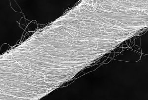
Microscopic image of carbon nanotube yarn.

Tapping into the autonomous nervous system – the control center for things like heartbeat and breathing – is a relatively new part of neurostimulation technologies to both record and direct organ function. Implants designed for stimulating peripheral nerves often fail because the protective tissue surrounding nerve bundles (the perineurium) is difficult to penetrate, and the body’s immune response often builds a scar around the implanted device.

Now, a team of scientists from Case Western Reserve University (CWRUL) has used carbon nanotubes to overcome these obstacles, reporting their findings in Scientific Reports. The authors, led by Dominique M. Durand, Ph.D., Director of the Neural Engineering Center and El Lindseth Professor of Biomedical Engineering, Neurosciences, Physiology and Biophysics at CWRU, fabricated yarn made of carbon nanotubes that was 10 to 20 µm in diameter. The yarn was then used to create electrodes, which were implanted into rats to monitor activity of the glossopharyngeal and vagus nerves.

The authors found that they could use the implants to monitor nerve activity under conditions of hypoxia and stomach distention. They report that the success of their experiments likely derives from the similarity of the nanotube yarn to the actual neural tissue surrounding the implant. The implants are a long way from being tried in humans, but the large number of functions controlled by just these two nerves indicates that such implants could find use in an enormous number of diseases.

Better Screening of Nanoparticle Delivery

As we discussed last week, the development of gene-based therapies is hindered by the sheer size of the human genome. The immense volume of information involved can quickly become difficult to manage, so one way in which scientists “keep track” of genetic information during the process of introducing new genetic material into an organism is DNA barcoding. This process attaches a small piece of DNA to the gene being studied; if and when the gene causes cells to replicate, these cells will bear the barcode, thus allowing the observer to be certain of the gene identities the whole time.

Seeking to determine whether DNA barcoding of lipid nanoparticles for injection into living models would outperform in vitro testing, a team of investigators at Georgia Tech and Emory University conducted a comparison of the two techniques, reporting their findings in Nano Letters. The authors, led by James Dahlman, Ph.D., Assistant Professor of Biomedical Engineering at GT/Emory, found that in vitro testing did not predict in vivo delivery. Further, they were able to track several dozen barcodes delivered by nanoparticles to eight different cell lines.

The authors believe that their technique, which they call JOint Rapid DNA Analysis of Nanoparticles (JORDAN), is superior to in vitro screening of nanoparticles to predict successful transplantation. They are offering JORDAN online as open source software so other scientists can use the technology to more accurately screen nanomaterials.

Synthetic Biologists Create Gene Circuits

Among the many types of molecules that regulate genetic expression in the body are microRNAs, non-coding strands of RNA that are responsible for gene silencing and other forms of gene expression regulation. The ability to harness and control the functions of microRNAs could have important implications for disease prevention and treatment.

In a recent article in Systems Biology and Applications, researchers at the University of Texas, Dallas, report on their engineering of a microRNA-based genetic circuit and its deployment in living cells. They created the circuit using strands of RNA from a variety of organisms, including viruses and jellyfish. The authors, led by Leonidas Bleris, Ph.D., Associate Professor of Bioengineering at UT Dallas, used the circuits to better understand how microRNAs change gene expression under different conditions.

More importantly, the authors found that their circuit had the ability to outproduce types of gene expression, which decreased as the number of gene replications increased.  The authors believe that their discoveries could have applications in a number of genetic disorders.

Discouraging Smoking at the Level of the Brain

Cigarette smoking is the single greatest contributor to negative health outcomes in the population.  Nicotine addiction often appears during the teenage years, and aggressive advertising has been used for the last couple of decades to encourage people to quit smoking and younger people not to start. Despite the widespread use of advertising to change human behavior, remarkably little is known on how the brain responds to advertising messages.

Danielle S. Bassett, Ph.D., Eduardo D. Glandt Faculty Fellow and Associate Professor of Bioengineering at Penn, recently collaborated with faculty from Penn’s Annenberg School of Communication to determine the neuroscience underlying this outcome. The collaborators showed graphic warning labels to a cohort of smokers while they were subjected to functional magnetic resonance imaging, which images brain activity during specific tasks. They found that smokers whose brains showed greater coherence between regions in the valuation network were more likely to quit smoking. Determining why these brain regions acted as they did could yield even more effective smoking-cessation messaging.

Purdue Startup Working to Expand MRI

Engineers at Purdue, including Zhongming Liu, Ph.D., Assistant Professor of Biomedical Engineering and Electrical and Computer Engineering, have cofounded at startup company, called MR-Link, to develop and produce a coin-sized device that can be inserted into MRI machines, allowing them to perform multiple scans simultaneously.

The device could be useful in reducing the amount of electromagnetic force to which patients are exposed during an MRI scan. In addition, Dr. Liu and his colleagues believe the device will cost perhaps less than a tenth of what similar devices currently cost. Given the widespread use of MRI, the device could ultimately impact how a number of diseases and disorders are diagnosed and tracked.