Understanding the Physics of Kidney Development

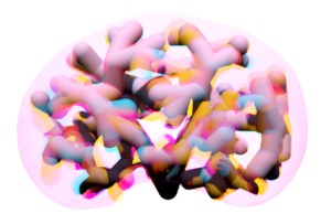
Abstract image of tubules repelling each other and shifting around.
The model of tubule packing developed by the Hughes Lab shows the tubules repelling each other and shifting around.

A recent study by Penn Bioengineering researchers sheds new light on the role of physics in kidney development. The kidney uses structures called nephrons and tubules to filter blood and pass urine to the bladder. Nephron number is set at birth and can vary over an order of magnitude (anywhere from 100,000 to over a million nephrons in an individual kidney). While the reasons for this variability remain unclear, low numbers of nephrons predispose patients to hypertension and chronic kidney disease. 

Now, research published in Developmental Cell led by Alex J. Hughes, Assistant Professor in the Department of Bioengineering, demonstrates a new physics-driven approach to better visualize and understand how a healthy kidney develops to avoid organizational defects that would impair its function. While previous efforts have typically approached this problem using molecular genetics and mouse models, the Hughes Lab’s physics-based approach could link particular types of defects to this genetic information and possibly highlight new treatments to prevent or fix congenital defects.

During embryonic development, kidney tubules grow and the tips divide to make a branched tree with clusters of nephron stem cells surrounding each branch tip. In order to build more nephrons, the tree needs to grow more branches. To keep the branches from overlapping, the kidney’s surface grows more crowded as the number of branches increase. “At this point, it’s like adding more people to a crowded elevator,” says Louis Prahl, first author of the paper and Postdoctoral Fellow in the Hughes Lab. “The branches need to keep rearranging to accommodate more until organ growth stops.”

To understand this process, Hughes, Prahl and their team investigated branch organization in mouse kidneys as well as using computer models and a 3D printed model of tubules. Their results show that tubules have to actively restructure – essentially divide at narrower angles – to accommodate more tubules. Computer simulations also identified ‘defective’ packing, in which the simulation parameters caused tubules to either overlap or be forced beneath the kidney surface. The team’s experimentation and analysis of published studies of genetic mouse models of kidney disease confirmed that these defects do occur.

This study represents a unique synthesis of different fields to understand congenital kidney disease. Mathematicians have studied geometric packing problems for decades in other contexts, but the structural features of the kidney present new applications for these models. Previous models of kidney branching have approached these problems from the perspective of individual branches or using purely geometric models that don’t account for tissue mechanics. By contrast, The Hughes Lab’s computer model demonstrates the physics of how tubule families interact with each other, allowing them to identify ‘phases’ of kidney organization that either relate to normal kidney development or organizational defects. Their 3D printed model of tubules shows that these effects can occur even when one sets the biology aside.

Hughes has been widely recognized for his research in the understanding of kidney development. This new publication is the first fruit of his 2021 CAREER Award from the National Science Foundation (NSF) and he was recently named a 2023 Rising Star by the Cellular and Molecular Bioengineering (CMBE) Special Interest Group. In 2020 he became the first Penn Engineering faculty member to receive the Maximizing Investigators’ Research Award (MIRA) from the National Institutes of Health (NIH) for his forward-thinking work in the creation of new tools for tissue engineering.

Pediatric nephrologists have long worked to understand the cause of these childhood kidney defects. These efforts are often confounded by a lack of evidence for a single causative mutation. The Hughes Lab’s approach presents a new and different application of the packing problem and could help answer some of these unsolved questions and open doors to prevention of these diseases. Following this study, Hughes and his lab members will continue to explore the physics of kidney tubule packing, looking for interesting connections between packing organization, mechanical stresses between neighboring tubule tips, and nephron formation while attempting to copy these principles to build stem cell derived tissues to replace damaged or diseased kidney tissue. Mechanical forces play an important role in developmental biology and there is much scope for Hughes, Prahl and their colleagues to learn about these properties in relation to the kidney.

Read The developing murine kidney actively negotiates geometric packing conflicts to avoid defects” in Developmental Cell.

Other authors include Bioengineering Ph.D. students and Hughes Lab members John Viola and Jiageng Liu.

This work was supported by NSF CAREER 2047271, NIH MIRA R35GM133380, Predoctoral Training Program in Developmental Biology T32HD083185, and NIH F32 fellowship DK126385.

New Single Cell Analysis Tool

by Nathi Magubane

Researchers at Penn and colleagues have developed a tool to analyze single cells that assesses both the patterns of gene activation within a cell and which sibling cells shared a common progenitor.

3D illustration of a cell held by a pipet and a needle
Arjun Raj of the School of Engineering and Applied Science and the Perelman School of Medicine, former postdoc Lee Richman, now of Brigham and Women’s Hospital, and colleagues have developed a new analysis tool that combines a cell’s unique gene expression data with information about the cell’s origins. The method can be applied to identify new cell subsets throughout development and better understand drug resistance.

Recent advances in analyzing data at the single-cell level have helped biologists make great strides in uncovering new information about cells and their behaviors. One commonly used approach, known as clustering, allows scientists to group cells based on characteristics such as the unique patterns of active or inactive genes or by the progeny of duplicating cells, known as clones, over several generations.

Although single-cell clustering has led to many significant findings, for example, new cancer cell subsets or the way immature stem cells mature into “specialized” cells, researchers to this point had not been able to marry what they knew about gene-activation patterns with what they knew about clone lineages.

Now, research published in Cell Genomics led by University of Pennsylvania professor of bioengineering Arjun Raj has resulted in the development of ClonoCluster, an open-source tool that combines unique patterns of gene activation with clonal information. This produces hybrid cluster data that can quickly identify new cellular traits; that can then be used to better understand resistance to some cancer therapies.

“Before, these were independent modalities, where you would cluster the cells that express the same genes in one lot and cluster the others that share a common ancestor in another,” says Lee Richman, first paper author and a former postdoc in the Raj lab who is now at Brigham and Women’s Hospital in Boston. “What’s exciting is that this tool allows you to draw new lines around your clusters and explore their properties, which could help us identify new cell types, functions, and molecular pathways.”

Researchers in the Raj Lab use a technique known as barcoding to assign labels to cells they are interested in studying, particularly useful for tracking cells, clustering data based on cells’ offspring, and following lineages over time. Believing they could parse more valuable information out of this data by incorporating the cell’s unique patterns of gene activation, the researchers applied ClonoCluster to six experimental datasets that used barcoding to track dividing cells’ offspring. Specifically, they looked at the development of chemotherapy resistance and of stem cells into specialized tissue types.

Read the full story in Penn Today.

Alex Hughes Named CMBE Rising Star

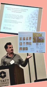
A collage of photos: Alex Hughes presenting, the title slide of his presentation with the title "Interpreting geometric rules of early kidney formation for synthetic morphogenesis," and his acknowledgements slides.
Alex J. Hughes presents at the BMES CMBE conference in January 2023. (Image credit: Riccardo Gottardi, Assistant Professor in Pediatrics and Bioengineering)

Alex J. Hughes, Assistant Professor in the Department of Bioengineering, was one of thirteen recipients of the 2023 Rising Star Award for Junior Faculty by the Cellular and Molecular Bioengineering (CMBE) Special Interest Group. The Rising Star Award recognizes a CMBE member in their early independent career stage that has made an outstanding impact on the field of cellular and molecular bioengineering. CMBE is a special interest group of the Biomedical Engineering Society (BMES), the premier professional organization of bioengineers.

The Hughes Lab in Penn Bioengineering works to “bring developmental processes that operate in vertebrate embryos and regenerating organs under an engineering control framework” in order to “build better tissues.” Hughes’s research interest is in harnessing the developmental principles of organs, allowing him to design medically relevant scaffolds and machines. In 2020 he became the first Penn Engineering faculty member to receive the Maximizing Investigators’ Research Award (MIRA) from the National Institutes of Health (NIH), and he was awarded a prestigious CAREER Award from the National Science Foundation (NSF) in 2021. Most recently, Hughes’s work has focused on understanding the development of cells and tissues in the human kidney via the creation of “organoids”: miniscule organ models that can mimic the biochemical and mechanical properties of the developing kidney. Understanding and engineering how the kidney functions could open doors to more successful regenerative medicine strategies to address highly prevalent congenital and adult diseases.

Hughes and his fellow award recipients were recognized at the annual BMES CBME conference in Indian Wells, CA in January 2023.

Read the full list of 2023 CMBE Award Winners.

OCTOPUS, an Optimized Device for Growing Mini-Organs in a Dish

by Devorah Fischler

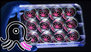
With OCTOPUS, Dan Huh’s team has significantly advanced the frontiers of organoid research, providing a platform superior to conventional gel droplets. OCTOPUS splits the soft hydrogel culture material into a tentacled geometry. The thin, radial culture chambers sit on a circular disk the size of a U.S. quarter, allowing organoids to advance to an unprecedented degree of maturity.

When it comes to human bodies, there is no such thing as typical. Variation is the rule. In recent years, the biological sciences have increased their focus on exploring the poignant lack of norms between individuals, and medical and pharmaceutical researchers are asking questions about translating insights concerning biological variation into more precise and compassionate care.

What if therapies could be tailored to each patient? What would happen if we could predict an individual body’s response to a drug before trial-and-error treatment? Is it possible to understand the way a person’s disease begins and develops so we can know exactly how to cure it?

Dan Huh, Associate Professor in the Department of Bioengineering at the University of Pennsylvania’s School of Engineering and Applied Science, seeks answers to these questions by replicating biological systems outside of the body. These external copies of internal systems promise to boost drug efficacy while providing new levels of knowledge about patient health.

An innovator of organ-on-a-chip technology, or miniature copies of bodily systems stored in plastic devices no larger than a thumb drive, Huh has broadened his attention to engineering mini-organs in a dish using a patient’s own cells.

A recent study published in Nature Methods helmed by Huh introduces OCTOPUS, a device that nurtures organs-in-a-dish to unmatched levels of maturity. The study leaders include Estelle Park, doctoral student in Bioengineering, Tatiana Karakasheva, Associate Director of the Gastrointestinal Epithelium Modeling Program at Children’s Hospital of Philadelphia (CHOP), and Kathryn Hamilton, Assistant Professor of Pediatrics in Penn’s Perelman School of Medicine and Co-Director of the Gastrointestinal Epithelial Modeling Program at CHOP.

Read the full story in Penn Engineering Today.

CAR T Cell Therapy Reaches Beyond Cancer

Penn Medicine researchers laud the early results for CAR T therapy in lupus patients, which point to broader horizons for the use of personalized cellular therapies.

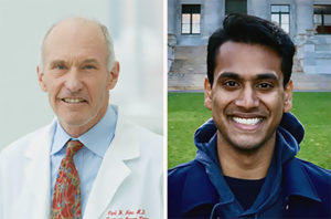
Penn Medicine’s Carl June and Daniel Baker.

Engineered immune cells, known as CAR T cells, have shown the world what personalized immunotherapies can do to fight blood cancers. Now, investigators have reported highly promising early results for CAR T therapy in a small set of patients with the autoimmune disease lupus. Penn Medicine CAR T pioneer Carl June and Daniel Baker, a doctoral student in cell and molecular biology in the Perelman School of Medicine, discuss this development in a commentary published in Cell.

“We’ve always known that in principle, CAR T therapies could have broad applications, and it’s very encouraging to see early evidence that this promise is now being realized,” says June, who is the Richard W. Vague Professor in Immunotherapy in the department of Pathology and Laboratory Medicine at Penn Medicine and director of the Center for Cellular Immunotherapies at the Abramson Cancer Center.

T cells are among the immune system’s most powerful weapons. They can bind to, and kill, other cells they recognize as valid targets, including virus-infected cells. CAR T cells are T cells that have been redirected, through genetic engineering, to efficiently kill specifically defined cell types.

CAR T therapies are created out of each patient’s own cells—collected from the patient’s blood, and then engineered and multiplied in the lab before being reinfused into the patient as a “living drug.” The first CAR T therapy, Kymriah, was developed by June and his team at Penn Medicine, and received Food & Drug Administration approval in 2017. There are now six FDA-approved CAR T cell therapies in the United States, for six different cancers.

From the start of CAR T research, experts believed that T cells could be engineered to fight many conditions other than B cell cancers. Dozens of research teams around the world, including teams at Penn Medicine and biotech spinoffs who are working to develop effective treatments from Penn-developed personalized cellular therapy constructs, are examining these potential new applications. Researchers say lupus is an obvious choice for CAR T therapy because it too is driven by B cells, and thus experimental CAR T therapies against it can employ existing anti-B-cell designs. B cells are the immune system’s antibody-producing cells, and, in lupus, B cells arise that attack the patient’s own organs and tissues.

This story is by Meagan Raeke. Read more at Penn Medicine News.

Carl June is a member of the Penn Bioengineering Graduate Group. Read more stories featuring June’s research here.

Bushra Raj Receives NIH Grant Through High-risk, High-reward Research Program

Bushra Raj, Ph.D.

Eight researchers from the Perelman School of Medicine have received research grants designed to invest in high-risk, high-reward projects.

Bushra Raj, Assistant Professor of Cell and Developmental Biology in the Perelman School of Medicine and member of the Penn Bioengineering Graduate Group, was one of three Penn winners of the NIH Director’s New Innovator Award for independent projects developed by early-career investigators. More additional Penn scientists who received NIH Director’s Transformative Research Award for a project focusing on cancer research.

Raj’s project focuses on “testing a novel technology that uses CRISPR/Cas gene-editing tools to genomically record inputs from two signaling pathways in the developing zebrafish brain.”

Established in 2009, the Transformative Research Award promotes cross-cutting, interdisciplinary science and is open to individuals and teams of investigators who propose research that could potentially create or challenge existing paradigms.

Read the full list of grant recipients in Penn Medicine News.

The Penn Center for Precision Engineering for Health Announces First Round of Seed Funding

by Melissa Pappas

CPE4H is one of the focal points of Penn Engineering signature initiative on Engineering Health.

The Penn Center for Precision Engineering for Health (CPE4H) was established late last year to accelerate engineering solutions to significant problems in healthcare. The center is one of the signature initiatives for Penn’s School of Engineering and Applied Science and is supported by a $100 million commitment to hire faculty and support new research on innovative approaches to those problems.

Acting on that commitment, CPE4H solicited proposals during the spring of 2022 for seed grants of $80K per year for two years for research projects that address healthcare challenges in several key areas of strategic importance to Penn: synthetic biology and tissue engineering, diagnosis and drug delivery, and the development of innovative devices. While the primary investigators (PIs) for the proposed projects were required to have a primary faculty appointment within Penn Engineering, teams involving co-PIs and collaborators from other schools were eligible for support. The seed program is expected to continue for the next four years.

“It was a delight to read so many novel and creative proposals,” says Daniel A. Hammer, Alfred G. and Meta A. Ennis Professor in Bioengineering and the Inaugural Director of CPE4H. “It was very hard to make the final selection from a pool of such promising projects.”

Judged on technical innovation, potential to attract future resources, and ability to address a significant medical problem, the following research projects were selected to receive funding.

Evolving and Engineering Thermal Control of Mammalian Cells

Led by Lukasz Bugaj, Assistant Professor in Bioengineering, this project will engineer molecular switches that can be toggled on and off inside mammalian cells at near-physiological temperatures. Successful development of these switches will provide new ways to communicate with cells, an advance that could be used to make safer and more effective cellular therapies.  The project will use directed evolution to generate and find candidate molecular tools with the desired properties. Separately, the research will also develop new technology for manipulating cellular temperature in a rapid and programmable way. Such devices will enhance the speed and sophistication of studies of biological temperature regulation.

A Quantum Sensing Platform for Rapid and Accurate Point-of-Care Detection of Respiratory Viral Infections

Combining microfluidics and quantum photonics, PI Liang Feng, Professor in Materials Science and Engineering and Electrical and Systems Engineering, Ritesh Agarwal, Professor in Materials Science Engineering, and Shu Yang, Joseph Bordogna Professor in Materials Science and Engineering and Chemical and Biomolecular Engineering, are teaming up with Ping Wang, Professor of Pathology and Laboratory Medicine in Penn’s Perelman School of Medicine, to design, build and test an ultrasensitive point-of-care detector for respiratory pathogens. In light of the COVID-19 pandemic, a generalizable platform for rapid and accurate detection of viral pathogenesis would be extremely important and timely.

Versatile Coacervating Peptides as Carriers and Synthetic Organelles for Cell Engineering

PI Amish Patel, Associate Professor in Chemical and Biomolecular Engineering, and Matthew C. Good, Associate Professor of Cell and Developmental Biology in the Perelman School of Medicine and in Bioengineering, will design and create small proteins that self-assemble into droplet-like structures known as coacervates, which can then pass through the membranes of biological cells. Upon cellular entry, these protein coacervates can disassemble to deliver cargo that modulates cell behavior or be maintained as synthetic membraneless organelles. The team will design new chemistries that will facilitate passage across cell membranes, and molecular switches to sequester and release protein therapeutics. If successful, this approach could be used to deliver a wide range of macromolecule drugs to cells.

Towards an Artificial Muscle Replacement for Facial Reanimation

Cynthia Sung, Gabel Family Term Assistant Professor in Mechanical Engineering and Applied Mechanics and Computer Information Science, will lead a research team including Flavia Vitale, Assistant Professor of Neurology and Bioengineering, and Niv Milbar, Assistant Instructor in Surgery in the Perelman School of Medicine. The team will develop and validate an electrically driven actuator to restore basic muscle responses in patients with partial facial paralysis, which can occur after a stroke or injury. The research will combine elements of robotics and biology, and aims to produce a device that can be clinically tested.

“These novel ideas are a great way to kick off the activities of the center,” says Hammer. “We look forward to soliciting other exciting seed proposals over the next several years.”

This article originally appeared in Penn Engineering Today.

Penn Startup Vittoria Biotherapeutics Raises $10M in Seed Funding

Marco Ruella, MD

A Philadelphia life sciences company spun out of Penn is emerging from stealth mode with nearly $10 million from a seed funding round. Vittoria Biotherapeutics’ mission is to overcome limitations of CAR T cell therapy by using unique cell engineering and gene editing technologies to create new therapies that address unmet clinical needs. The technology the company is attempting to commercialize was developed by Marco Ruella, M.D., Assistant Professor of Medicine in the Perelman School of Medicine and member of the Penn Bioengineering Graduate Group, who is the company’s scientific founder.

Read “Penn spinout Vittoria Biotherapeutics emerges from stealth mode with $10M seed round” in the Philadelphia Business Journal.

Taimoor Qazi Appointed Assistant Professor at Purdue University

Taimoor H. Qazi, Ph.D.

The Department of Bioengineering is proud to congratulate Taimoor H. Qazi, Ph.D. on his appointment as Assistant Professor in the Weldon School of Biomedical Engineering at Purdue University. Qazi’s appointment will begin in Fall 2022.

Qazi obtained his Ph.D. at the Technical University of Berlin and the Charité Hospital in Berlin, Germany working on translational approaches for musculoskeletal tissue repair using biomaterials and stem cells under the co-advisement of Georg Duda, Director of the Berlin Institute of Health and David Mooney, Mercator Fellow at Charité – Universitätsmedizin Berlin. After arriving at Penn in 2019, Qazi performed research on microscale granular hydrogels in the Polymeric Biomaterials Laboratory of Jason Burdick, Adjunct Professor in Bioengineering at Penn and Bowman Endowed Professor in Chemical and Biological Engineering at the University of Colorado, Boulder. While conducting postdoctoral research, Qazi also collaborated with the groups of David Issadore, Associate Professor in Bioengineering and in Electrical and Systems Engineering, and Daeyeon Lee, Professor and Evan C. Thompson Term Chair for Excellence in Teaching in Chemical and Biomolecular Engineering and member of the Penn Bioengineering Graduate Group. Qazi’s postdoctoral research was supported through a fellowship from the German Research Foundation, and resulted in several publications in high-profile journals, including Advanced Materials, Cell Stem Cell, Small, and ACS Biomaterials Science and Engineering.

“Taimoor has done really fantastic research as a postdoctoral fellow in the group,” says Burdick. “Purdue has a long history of excellence in biomaterials research and will be a great place for him to build a strong research program.”

Qazi’s future research program will engineer biomaterials to make fundamental and translational advances in musculoskeletal tissue engineering, including the study of how rare tissue-resident cells respond to spatiotemporal signals and participate in tissue repair, and developing modular hydrogels that permit minimally invasive delivery for tissue regeneration. The ultimate goal is to create scalable, translational, and biologically inspired healthcare solutions that benefit a patient population that is expected to grow manifold in the coming years.

Qazi is looking to build a strong and inclusive team of scientists and engineers with diverse backgrounds interested in tackling problems at the interface of translational medicine, materials science, bioengineering, and cell biology, and will be recruiting graduate students immediately. Interested students can contact him directly at thqazi@seas.upenn.edu.

“I am excited to launch my independent research career at a prestigious institution like Purdue,” says Qazi. “Being at Penn and particularly in the Department of Bioengineering greatly helped me prepare for the journey ahead. I am grateful for Jason’s mentorship over the years and the access to resources provided by Jason, Dave Issadore, Ravi, Dave Meany and other faculty which support the training and professional development of postdoctoral fellows in Penn Bioengineering.”

Congratulations to Dr. Qazi from everyone at Penn Bioengineering!

Erin Berlew and Rhea Chitalia Receive Solomon R. Pollack Awards for Excellence in Graduate Bioengineering Research

The Solomon R. Pollack Award for Excellence in Graduate Bioengineering Research is given annually to the most deserving Bioengineering graduate students who have successfully completed research that is original and recognized as being at the forefront of their field. This year Penn Bioengineering recognizes the outstanding work of two graduate students in Bioengineering: Erin Berlew and Rhea Chitalia.

Erin Berlew, Ph.D. candidate in Bioengineering

Erin Berlew is a Ph.D. candidate in the lab of Brian Chow, Associate Professor in Bioengineering. She successfully defended her thesis, titled “Single-component optogenetic tools for cytoskeletal rearrangements,” in December 2021. In her research, she used the BcLOV4 optogenetic platform discovered/developed in the Chow lab to control RhoGTPase signaling. Erin earned a B.S. in Chemistry from Haverford College in 2015 and was an Americorps member with City Year Philadelphia from 2015-2016. “Erin is a world-class bioengineering with an uncommon record of productivity gained through her complementary expertise in molecular, cellular, and computational biology,” says Chow. “She embodies everything wonderful, both academically and culturally, about our graduate program and its distinguished history.” Erin’s hobbies outside the lab include spending time with family, reading mystery novels, enjoying Philadelphia, and crossword puzzles. In the future, she hopes to continue to teach for the BE department (she has already taught ENGR 105 and served as a TA for undergraduate and graduate courses) and to conduct further research at Penn.

Rhea Chitalia, Ph.D. candidate in Bioengineering

Rhea Chitalia is a Ph.D. candidate in Bioengineering and a member of the Computational Biomarker Imaging Group (CBIG), advised by Despina Kontos, Matthew J. Wilson Associate Professor of Research Radiology II in the Perelman School of Medicine. Rhea completed her B.S.E. in Biomedical Engineering at Duke University in 2015. Her doctoral research concerns leveraging machine learning, bioinformatics, and computer vision to develop computational imaging biomarkers for improved precision cancer care. In December 2021 she successfully defended her thesis titled “Computational imaging biomarkers for precision medicine: characterizing intratumor heterogeneity in breast cancer.” “It has been such a privilege to mentor Rhea on her dissertation research,” says Kontos. “Rhea has been a star graduate student. Her work has made fundamental contributions in developing computational methods that will allow us to gain important insight into tumor heterogeneity by utilizing a multi-modality imaging approach.” David Mankoff, Matthew J. Wilson Professor of Research Radiology in the Perelman School of Medicine, served as Rhea’s second thesis advisor. “It was a true pleasure for me to work with Rhea and to Chair her BE Thesis Committee,” Mankoff adds. “Rhea’s Ph.D. thesis and thesis presentation was one of the best I have had the chance to be involved with in my graduate mentoring career.” After graduation, Rhea hopes to further precision medicine initiatives through the use of real world, multi-omic data in translational industry settings. She will be joining Invicro as an Imaging Scientist. In her spare time, Rhea enjoys trying new restaurants, reading, and spending time with friends and family.