2023 Graduate Research Fellowships for Bioengineering Students

Congratulations to the fourteen Bioengineering students to receive 2023  National Science Foundation Graduate Research Fellowship Program (NSF GRFP) fellowships. The prestigious NSF GRFP program recognizes and supports outstanding graduate students in NSF-supported fields. The recipients honorees were selected from a highly-competitive, nationwide pool. Further information about the program can be found on the NSF website.

Carlos Armando Aguila, Ph.D. student in Bioengineering, is a member of the Center of Neuroengineering and Therapeutics, advised by Erin Conrad, Assistant Professor in Neurology, and Brian Litt, Professor in Bioengineering and Neurology. His research focuses on analyzing electroencephalogram (EEG) signals to better understand epilepsy.

Joseph Lance Victoria Casila is a Ph.D. student in Bioengineering in the lab of Riccardo Gottardi, Assistant Professor in Pediatrics and Bioengineering. His research focuses on probing environmental factors that influence stem cell differentiation towards chondrogenesis for cartilage engineering and regeneration.

Trevor Chan is a Ph.D. student in Bioengineering in the lab of Felix Wehrli, Professor of Radiologic Science. His research is in developing computational methods for medical image refinement and analysis. Two ongoing projects are: self-supervised methods for CT super-resolution and assessment of osteoporosis, and semi-supervised segmentation of 3D and 4D echocardiograms for surgical correction of congenital heart-valve defects.

Rakan El-Mayta is an incoming Ph.D. student in the lab of Drew Weissman, Roberts Family Professor in Vaccine Research. Rakan studies messenger RNA-lipid nanoparticle vaccines for the treatment and prevention of infectious diseases. Prior to starting in the Bioengineering graduate program, he worked as a Research Assistant in Weissman lab and in the lab of Michael Mitchell, Associate Professor in Bioengineering.

Austin Jenk is a Ph.D. student in the lab of Robert Mauck, Mary Black Ralston Professor in Orthopaedic Surgery and Bioengineering. Austin aims to develop early intervention, intra-articular therapeutics to combat the onset of post-traumatic osteoarthritis following acute joint injuries. His work focuses on developing a therapeutic that can be employed not only in conventional healthcare settings, but also emergency and battlefield medicine.

Jiageng Liu is a Ph.D. student in the lab of Alex Hughes, Assistant Professor in Bioengineering. His work aims to precisely control the bio-physical/chemical properties of iPSC-derived organoids with advanced synthetic biology approaches to create functional replacement renal tissues.

Alexandra Neeser is a Ph.D. student in the lab of Leyuan Ma, Assistant Professor of Pathology and Laboratory Medicine. Her research focuses on solid tumor microenvironment delivery of therapeutics.


William Karl Selboe Ojemann, a Ph.D. Student in Bioengineering, is a member of the Center for Neuroengineering and Therapeutics directed by Brian Litt, Professor in Bioengineering and Neurology. His research is focused on developing improved neurostimulation therapies for epilepsy and other neurological disorders.

Savan Patel (BSE Class of 2023) conducted research in the lab of Michael Mitchell, Associate Professor in Bioengineering, where he worked to develop lipid nanoparticle formulations for immunotherapy and extrahepatic delivery of mRNA. He will be joining the Harvard-MIT HST MEMP Ph.D. program in the fall of 2023.

David E. Reynolds, a Ph.D. student in Bioengineering, is a member of the lab of Jina Ko, Assistant Professor in Bioengineering and Pathology and Laboratory Medicine. His research focuses on developing novel and translatable technologies to address currently intractable diagnostic challenges for precision medicine.

Andre Roots is a Ph.D. student in the lab of Christopher Madl, Assistant Professor in Materials Science and Engineering. His research focuses on the use of protein engineering techniques and an optimized 3D human skeletal muscle microtissue platform to study the effects of biophysical material properties on cells.

Emily Sharp, a second year Ph.D. student in Bioengineering, is a member of the lab of Robert Mauck, Mary Black Ralston Professor in Orthopaedic Surgery and Bioengineering, part of the McKay Orthopaedic Research Laboratories. Her research focuses on designing multi-functional biomaterials to enhance tissue repair, specifically intervertebral disc repair following herniation and discectomy.

Nat Thurlow is a Ph.D. student in the lab of Louis J. Soslowsky, Fairhill Professor in Orthopedic Surgery and Bioengineering. Their current work focuses on delineating the roles of collagens V and XI in tendon mechanics, fibril structure, and gene expression during tendon development and healing.

Maggie Wagner, Ph.D. student in Bioengineering, is a member in the labs of Josh Baxter, Assistant Professor of Orthopaedic Surgery, and Flavia Vitale, Assistant Professor in Neurology and Bioengineering. Her research focuses on the development of novel sensors to record and monitor muscle neuromechanics.

César de la Fuente Receives 2023 Rao Makineni Lectureship Award

by

César de la Fuente
César de la Fuente

The American Peptide Society has selected César de la Fuente, Presidential Assistant Professor in Psychiatry, Microbiology, Bioengineering and in Chemical and Biomolecular Engineering, as the recipient of the prestigious 2023 Rao Makineni Lectureship Award.

Presented at the biennial American Peptide Symposium, the Makineni Lectureship Award recognizes an individual who has made a recent contribution of unusual merit to research in the field of peptide science, and is intended to acknowledge original and singular discoveries.

Established in 2003 by an endowment by PolyPeptide Laboratories and Murray and Zelda Goodman, this lectureship honors Rao Makineni, a long-time supporter of peptide science, peptide scientists, and the American Peptide Society.

This story originally appeared in Penn Engineering Today.

Breaking Down Barriers to Blood Donation for LGBTQ+ People

by Meredith Mann

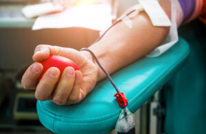
Close-up of a person's arm and hand as they donate blood.
(Image: iStock/hxdbzxy)

For decades, LGBTQ+ patients have faced stringent requirements to donate blood—most gay and bisexual men were not allowed to donate at all. Now, however, many more of them will be able to give this selfless gift. The U.S. Food and Drug Administration, which regulates blood donation in this country, has reworked the donor-screening criteria, and in the process opened the door to donation for more Americans.

The previous restriction on accepting blood from men who have sex with men (MSM) dates back to the early days of the AIDS epidemic, when blood donations weren’t able to be screened for HIV, leading to cases of transfusion-transmitted HIV. In 1985, the FDA instituted a lifetime ban on blood donation for MSM, effectively preventing gay and bisexual men from donating. (Also included were women who have sex with MSM.)

Twenty years later, the agency rescinded the ban—but added a restriction that only MSM who had been abstinent from sex for at least one year could donate. In 2020, the FDA shortened the “deferral” period to 90 days of abstinence. While the changes were welcome news for those who had been unable to donate, they still prevented many MSM from giving blood. As he wrote in an op-ed for the Philadelphia Inquirer last year, Kevin B. Johnson, the David L. Cohen University Professor with appointments in the School of Engineering and Applied Science, the Perelman School of Medicine, and Annenberg School for Communication, was one of them. He and his husband were shocked to learn when they went to donate blood during a shortage early in the COVID-19 pandemic, that despite being married and monogamous for close to 17 years, they could not donate unless they were celibate for three months.

“It is time to move quickly to a policy under which all donors are evaluated equally and fairly, and to encourage local blood collection facilities to comply with that policy,” Johnson wrote last year.

Now, such changes are underway. As the pandemic wound down, the FDA moved forward with plans to re-evaluate its donation criteria. The first big change was removal of an indefinite ban on people who lived in or spent significant amounts of time in the United Kingdom, Ireland, and France, a measure that aimed to protect the U.S. blood supply against Creutzfeldt-Jakob disease (CJD; also known as “mad cow disease”), a terminal brain condition caused by hard-to-detect prions that occurred in those countries in the 1980s and 1990s.

Extensive and careful evaluation of epidemiological studies and statistical analysis has shown that the risk of CJD transmission is no longer a concern. The changes to eligibility for LGBTQ+ patients are related to advances in medical and social science, and have also been very thoroughly studied to ensure that the changes will maintain the safety of the blood supply without being discriminatory.

“In the decades since HIV was first recognized, there have been advances in testing methods for detection of the virus, changes in how we process blood products, public health advances, and extensive study of the evolving risk of disease transmission given these advances,” says Sarah Nassau, vice chair of pathology and laboratory medicine at Lancaster General Hospital.

They also draw on rethinking the reliability of the guidelines. For example, while the rules partially or fully prevented gay and bisexual men from donating blood, they did not erect similar barriers to other people engaging in anal sex, or people who have multiple partners.

“Specifying the sexual orientation of the person rather than a behavior in which they engaged was discriminatory and not evidence based,” points out Judd David Flesch, vice chief of inpatient operations in the Department of Medicine at Penn Presbyterian Medical Center and co-director of the Penn Medicine Program for LGBT Health.

Read the full story in Penn Medicine News.

Kevin Johnson is the David L. Cohen University of Pennsylvania Professor in the Departments of Biostatistics, Epidemiology and Informatics and Computer and Information Science. As a Penn Integrates Knowlegde (PIK) University Professor, Johnson also holds appointments in the Departments of Bioengineering and Pediatrics, as well as in the Annenberg School of Communication.

The Art and Science of Living-Like Architecture

by

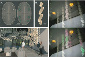
Collaborators from Penn Engineering and the Stuart Weitzman School of Design have created “living-like” bioactive interior architecture designed to one day protect us from hidden airborne threats. The figure above demonstrates (A) design for support lattices for the team’s innovative bioactive sites, (B) a ribbon-like geometry for hanging and (C, D) how these structures may be integrated into indoor environments to biologically sense and react to air.

“This technology is not alive,” says Laia Mogas-Soldevila. “It is living-like.”

The distinction is an important one for the assistant professor at the Stuart Weitzman School of Design, for reasons both scientific and artistic. With a doctorate in biomedical engineering, several degrees in architecture, and a devotion to sustainable design, Mogas-Soldevila brings biology to everyday life, creating materials for a future built halfway between nature and artifice.

The architectural technology she describes is unassuming at first look: A freeze-dried pellet, small enough to get lost in your pocket. But this tiny lump of matter, the result of more than a year’s collaboration between designers, engineers and biologists, is a biomaterial that contains a “living-like” system.

When touched by water, the pellet activates and expresses a glowing protein, its fluorescence demonstrating that life and art can harmonize into a third and very different thing, as ready to please as to protect. Woven into lattices made of flexible natural materials promoting air and moisture flow, the pellets form striking interior design elements that could one day keep us healthy.

“We envision them as sensors,” explains Mogas-Soldevila. “They may detect pathogens, such as bacteria or viruses, or alert people to toxins inside their home. The pellets are designed to interact with air. With development, they could monitor or even clean it.”

For now, they glow, a triumphant first stop on the team’s roadmap to the future. The fluorescence establishes that the lab’s biomaterial manufacturing process is compatible with the leading-edge cell-free engineering that gives the pellets their life-like properties.

A rapidly expanding technology, cell-free protein expression systems allow researchers to manufacture proteins without the use of living cells.

Gabrielle Ho, Ph.D. candidate in the Department of Bioengineering and co-leader of the project, explains how the team’s design work came to be cell-free, a technique rarely explored outside of lab study or medical applications.

“Typically, we’d use living E. coli cells to make a protein,” says Ho. “E. coli is a biological workhorse, accessible and very productive. We’d introduce DNA to the cell to encourage expression of specific proteins. But this traditional method was not an option for this project. You can’t have engineered E. coli hanging on your walls.”

Cell-free systems contain all the components a living cell requires to manufacture protein —energy, enzymes and amino acids — and not much else. These systems are therefore not alive. They do not replicate, and neither can they cause infection. They are “living-like,” designed to take in DNA and push out protein in ways that previously were only possible using living cells.

“One of the nicest things about these materials not being alive,” says Mogas-Soldevila, “is that we don’t need to worry about keeping them that way.”

Unlike living cells, cell-free materials don’t need a wet environment or constant monitoring in a lab. The team’s research has established a process for making these dry pellets that preserves bioactivity throughout manufacturing, storage and use.

Bioactive, expressive and programmable, this technology is designed to capitalize on the unique properties of organic materials.

Mogas-Soldevila, whose lab focuses exclusively on biodegradable architecture, understands the value of biomaterials as both environmentally responsible and aesthetically rich.

“Architects are coming to the realization that conventional materials — concrete, steel, glass, ceramic, etc. — are environmentally damaging and they are becoming more and more interested in alternatives to replace at least some of them. Because we use so much, even being able to replace a small percentage would result in a significant reduction in waste and pollution.”

Her lab’s signature materials — biopolymers made from shrimp shells, wood pulp, sand and soil, silk cocoons, and algae gums — lend qualities over and above their sustainable advantages.

“My obsession is diagnostic, but my passion is playfulness,” says Mogas-Soldevila. “Biomaterials are the only materials that can encapsulate this double function observed in nature.”

This multivalent approach benefited from the help of Penn Engineering’s George H. Stephenson Foundation Educational Laboratory & Bio-MakerSpace, and the support of its director, Sevile Mannickarottu. In addition to contributing essential equipment and research infrastructure to the team, Mannickarottu was instrumental in enabling the interdisciplinary relationships that led the team to success, introducing Ho to the DumoLab Research team collaborators. These include Mogas-Soldevila, Camila Irabien, a Penn Biology major who provided crucial contributions to experimental work, and Fulbright design fellow Vlasta Kubušová, who co-led the project during her time at Penn and who will continue fueling the project’s next steps.

Read the full story in Penn Engineering Today.

Penn Dental Medicine Collaboration Identifies New Bacterial Species Involved in Tooth Decay

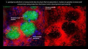
S. sputigena cells form a honeycomb-like structure that encapsulates S. mutans to greatly increase and concentrate acid production that boost caries development and severity.
Image: Courtesy of Penn Dental Medicine

Collaborating researchers from the University of Pennsylvania School of Dental Medicine and the Adams School of Dentistry and Gillings School of Global Public Health at the University of North Carolina have discovered that a bacterial species called Selenomonas sputigena can have a major role in causing tooth decay.

Scientists have long considered another bacterial species, the plaque-forming, acid-making Streptococcus mutans, as the principal cause of tooth decay—also known as dental caries. However, in the study, published in Nature Communications, the Penn Dental Medicine and UNC researchers showed that S. sputigena, previously associated only with gum disease, can work as a key partner of S. mutans, greatly enhancing its cavity-making power.

“This was an unexpected finding that gives us new insights into the development of caries, highlights potential future targets for cavity prevention, and reveals novel mechanisms of bacterial biofilm formation that may be relevant in other clinical contexts,” says study co-senior author Hyun (Michel) Koo, a professor in the Department of Orthodontics and Divisions of Pediatrics and Community Oral Health and co-director of the Center for Innovation & Precision Dentistry at Penn Dental Medicine.

The other two co-senior authors of the study were Kimon Divaris, professor at UNC’s Adams School of Dentistry, and Di Wu, associate professor at the Adams School and at the UNC Gillings School of Global Public Health.

“This was a perfect example of collaborative science that couldn’t have been done without the complementary expertise of many groups and individual investigators and trainees,” Divaris says.

Read the full story in Penn Today.

Michel Koo is a professor in the Department of Orthodontics and divisions of Community Oral Health and Pediatric Dentistry in Penn Dental Medicine and co-director of the Center for Innovation & Precision Dentistry. He is a member of the Penn Bioengineering Graduate Group.

Wandering and Wondering

Wangari Mbuthia at Marina Bay, Singapore (photo credit: Wangari Mbuthia)

Wangari Mbuthia, Penn Bioengineering Class of 2025, shares her experience in Singapore while studying abroad with the Global Research and Internship Program (GRIP) at Penn. GRIP provides outstanding undergraduate and graduate students the opportunity to intern or conduct research abroad for 8 to 12 weeks over the summer. Participants gain career-enhancing experience and global exposure that is essential in a global workforce.

Engineering Research in Singapore

If someone would have told me this time last year that I would be doing an engineering research program in Singapore, I wouldn’t have believed it. But rest assured here I am, two weeks in, and it has been an incredible experience.

Admittedly before coming to Singapore, basically everything I knew about this country could somewhat be summarized in that it was hot, beautiful and diverse. Before this I had never traveled to an Asian country and I was both excited and nervous about taking this trip. I was excited for food, sights and new experiences but I was also particularly nervous about being in a country where almost no one looks like me. Nevertheless, I decided to travel with an open mind, letting myself wander and wonder as I went and I thought I’d share some of my initial discoveries here.

Walking around Singapore it is clear that it is a place where many cultures have come together – Chinese, Malay, Indian and more – but I could probably count the number of Black people I saw on my two hands. This cultural landscape left me feeling very visible everywhere I went. But at the same time also somewhat invisible because for the most part, no one really made me feel like the odd one out. Rather, my presence only seemed to spark harmless (and sometimes comical) curiosity about where I was from or how I do my braids.

To my delight, the cultural diversity of Singapore is equally reflected in the food options. I can easily have access to almost any type of Asian cuisine at any given time and even quite a lot Western varieties too. I have eagerly been documenting the foods I try and rating them. One of my favorites has been a kaya (a type of sweet coconut spread) toast breakfast with soft-boiled eggs and teh-c (tea with evaporated milk). I also still need to try the unique, smelly fruit (so smelly it is not allowed on public transport), durian.

Another wonderful discovery was to see how Singapore lives up to its name, “garden city”. Not only is the city filled with beautiful buildings each with their own personality, but the city landscape is so artfully integrated with nature inside and out. I have seen indoor gardens and waterfalls but also gorgeous waterfront and outdoor spaces that I could sit in for hours.

It’s hard to believe how a country with such little land area and no natural resources has grown to be one of the richest cities in the world. Singapore truly feels like a place where so much is possible and that has been really special to see.

Cesar de la Fuente On the “Next Frontier” of Antibiotics

César de la Fuente
César de la Fuente

In a recent CNN feature, César de la Fuente, Presidential Assistant Professor in Bioengineering, Psychiatry, Microbiology, and in Chemical and Biomolecular Engineering commented on a study about a new type of antibiotic that was discovered with artificial intelligence:

“I think AI, as we’ve seen, can be applied successfully in many domains, and I think drug discovery is sort of the next frontier.”

The de la Fuente lab uses machine learning and biology to help prevent, detect, and treat infectious diseases, and is pioneering the research and discovery of new antibiotics.

Read “A new antibiotic, discovered with artificial intelligence, may defeat a dangerous superbug” in CNN Health.

RNA Nanoparticle Therapy Stops the Spread of Incurable Bone Marrow Cancer

by

Myeloma cells producing monoclonal proteins of varying types, created by Scientific Animations under the Creative Commons Attributions-Share Alike International 4.0 License

Multiple myeloma is an incurable bone marrow cancer that kills over 100,000 people every year. Known for its quick and deadly spread, this disease is one of the most challenging to address. As these cancer cells move through different parts of the body, they mutate, outpacing possible treatments. People diagnosed with severe multiple myeloma that is resistant to chemotherapy typically survive for only three to six months. Innovative therapies are desperately needed to prevent the spread of this disease and provide a fighting chance for those who suffer from it.

Michael Mitchell, J. Peter and Geri Skirkanich Assistant Professor of Innovation in Bioengineering (BE), and Christian Figueroa-Espada, doctoral student in BE at the University of Pennsylvania School of Engineering and Applied Science, created an RNA nanoparticle therapy that makes it impossible for multiple myeloma to move and mutate. The treatment, described in their study published in PNAS, turns off a cancer-attracting function in blood vessels, disabling the pathways through which multiple myeloma cells travel.

By shutting down this “chemical GPS” that induces the migration of cancer cells, the team’s therapy stops the spread of multiple myeloma, helping to eliminate it altogether.

Read the full story in Penn Engineering Today.

Engineered White Blood Cells Eliminate Cancer

by

“Macrophages killing cancer cell” photographed by Susan Arnold.

By silencing the molecular pathway that prevents macrophages from attacking our own cells, Penn Engineers have manipulated these white blood cells to eliminate solid tumors.

Cancer remains one of the leading causes of death in the U.S. at over 600,000 deaths per year. Cancers that form solid tumors such as in the breast, brain or skin are particularly hard to treat. Surgery is typically the first line of defense for patients fighting solid tumors. But surgery may not remove all cancerous cells, and leftover cells can mutate and spread throughout the body. A more targeted and wholistic treatment could replace the blunt approach of surgery with one that eliminates cancer from the inside using our own cells.

Dennis Discher, Robert D. Bent Professor in Chemical and Biomolecular Engineering, Bioengineering, and Mechanical Engineering and Applied Mechanics, and postdoctoral fellow, Larry Dooling, provide a new approach in targeted therapies for solid tumor cancers in their study, published in Nature Biomedical Engineering. Their therapy not only eliminates cancerous cells, but teaches the immune system to recognize and kill them in the future.

“Due to a solid tumor’s physical properties, it is challenging to design molecules that can enter these masses,” says Discher. “Instead of creating a new molecule to do the job, we propose using cells that ‘eat’ invaders – macrophages.”

Macrophages, a type of white blood cell, immediately engulf and destroy – phagocytize – invaders such as bacteria, viruses, and even implants to remove them from the body. A macrophage’s innate immune response teaches our bodies to remember and attack invading cells in the future. This learned immunity is essential to creating a kind of cancer vaccine.

But, a macrophage can’t attack what it can’t see.

“Macrophages recognize cancer cells as part of the body, not invaders,” says Dooling. “To allow these white blood cells to see and attack cancer cells, we had to investigate the molecular pathway that controls cell-to-cell communication. Turning off this pathway – a checkpoint interaction between a protein called SIRPa on the macrophage and the CD47 protein found on all ‘self’ cells – was the key to creating this therapy.”

Read the full story in Penn Engineering Today.

Multiple members in the biophysical engineering lab lead by Dennis Discher, including co-lead author and postdoctoral fellow and Penn Bioengineering alumnus Jason Andrechak and Bioengineering Ph.D. student Brandon Hayes, contributed to this study. The research was funded by grants from the National Heart, Lung, and Blood Institute and the National Cancer Institute, including the Physical Sciences Oncology Network, of the US National Institutes of Health.

Mustafa Mir Named HHMI Freeman Hrabowski Scholar

Mustafa Mir

Mustafa Mir, Assistant Professor of Cell and Developmental Biology in the Perelman School of Medicine and member of the Penn Bioengineering Graduate Group, was selected as one of Howard Hughes Medical Institute’s 31 new Freeman Hrabowski Scholars. The group consists of outstanding early career faculty in science who have potential to become leaders in their research fields and to create diverse and inclusive lab environments in which everyone can thrive. Mir and his lab develop and apply new microscopes to directly visualize the molecular scale events that underlie gene expression within live embryos.

Read a Q&A with Mir in the Children’s Hopsital of Philadelphia (CHOP)’s Cornerstone Blog: “New Technologies Lead to New Discoveries’: Q&A With HHMI Scholar Mustafa Mir, PhD.

This announcement originally appeared in Penn Medicine News.