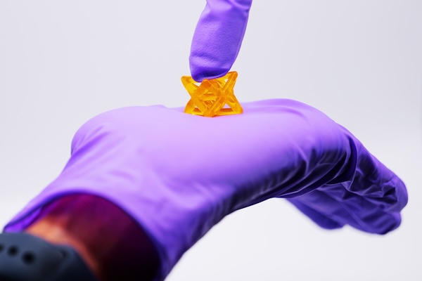
Biomaterials 3D-printed with the new method can be used inside the body and could even serve as bandages on a beating human heart. (Photo by Casey A. Cass/University of Colorado)
In the quest to develop life-like materials to replace and repair human body parts, scientists face a formidable challenge: Real tissues are often both strong and stretchable and vary in shape and size.
A CU Boulder-led team, in collaboration with researchers at the University of Pennsylvania, has taken a critical step toward cracking that code. They’ve developed a new way to 3D print material that is at once elastic enough to withstand a heart’s persistent beating, tough enough to endure the crushing load placed on joints, and easily shapeable to fit a patient’s unique defects.
Their breakthrough, described in the Aug. 2 edition of the journal Science, helps pave the way toward a new generation of biomaterials, from internal bandages that deliver drugs directly to the heart to cartilage patches and needle-free sutures.
“This is a simple 3D processing method that people could ultimately use in their own academic labs as well as in industry to improve the mechanical properties of materials for a wide variety of applications,” says first author Abhishek Dhand, a researcher in the Burdick Lab and doctoral candidate in the Department of Bioengineering at the University of Pennsylvania. “It solves a big problem for 3D printing.”
Jason Burdick is Bowman Endowed Professor in Chemical and Biological Engineering at the University of Colorado Boulder and Adjunct Professor in Bioengineering at Penn Engineering.
A team of Penn Bioengineering Senior Design students was featured as the 3D print of the week by the Penn Biomedical Library’s Biomeditations blog.
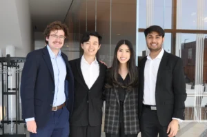
The StablEyes team. From left to right, Jake Becker (BE ’23), Ruoming Fan (BE ’23), Ella Atsavapranee (BE ’23), and Savan Patel (M&T ’23).
Fourth-year undergraduate students Ella Atsavapranee, Jake Becker, Ruoming Fan, and Savan Patel created StablEyes, “a stabilization mount that provides fine, motorized control of the handheld OCT to improve ease of use for physicians and machine learning-based software to aid in diagnosis from retinal images.” The team made use of 3D printing services, laboratory space, and expertise across Penn’s campus to create their innovative design, including the Bollinger Digital Fabrication Lab in the Holman Biotech Commons, the Fisher Fine Arts Library, the Children’s Hospital of Philadelphia (CHOP), and the George H. Stephenson Foundation Educational Laboratory & Bio-MakerSpace (aka the Penn BE Labs).
Congratulations to recent Penn Bioengineering graduate Gabriel DeSantis on being awarded a Fulbright grant for the 2021-22 academic year:
“The Fulbright Program is the United States government’s flagship international educational exchange program, awarding grants to fund as long as 12 months of international experience.
‘As an avenue for building cross-cultural understanding, the U.S. Student Fulbright Program is an unparalleled opportunity for American students to represent our country and our University across the world,’ says Jane Morris, executive director of Penn’s Center for Undergraduate Research and Fellowships, which supports applicants. ‘We are so proud of all our Penn Fulbright students who will be contributing to this important mission through their study, research, and English teaching as Fulbrighters.’
Gabriel DeSantis, from Wellesley, Massachusetts, received his bachelor’s degree from Penn Bioengineering in 2020 and will graduate in May with a master’s degree in bioengineering from the School of Engineering and Applied Science. He was awarded a Fulbright to conduct research in Portugal at the International Iberian Nanotechnology Laboratory. There he will be creating a 3D bio-printed model to optimize the texture and nutritional profiles of cultivated meat. At Penn his academic interests included biology, food science, and sustainability, which he hopes to use to develop new systems of food production. On campus, DeSantis was a Penn Abroad Leader and board member of the Graduate Association of Bioengineers. He is a past chair of the Mask and Wig Club. He currently works as a research assistant for Allevi, a Philadelphia-based bioprinting company at Pennovation Works.”
Read the full list of Fulbright awardees in Penn Today.
Some medical conditions, like diabetes or limb amputation, have the potential to result in wounds that never heal, affecting patients for the rest of their lives. Though normal wound-healing processes are relatively understood by medical professionals, the complications that can lead to chronic non-healing wounds are often varied and complex, creating a gap in successful treatments. But biomedical engineering faculty from the University of Connecticut want to change that.
Ali Tamayol, Ph.D., an Associate Professor in UConn’s Biomedical Engineering Department, developed what he’s calling a “smart” bandage in collaboration with researchers from the University of Nebraska-Lincoln and Harvard Medical School. The bandage, paired with a smartphone platform, has the ability to deliver medications to the wound via wirelessly controlled mini needles. The minimally invasive device thus allows doctors to control medication dosages for wounds without the patient even having to come in for an appointment. Early tests of the device on mice showed success in wound-healing processes, and Tamayol hopes that soon, the technology will be able to do the same for humans.
A New Patch Could Fix Broken Hearts
Heart disease is by far one of the most common medical conditions in the world, and has a high risk of morbidity. While some efforts in tissue engineering have sought to resolve cardiac tissue damage, they often require the use of existing heart cells, which can introduce a variety of complications to its integration into the human body. So, a group of bioengineers at Trinity College in Dublin sought to eliminate the need for cells by creating a patch that mimics both the mechanical and electrical properties of cardiac tissue.
Using thermoelastic polymers, the engineers, led by Ussher Assistant Professor in Biomedical Engineering Michael Monaghan, Ph.D., created a patch that could withstand multiple rounds of stretching and exhibited elasticity: two of the biggest challenges in designing synthetic cardiac tissues. With the desired mechanical properties working, the team then coated the patches with an electroconductive polymer that would allow for the necessary electrical signaling of cardiac tissue without decreasing cell compatibility in the patch. So far, the patch has demonstrated success in both mechanical and electrical behaviors in ex vivo models, suggesting promise that it might be able to work in the human body, too.
3-D Printing a New Tissue Engineering Scaffold
While successful tissue engineering innovations often hold tremendous promise for advances in personalized medicine and regeneration, creating the right scaffold for cells to grow on either before or after implantation into the body can be tricky. One common approach is to use 3-D printers to extrude scaffolds into customizable shapes. But the problem is that not all scaffold materials that are best for the body will hold up their structure in the 3-D printing process.
A team of biomedical engineers at Rutgers University led by Chair of Biomedical Engineering David I. Schreiber, Ph.D., hopes to apply the use of hyaluronic acid — a common natural molecule throughout the human body — in conjunction with polyethylene glycol to create a gel-like scaffold. The hope is that the polyethylene glycol will improve the scaffold’s durability, as using hyaluronic acid alone creates a substance that is often too weak for tissue engineering use. Envisioning this gel-like scaffold as a sort of ink cartridge, the engineers hope that they can create a platform that’s customizable for a variety of different cells that require different mechanical properties to survive. Notably, this new approach can specifically control both the stiffness and the ligands of the scaffold, tailoring it to a number of tissue engineering applications.
A New Portable Chip Can Track Wide Ranges of Brain Activity
Understanding the workings of the human brain is no small feat, and neuroscience still has a long way to go. While recent technology in brain probes and imaging allows for better understanding of the organ than ever before, that technology often requires immense amounts of wires and stationary attachments, limiting the scope of brain activity that can be studied. The answer to this problem? Figure out a way to implant a portable probe into the brain to monitor its everyday signaling pathways.
That’s exactly what researchers from the University of Arizona, George Washington University, and Northwestern University set out to do. Together, they created a small, wireless, and battery-free device that can monitor brain activity by using light. The light-sensing works by first tinting some neurons with a dye that can change its brightness according to neuronal activity levels. Instead of using a battery, the device relies on energy from oscillating magnetic fields that it can pick up with a miniature antenna. Led in part by the University of Arizona’s Gutruf Lab, the new device holds promise for better understanding how complex brain conditions like Alzheimer’s and Parkinson’s might work, as well as what the mechanisms of some mental health conditions look like, too.
People & Places
Each year, the National Academy of Engineering (NAE) elects new members in what is considered one of the highest professional honors in engineering. This year, NAE elected 87 new members and 18 international members, including a former Penn faculty member and alumna Susan S. Margulies, Ph.D. Now a professor of Biomedical Engineering at Georgia Tech and Emory University, Margulies was recognized by the NAE for her contributions to “elaborating the traumatic injury thresholds of brain and lung in terms of structure-function mechanisms.” Congratulations, Dr. Margulies!
Nimmi Ramanujam, Ph.D., a Distinguished Professor of Bioengineering at Duke University, was recently announced as having one of the highest-scoring proposals for the MacArthur Foundation’s 100&Change competition for her proposal “Women-Inspired Strategies for Health (WISH): A Revolution Against Cervical Cancer.” Dr. Ramanujam’s proposal, which will enter the next round of competition for the grant, focuses on closing the cervical cancer inequity gap by creating a new model of women-centered healthcare.
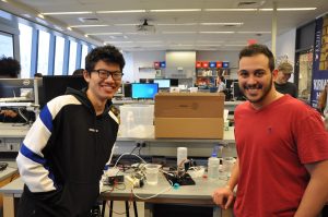
Andrew Chan (left, M.S.E. in Robotics ‘19) and Omar Abdoun (right, BE M.D./Ph.D. student) present “Cryogripper”
Almost every engineering school in the country offers a course in mechatronics — the overlap of mechanical, electrical, and computer engineering in electromechanical system design — but how many offer a course in biomechatronics? Taught by LeAnn Dourte, Ph.D., a Practice Associate Professor in Bioengineering, Penn Engineering’s Biomechatronics course (BE 570) gives students the chance to think about how the principles of mechatronic design can be used in biological settings involving orthopaedics, cardiovascular systems, and respiration, to name a few.
Throughout the course, students engage in different projects related to circuitry, signal processing, mechanics, motors, and analog controls, eventually applying all of these to biological examples before working on a final culminating project in design teams of two. In a simulation meant to mimic the sort of thinking and design processes that go behind innovations in robotic surgery, students create an electromechanical device that acts as a robotic hand. The catch? The “hand” has to have enough dexterity to pick up a water bead with a slipperiness similar to that of human tissue.
In addition to successfully performing this mechanical task using skills that the students learned throughout the semester, design teams also have to incorporate biological interfaces into the final project, such as using EMG signals to move part of the robotic hand, to give one example. Furthermore, each team needs to have a unique element to their design, whether in the use of a second biological interface, the application of Bluetooth to the system, or even a physical extension of the robotic hand to include the electromechanical equivalents of a shoulder, elbow, or wrist joint.
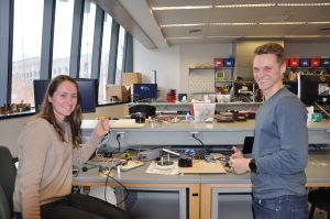
Carolyn Godone and Mike Furr (both M.S.E. in Bioengineering ‘19) model their design
Students Carolyn Godone and Mike Furr (both M.S.E. in Bioengineering ‘19) created a design inspired by the mechanical iris of a camera lens, using gears to push 3-D printed slices together in a symmetrical pattern to close around an object for pickup. They controlled their unique gripper with a thermal sensing camera that could employ a heat map of the device’s user to rotate, raise, and lower the gripper. Another pair of students, Omar Abdoun (BE M.D./Ph.D. student) and Andrew Chan (M.S.E. in Robotics ‘19), made what they called a “cryogripper”: a tissue moistened with water that freezes on demand when it contacts its target hydrogel. The ice allows the target to be lifted without falling, and the tissue can later be thawed with pumps of warm water to release hydrogel.
After weeks of working on their projects in the George H. Stephenson Foundation Educational Laboratory and Bio-MakerSpace, the class presented their final robotic hands during an open demonstration day (or Demo Day) in the lab. To see all the devices live and in action, watch the Facebook video below!
Innovations in Vascularization Could Lead to a New Future in Bioprinting
We may be one step closer to 3D-printing organs for transplants thanks to innovations in vascularization from researchers at Rice University and Washington University. Jordan Miller, Ph.D., a Penn Bioengineering alumnus, now an assistant professor of bioengineering at Rice, worked with his colleague Kelly Stevens, Ph.D., an assistant professor of the bioengineering department at Washington, to develop 3D-printed networks that mimicked the vascularized pathways for the transport of blood, lymph, and other fluids in the body. Their work appeared on a recent cover of Science, featuring a visual representation of the 3D-printed vessels in vasculature meant to mirror that of the human lung.
Relying heavily on open source 3D-printing, Miller and Stevens, along with collaborators from a handful of other institutions and start-ups, found ways to model dynamic vasculature systems similar to heart valves, airways systems, and bile ducts to keep 3D-printed tissue viable. The video below demonstrates the way the team successfully modeled vasculature in a small portion of the lung by designing a net-like structure around a sack of air. But Miller, a long-time supporter of open source printing and bioprinting, hopes that this is merely one step closer to what he sees as the ultimate goal of allowing for all organs to be bioprinted. Having that sort of power would reduce the complex issues that come with organ transplants, from organ availability to compatibility, and bring an end to a health issue that affects the over 100,000 people on the organ transplant waiting list.
A Combination of Protein Synthesis and Spectrometry Improve Cell Engineering
One goal of modern medicine is to create individualized therapeutics by figuring out a way to control cell function to perform specific tasks for the body without disrupting normal cell function. Balancing these two goals often proves to be one of the greatest difficulties of this endeavor in the lab, but researchers at Northwestern University found a way to combine the two functions at once in methods they’re calling cell-free protein synthesis and self-assembled monolayer desorption ionization (SAMDI) mass spectrometry. This innovation in the combination of the two methods accelerates the trial and error process that comes with engineering cells for a specific need, allowing researchers to cover a lot more ground in determining what works best in a smaller amount of time.
Leading the study are Milan Mrksich, Ph.D., a Henry Wade Rogers Professor of Biomedical Engineering at Northwestern, and Michael Jewett, Ph.D., a Charles Deering McCormick Professor of Teaching Excellence and co-director of the Center for Synthetic Biology at Northwestern. Together, they hope to continue to take advantage of the factory-like qualities of cell operations in order to use cells from any organisms to our advantage as needed. By helping to reduce the amount of time spent on trial and error, this study brings us one step closer to a world of efficient and individualized medicine.
Non-Invasive Sensory Stimulation as New Way of Treating Alzheimer’s
What if we could reduce the effects of Alzheimer’s disease with a non-invasive therapy comprised of only sensory inputs of light and sound? A recent study between Georgia Tech and MIT tries to make that possible. Alzheimer’s patients often have a larger than normal number of amyloid plaques in their brains, which is a naturally occurring protein that in excess can disrupt neurological function. The treatment — designed in part by Abigail Paulson, a graduate student in the lab of Annabelle Singer, Ph.D., assistant professor of Biomedical Engineering at Georgia Tech and Emory University — uses a combination of light and sound to induce gamma oscillations in brain waves of mice with high amounts of these amyloid plaques. Another lead author of the study is Anthony Martorell, a graduate student in the Tsai Lab at MIT, where Singer was a postdoctoral researcher.
This new approach is different from other non-invasive brain therapies for memory improvement, as tests demonstrated that it had the power to not only reach the visual cortex, but that it also had an effect on the memory centers in the hippocampus. An innovation like this could bring about a more widespread form of treatment for Alzheimer’s patients, as the lack of a need for surgery makes it far more accessible. Singer hopes to continue the project in the future by looking at how these sensory stimulations affect the brain throughout a variety of processes, and more importantly, if the therapy can be successfully applied to human patients.
NIH Grant Awarded to Marquette Biomedical Engineering Professor for Metal Artifact Reduction Techniques in CT Scans
Taly Gilat-Shmidt, Ph.D., an associate professor of biomedical engineering at Marquette University, recently received a $1.4 million grant from the National Institute of Health to improve methods for radiation treatment through metal artifact reduction techniques. When patients have some sort of metal that can’t be removed, such as an orthopaedic implant like a hip or knee replacement, it can interfere with the imaging process for CT scans and lead to inaccuracies by obscuring some tissue in the final images. These inaccuracies can lead to difficulty in devising treatment plans for patients who require radiation, as CT scans are often used to assess patients and determine which line of treatment is most appropriate. Gilat-Schmidt hopes to use the grant to implement tested algorithms to help reduce this variability in imaging that comes from metal implants.
People and Places
Activities for Community Education in Science (ACES), founded by Penn chemistry graduate students in 2014, aims to inspire interest and provide a positive outlook in STEM for kids and their families. The biannual event provides students grades 3–8 with an afternoon of demonstrations, experiments, and hands-on activities focused on physics and chemistry.
After an explosive opening demonstration, more than 70 students made their way between experiments in small groups, each participating in different experiments based on their age.
The Society of Women Engineers (SWE) is a non-profit organization serving as one of the world’s largest advocates for women in engineering and technology over the past six decades. With a mission to empower women to become the next leading engineers of the world, SWE is just one of many agents hoping to bring more diversity to the field. Our chapter of SWE at Penn focuses particularly on professional development, local educational outreach, and social activities across all general body members. In a new article from SWE Magazine, the organization collected social media responses from the public on the women engineers we should all know. With a diverse list of engineers from both the past and present, the article helps bring to light just how much even a handful of women contributed to the field of engineering already.
Synthetic Spinal Discs from a Penn Research Team Might Be the Solution to Chronic Back Pain
Spinal discs, the concentric circles of collagen fiber found between each vertebra of the spine, can be the source of immense back pain when ruptured. Especially for truck and bus drivers, veterans, and cigarette smokers, there is an increased risk in spinal disc rupture due to overuse or deterioration over time. But these patients aren’t alone. In fact, spinal discs erode over time for almost everyone, and are one of the sources of back pain in older patients, especially when the discs erode so much that they allow direct bone-to-bone contact between two or more vertebrae.
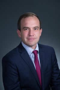
Robert Mauck, Ph.D.
Robert Mauck, Ph.D., who is the director of the McKay Orthopaedic Research Laboratory here at Penn and a member of the Bioengineering Graduate Group Faculty, led a research team in creating artificial spinal discs, with an outer layer made from biodegradable polymer and an inner layer made with a sugar-like gel. Their findings appear in Science Translational Medicine. These synthetic discs are also seeded with stem cells that produce collagen over time, meant to replace the polymer as it degrades in vivo over time. Though Mauck and his time are still far from human clinical trials for the discs, they’ve shown some success in goat models so far. If successful, these biodegradable discs could lead to a solution for back pain that integrates itself into the human body over time, potentially eliminating the need of multiple invasive procedures that current solutions require. Mauck’s work was recently featured in Philly.com.
An Untethered, Light-Activated Electrode for Innovations in Neurostimulation
Neurostimulation, a process by which nervous system activity can be purposefully modulated, is a common treatment for patients with some form of paralysis or neurological disorders like Parkinson’s disease. This procedure is typically invasive, and because of the brain’s extreme sensitivity, even the slightest involuntary movement of the cables, electrodes, and other components involved can lead to further brain damage through inflammation and scarring. In an effort to solve this common problem, researchers from the B.I.O.N.I.C. Lab run by Takashi D.Y. Kozai, Ph. D., at the University of Pittsburgh replaced long cables with long wavelength light and a formerly tethered electrode with a smaller, untethered one.
The research team, which includes Pitt senior bioengineering and computer engineering student Kaylene Stocking, centered the device on the principle of the photoelectric effect – a concept first described in a publication by Einstein as the local change in electric potential for an object when hit with a photon. Their design, which includes a 7-8 micron diameter carbon fiber implant, is now patent pending, and Kozai hopes that it will lead to safer and more precise advancements in neurostimulation for patients in the future.
A New Microfluidic Chip Can Detect Cancer in a Drop of Blood
Many forms of cancer cannot be detected until the disease has progressed past the point of optimum treatment time, increasing the risk for patients who receive late diagnoses of these kinds of cancer. But what if the diagnostic process could be simplified and made more efficient so that even a single drop of blood could be enough input to detect the presence of cancer in a patient? Yong Zeng, Ph.D., and his team of researchers at the University of Kansas in Lawrence sought to answer that question.
They designed a self-assembled 3D-nanopatterned microfluidic chip to increase typical microfluidic chip sensitivity so that it can now detect lower levels of tumor-associated exosomes in patient blood plasma. This is in large part due to the nanopatterns in the structure of the chip, which promote mass transfer and increase surface area, which in turn promotes surface-particle interactions in the device. The team applied the device to their studies of ovarian cancer, one of the notoriously more difficult kinds of cancer to detect early on in patients.
A Wearable Respiration Monitor Made from Shrinky Dinks
Michelle Khine, Ph. D., a professor of biomedical engineering at the University of California, Irvine incorporates Shrinky Dinks into her research. After using them once before in a medical device involving microfluidics, her lab recently worked to incorporate them into a wearable respiration monitor – a device that would be useful for patients with asthma, cystic fibrosis, and other chronic pulmonary diseases. The device has the capability to track the rate and volume of its user’s respiration based on measurements of the strain at the locations where the device makes contact with the user’s abdomen.
Paired with Bluetooth technology, this sensor can feed live readings to a smartphone app, giving constant updates to users and doctors, as opposed to the typical pulmonary function test, which only provides information from the time at which the test takes a reading. Though Khine and her team have only tested the device on healthy patients so far, they look forward to testing with patients who have pulmonary disorders, in hopes that the device will provide more comprehensive and accessible data on their respiration.
People and Places
Ashley Kimbel, a high school senior from Grissom High School in Huntsville, Alabama, designed a lightweight prosthetic leg for local Marine, Kendall Bane, after an attack in Afghanistan led him to amputate one of his legs below the knee. Bane, who likes to keep as active as possible, said the new lighter design is more ideal for activities like hiking and mountain biking, especially as any added weight makes balance during these activities more difficult. Kimbel used a CAD-modeling software produced by Siemens called Solid Edge, which the company hopes to continue improving in accessibility so that more students can start projects like Kimbel’s.
This week, we would like to congratulate Angela Belcher, Ph.D., on being named the new head of the Department of Biological Engineering at the Massachusetts Institute of Technology (MIT). With her appointment to this role, now half of the MIT engineering department heads are women. Belcher’s research is in the overlap of materials science and biological engineering, with a particular focus on creating nanostructures based on the evolution of ancient organisms for applications in medical diagnostics, batteries, solar cells, and more.
We would also like to congratulate Eva Dyer, Ph.D., and Chethan Pandarinath, Ph.D., both of whom are faculty members at the Walter H. Coulter Department of Biomedical Engineering at Georgia Tech and Emory University, on receiving research fellowships from the Alfred P. Sloan Foundation. Dr. Dyer, who formerly worked with Penn bioengineering faculty member Dr. Konrad Kording while he was at Northwestern University, leads research in the field of using data analysis methods to quantify neuroanatomy. Dr. Pandarinth leads the Emory and Georgia Tech Systems Neural Engineering Lab, where he works with a team of researchers to use properties of artificial intelligence and machine learning to better understand large neural networks in the brain.
Chondrus crispus, a common red algae from which carrageenan is extracted.
Medicine has made tremendous strides since the 1960s, as evidenced by the increased survival rates of combat soldiers since Vietnam. Nevertheless, blood loss remains the most common cause of death of soldiers on the battlefield. Finding a way for medics or soldiers to stop bleeding can significantly cut down on these deaths, but current approaches are either very expensive or not easy to use in combat.
According to a new paper published in Acta Biomaterialia, a solution to this problem could come from seaweed — or more precisely, from kappa-carrageenan, a type of polymeric carbohydrate produced by certain types of edible seaweed. Akhilesh K. Gaharwar, PhD, Assistant Professor of Biomedical Engineering at Texas A&M, led a study team who developed and tested an injectable hydrogel nanoengineered from kappa-carrageenan.
The authors combined kappa-carrageenan with clay-based nanoparticles to yield a hydrogel that can be injected into wounds. When the gel solidifies, it both stanches the flow of blood and helps to generate new tissue. The gel performed well in in vitro experiments. The next step will be to test the gel in animal models of wounds.
A New Understanding of Anatomy
A group of scientists collaborating among Mount Sinai Medical Center, NYU, Weill Cornell Medical Center, and the University of Pennsylvania, including Penn Bioengineering secondary faculty member Rebecca Wells, MD, published a paper in Scientific Reports detailing the heretofore unknown extent of the human interstitium and providing a new understanding of these fluid-filled compartments beneath the skin surface. The study used confocal laser endomicroscopy, which can examine structures at depths of 60-70 µm, to look at human hepatobiliary tissue. They found a reticular pattern of fluid-filled sinuses not detected before, which is connected to the lymph nodes and similar to structures found in other organs and organ systems.
On the basis of their findings, the authors suggest that our current understanding of the anatomy might be revised. Much more research is necessary, but they also believe that the fluid-filled spaces might play important roles in cancer metastasis and a number of other disease processes.
Bringing Bioprinting to the Masses
Three-dimensional printing is one of the great innovations of the last decade, and it has transformed numerous fields inside and outside of science. In the health sciences, the ability to manufacture 3D biomaterials holds enormous promise. Unfortunately, the costs of 3D printing remain prohibitive; the available models range between $10,000 and $200,000 in cost, not including the raw materials, software, etc. However, engineers at Carnegie Mellon University (CMU) might have devised a solution. In a paper published in HardwareX, Adam Feinberg, PhD, Associate Professor of Biomedical Engineering at CMU, and his coauthors describe their development of a syringe-pump large volume extruder (LVE).
Syringe pump extruders, which inject raw material into 3D printers, are already used to print biomaterials. However, achieving cheap, fast, and precise printing of 3D materials is a major technical challenge. The LVE, which is based on open-source hardware and software, significantly increases the size of the extruder without compromising speed, and it can print at sizes as small as 100 µm. The authors estimate that the materials necessary to build their bioprinter would cost less than $500 — orders of magnitude less than current models that are slower and unable to print using large volumes. Their source materials are online here.
People and Places
Missouri dominates this week’s news, with a new program at one institution and a symposium at another. At the University of Missouri, the College of Engineering has announced that it will begin offering an undergraduate program in biomedical engineering in the fall. Ninety miles away at Missouri University of Science and Technology, a symposium will be held this week — the first to be convened on the topic of biomedical humanities. The event is a collaboration between Missouri S&T’s Center for Science, Technology, and Society and the Center for Biomedical Research.
Colorado State University’s Department of Biomedical Engineering is celebrating its 10th anniversary. In that time, the department has added more than 20 faculty members to its original cohort of 29.
Finally, we offer our congratulations to Jelena Kovačević, PhD, who has been named the new dean at NYU’s Tandon School of Engineering. A graduate of Columbia and the University of Belgrade, Dr. Kovačević, who is an electrical engineer with broad interest in biomedical applications, moves to NYU from CMU and is the first-ever female dean of Tandon. Congratulations Jelena!


 Some medical conditions, like diabetes or limb amputation, have the potential to result in wounds that never heal, affecting patients for the rest of their lives. Though normal wound-healing processes are relatively understood by medical professionals, the complications that can lead to chronic non-healing wounds are often varied and complex, creating a gap in successful treatments. But biomedical engineering faculty from the University of Connecticut want to change that.