by Kaila Helm, Biological Basis of Behavior ’20; Kathleen Givan, Bioengineering and Political Science ’20; Katharine Cocherl, Bioengineering ’20; Hope McMahon, Chemical and Biomolecular Engineering ’18; and Dave Pontoriero, Biotechnology MS ’18
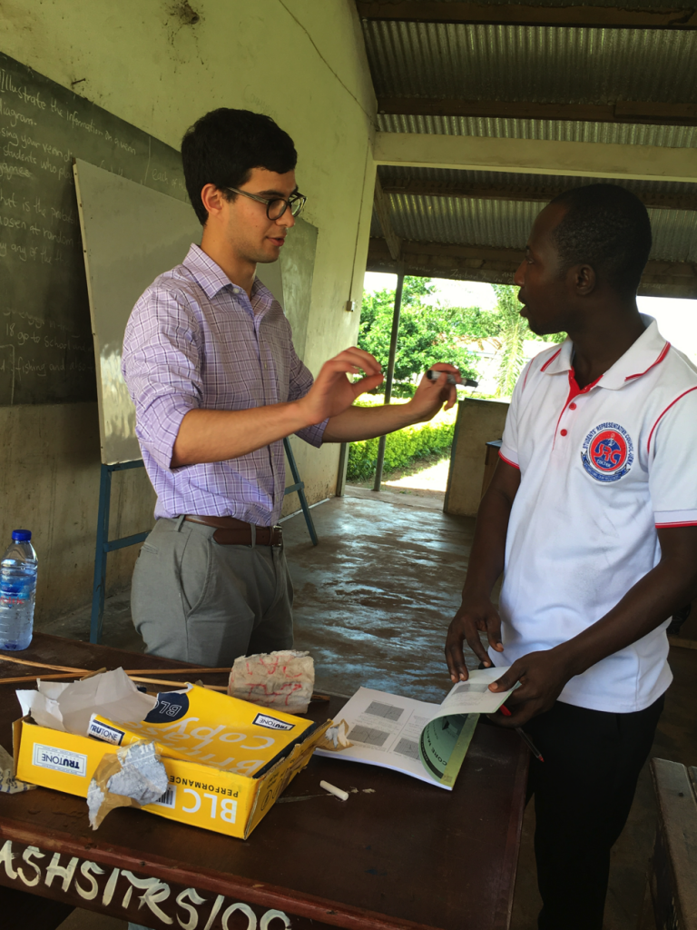
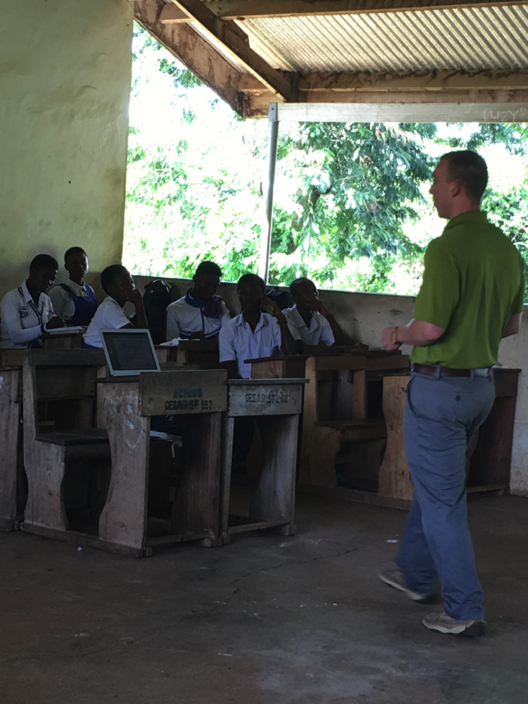
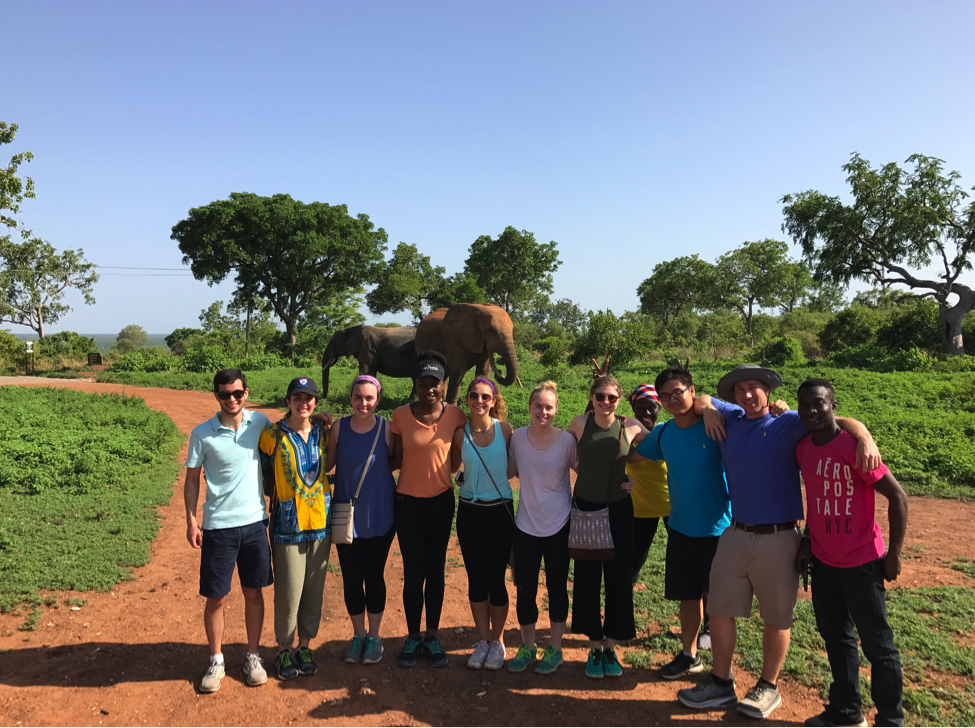
David Issadore, a faculty member in the Department of Bioengineering at the University of Pennsylvania teaches an engineering course ENGR566 – Appropriate Point of Care Diagnostics. As part of this course, he and Miriam Wattenberger from CBE, have taken nine Penn students, most of them majoring in Bioengineering, to Kumasi, Ghana, to study the diagnosis of pediatric tuberculosis. While in Ghana, these students are blogging daily on their experiences.
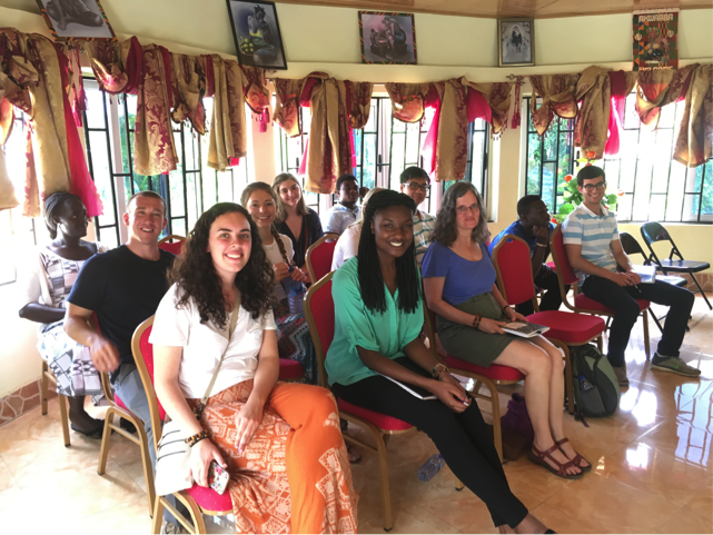
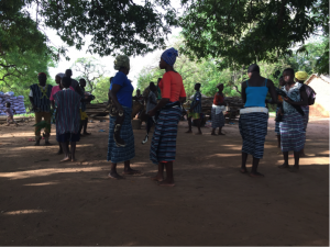
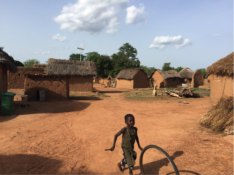
Our day started out early with a trip back to the fetish priestess’s house. Just as we were arriving at her compound, the rainy-season storm drops began to fall. Over breakfast (and for a few more hours after that), we chatted and watched both the rain and a group of small children running in, out, and around the area we were sitting.
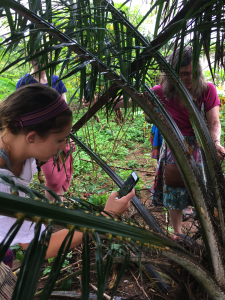
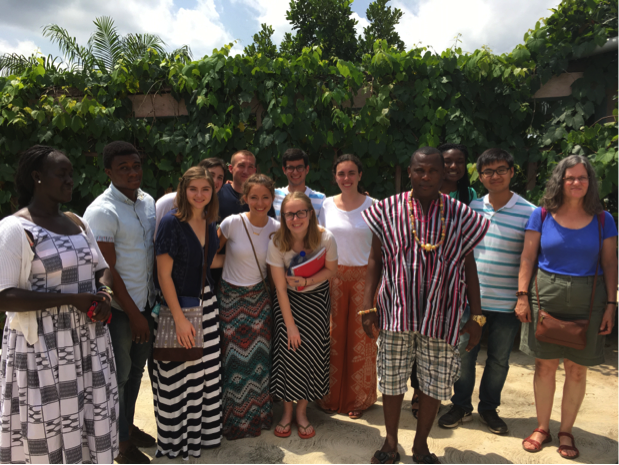
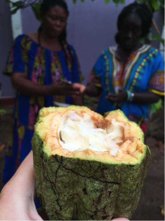
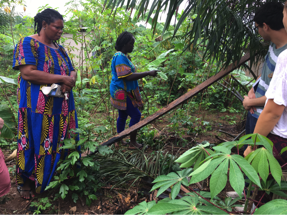
After exhausting our entire repertoire of team-bonding games and revisiting the implementation of our medical diagnostic tools with the Ghanaian program coordinator Nana Yaa, we began the serious portion of the day with a jungle tour that focused on the plants used by the priestess. She eased us in, warming us up to her knowledge by taking a strange fruit, banging it authoritatively on the wall, and offering the white flesh inside to us. Suck on the beans, she instructed. Don’t chew them. Later we learned that it was in fact the fruit of the cacao plant — yum. We give it an APOC 2017 official 5-star recommendation.
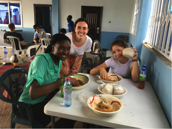
The rest of the plant tour led us into her enchanted forest of sorts. We loved hearing about how one plant could be used in so many ways. The uses ranged from curing an upset stomach to helping witches fly. She explained to us how she acquired her spiritual powers from a local river deity and how her spiritual “sight” helped her treat her patients. She even elaborated on which leaves to look for if you have a low red blood cell count. After we worked up an appetite hiking through her garden, we took a break for lunch, which consisted of yams and garden egg (eggplant) stew that the priestess herself made. She cooked all of the meals for us, and she was very welcoming to our group, greeting us hospitably.
When we arrived back at KCCR, we spent some time chatting in groups and meeting the newest resident of the guest house, a rising second-year Pitt medical school student. Others tested out the handmade slingshots gifted to them in the village we visited. After visiting one of the Ghanaian students’ dorm room, in what they call their hostels, we arrived back at KCCR to watch an episode of Black Mirror and to make some homemade chocolate-chip cookies — a perfect way to end our most jam-packed day of the trip.