Cataracts are a leading cause of vision loss worldwide. In the world’s more developed countries, laser is commonly used for cataract removal. However, in much of the developing world, lasers are expensive or difficult to acquire. In these countries, cataract surgeries are still largely performed freehand, with all of the attendant risks that such procedures involve.
One of this year’s senior design projects in the Department of Bioengineering at Penn was a surgical tool for ophthalmic surgeons to perform capsulorhexis, the fancy term for the circular incision necessary to remove a cataract. The team, which included seniors Akshatha Bhat, Nimay Kulkarni, Steven Polomski, and Ananya Sureshkumar, collaborated with the Aravind Eye Hospital in Puducherry, India, created a surgical device called the Rhex to create this round incision
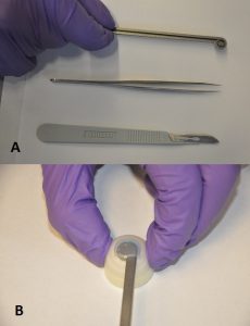
A) The Rhex (top), compared to forceps (center) and a scalpel (bottom); B) Rhex inserted into a model eye.
The Rhex (right) consists of a stem with a ring at the end, into which a blade is fitted. It can be inserted into a scleral incision, pressed, and rotated to perform the capsulorhexis. Initial testing indicated the Rhex could create a circulation incision approximately 6.7 mm in diameter, with eccentricity (indicating deviation from a perfect circle) of 0.254±0.08, which was within the acceptable range determined by the team.
The students designed the Rhex to be autoclavable and to use disposable blades. The next step will be to decrease the size of the instrument further and perhaps to use translucent or even transparent material to produce newer prototypes, which could be particularly useful, since cataract surgeries are open performed using backlighting.
With increasing age in the population, Parkinson’s disease has become increasingly common. One of the most frustrating effects of the disease is freezing of gait (FOG), in which a patient will suddenly stop while walking and find it difficult to begin again. Falls are a common consequence.
Despite intensive research, FOG is poorly understood. However, studies have shown that certain external stimuli, including metronomes and devices that provide visual cues, can be helpful. With this knowledge, a team of bioengineering students set to tackle this issue with their senior design project.
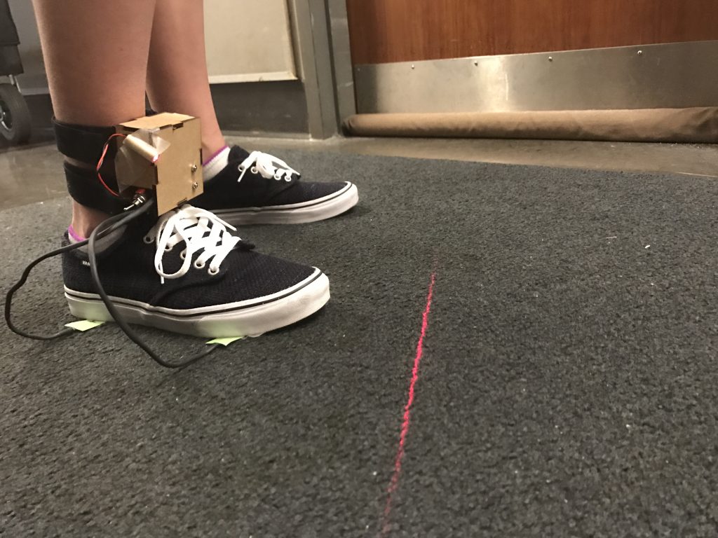
The team —whose members were Priyanka Ghosh, Fiona La, Laurel Leavitt, and Lia Lombardi — came up with ShuffleAssist, a wearable device that uses force sensors and an internal measurement unit to detect FOG and automatically provide a cue for the patient. The patient can choose a metronome beat or visual laser cue that can be provided either as determined by the device or continually, for patients who so choose.
ShuffleAssist tested well among normal subjects, detecting FOG correctly 98% of the time within approximate one second. In addition, the students were able to create their prototype for a cost of $107 per unit, compared to similarly intended products already on the market costing more than twice that much.
The next step for the team is to test the device in actual patients with Parkinson’s. The students have left the device with a faculty member in the Perelman School of Medicine who treats patients with motor disorders. This faculty member will offer the device to patients for testing.
See below for a video demonstration of ShuffleAssist.
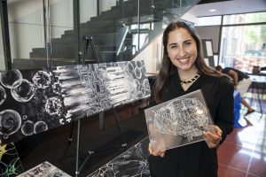
In high school, Rebecca Kellner (right) always had a dual love of art and science. When she entered the University of Pennsylvania as a freshman, she thought that her interest in art would always be separate from her pursuit of science. “I’ve always loved art and science and I wondered how I would integrate my passions into one area of study,” Rebecca says. “Then I heard about the Network Visualization Program run by Dr. Danielle Bassett . In this program, the intersection of art and science is celebrated, and this intersection is a place where I feel right at home.”
The Penn Network Visualization Program, begun in 2014, had long been a dream of Dr. Bassett. She wanted a forum where young artists and research scientists could interact with each other. “Science and art are often perceived to be at odds with each other, two fundamentally different ways of understanding the world. As a scientist, I’ve learned that the visual impact of the information I present is crucially important. Networks are visually intuitive,” says Bassett, “and represent an opportunity to foster a common language between scientists and artists.”
In this six-week summer program, young artists spend time with scientists at Penn who are performing cutting-edge research in network science as applied to social systems, human biology, and physical materials, with the underlying goal of advancing bioengineering. Faculty from the Warren Center for Network and Data Science who have volunteered their time and creativity to the project include Eleni Katifori, Erol Akcay, and Randy Kamien of the School of Arts and Sciences; Robert Ghrist and Victor Preciado of the School of Engineering and Applied Sciences; Sandra Gonzalez-Bailon of the Annenberg School of Communications; and Francis Diebold of the Wharton School of Business. During the course of the internship, the artists produce works of art interpreting and capturing the intricacies of these networks in novel ways. Artistic supervision and project advice are provided by local artists affiliated with the program. The goal of the internship is to provide scientists with new conceptualizations of their research and to provide the intern with new knowledge in scientific art applications.
Rebecca was thrilled when she was accepted into the program. During her internship she worked with a variety of scientists. Her final artwork focused on the research of Dr. Ann Hermundstad (Janelia), the postdoctoral researcher in the Physics of Living Matter Group, University of Pennsylvania Department of Physics and Astronomy. Dr. Hermundstad’s research focuses on what and how the brain sees. Fascinated by these networks, Rebecca created a painting and a laser-etched acrylic book.
Nicholas Hanchak
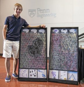
The program also invites six high school students who have exhibited creativity and academic achievement. Nicholas Hanchak (right) from Westtown School participated during the summer of 2016. “I love art, science and baseball and I am thinking about architecture as a possible career,” Nicholas says. “The Penn program challenged me to find new ways to combine these interests.” For his final project, Nicholas created a Plinko Game Board showing the difference between the networks in a healthy brain and in a brain damaged by stroke.
“Artists and scientists are kindred spirits because they both are interested in observing what is in front of them,” says Dr. Bassett. “The Network Visualization program offers an opportunity for scientists and artists to inform each other in very tangible ways.”
The program runs every other summer. During the fall, several of the artists’ pieces are showcased in Philadelphia-area middle and high schools, particularly in disadvantaged areas. These efforts are enabled by ongoing collaborations with the Netter Center for Community Partnerships and Penn’s Center for Curiosity, and they are partially funded by the National Science Foundation. Bassett hopes this outreach effort will encourage children to explore intersections between the arts and sciences, while instilling a growing appreciation of their networked world.
Brianna Wronko (left) and Guyrandy Jean-Gilles (right)
One of the Penn Bioengineering Department’s senior projects was the work of a two-person team: Brianna Wronko and Guyrandy Jean-Gilles. The result of their work was the MultiDiagnostic, a microfluidics platform that the two students describe as “A Fast, Inexpensive, and Accessible Diagnostic Solution.”
Brianna says that the project was originally conceived as a way for HIV clinics and treatment centers to test biological parameters such as viral load. However, the inability of Brianna and Guy to handle HIV-infected blood in the lab, as well as the desire to generate a product that could both serve patients directly and have a commercial focus. They decided their first offering would consist of liver function tests.
Manufactured by an automated process, the MultiDiagnostic is a paper microfluidics platform with a software component that can be run on a computer or cell phone. When a bodily fluid is placed into the platform, it diffuses into separate chambers of the platform, where colorimetric analysis is then conducted and data communicated via the software’s graphical user interface to the user.
The students currently have the platform in preclinical trials for the testing of aspartase aminotransferase and alkaline phosphatase; the ability to test alanine aminotransferase, bilirubin, and total protein are in the prototyping stage. Their current model is priced at a $10 customer price, which is considerably less expensive than competing technologies already on the market.
Among the most interesting aspects of this senior project team, other than the product itself, was that it had only two members. Asked how this fact affected their work, Brianna admitted that it posed a bit of an obstacle at first. However, she said, “we decided to break up the concept into parts, with me doing the wet lab parts, in which I have a background and Guy, whose background is in software, doing those parts.” In the end, they’re very happy with their final product.
Students in the BE Department have received several awards
Every year the Penn Bioengineering Department presents several awards to students. In addition to the Senior Design Awards, which will be featured over the course of the month, students were awarded for their service, originality, leadership, and scholarship.
The Hugo Otto Wolf Memorial Prize, endowed more than a century ago by the Philadelphia architect Otto Wolf, in memory of his son, was given to Margaret Nolan and Ingrid Lan. The Herman P. Schwan Award, named for a former faculty member in Bioengineering, was given to Elizabeth Kobe and Lucy Chai.
The Albert Giandomenico Award, presented to four students who “reflect several traits that include teamwork, leadership, creativity, and knowledge applied to discovery-based learning in the laboratory,” was given to Justin Averback, Jake Budlow, Justin Morena, and Young Shin.
In addition, Sushmitha Yarrabothula received the Bioengineering Student Leadership Award and four students — Hayley Williamson, Amey Vrudhula, Jane Shmushkis, and Ikshita Singh, won the Penn Engineering Exceptional Service Award.
Finally, the Biomedical Applied Science Senior Project Award, presented annually to the students who have “best demonstrated originality and creativity in the integration of knowledge,” was awarded to Derek Yee and Andrea Simi.
“These awards recognize many aspects of our students: their high academic achievement, exceptional collaborative spirit, and leadership abilities,” said BE department chair David Meaney. “However, these traits are not limited to the only these students. Every single one of our undergraduates at Penn pushes themselves well beyond the classroom and into the community to make a unique difference.”
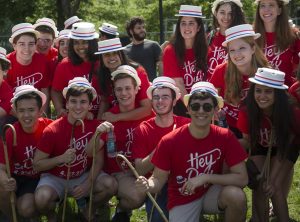
Since 1916, University of Pennsylvania undergraduates have celebrated their last class day as juniors to mark Hey Day. While initially conceived as something solemn and rather formal, today it is an opportunity for students to get decked out in red T-shirts and novelty straw hats and bamboo canes (fashions from 1916) and celebrate.
This year, Hey Day was on April 27, and it was no exception to previous years. Several of our rising seniors were celebrating with everyone on College Green.
Penn BE students celebrating Hey Day
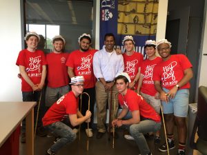
In addition to the gathering of students to be “officially” be made seniors by University President Amy Gutmann (see video here) and a passing of the gavel to next year’s junior class president, some students dropped in on their favorite teachers and staff members to say hello.
BE Lab director Sevile Mannickarottu with Penn juniors
When ABC premiered The Six Million Dollar Man more than 40 years ago, the idea of replacing or augmenting human limbs with fully functional biomechanical/biomechatronic versions probably seemed a distant possibility. In fact, the concept had already been in development for decades, but research in this area is only now coming to fruition. Three years ago, researchers in Chicago reported in the New England Journal of Medicine that they had fitted a 31-year-old amputee with a robotic leg that the patient could control with electromyographic, or EMG, signals from salvaged nerves.
Reflecting these developments, undergraduate students in the Department of Bioengineering (BE) have spent the last few weeks developing their own prosthetic devices, although both the mechanics and the “patient” are a bit cruder. Over the course of five lab sessions, these students are creating an “HCMI” — a human-cockroach machine interface that can translate an individual’s own nerve signals into ones that can control a cockroach leg.
The students performing these experiments are enrolled the first of two lab courses that BE students take as juniors. In the George H. Stephenson Foundation Undergraduate Bioengineering Laboratory, the students spend the first few sessions familiarizing themselves with cockroach anatomy. Each group then attaches an individual cockroach leg to a mechanical motor interface, creating a biomechatronic prosthesis, i.e., one that combines electronic, mechanical, and biological systems.
This part of the experiment was considered successful when the students were able to write the letters “BE” with the cockroach leg, using signals generated by computer. This is a more difficult task than it might seem, both because each cockroach leg responds at slightly different frequency-voltage ranges.
Why a cockroach leg?
“They’re easily attainable and easy to deal with,” says Sevile Mannickarottu, who is director of the Stephenson lab. “They’re also relatively large, which makes accessing their legs easy.”
The cockroach’s nervous system is also much simpler than those of birds or mammals, thus simplifying the process of creating the HCMI.
Once the students can write with the biomechantronic device, the final step of the experiment begins. Using human input, students are required to combine two devices to move the prosthetic. One of the devices is an EMG electrode; the other device is up to the student, and it can be a microphone, a motion sensor, or a range of other devices. Working directly with EMG signals is a challenge according to Mannickarottu, who described it as “incredibly noisy and difficult to interpret into meaningful data.”
After choosing their human input device, students send the signals from the device to a computer, which then converts the signal into an EMG signal, which is sent back out to the prosthetic leg. The students tried several different approaches to get the leg to move, including a musical keyboard, a force sensor, and a flex sensor. One group chose to use a Myo armband, a gesture recognition device produced by Thalmic Labs that is commonly used for video games.
With human prostheses and brain-machine interfaces rapidly advancing, overcoming a bit of entomophobia was a worthwhile endeavor for these undergrads.