Bioengineering Round-Up (December 2019)

by Sophie Burkholder

Positive results in first-in-U.S. trial of CRISPR-edited immune cells

3D render of the CRISPR-Cas9 genome editing system

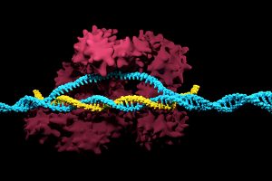
Genetically editing a cancer patient’s immune cells using CRISPR/Cas9 technology, then infusing those cells back into the patient appears safe and feasible based on early data from the first-ever clinical trial to test the approach in humans in the United States. Researchers from the Abramson Cancer Center have infused three participants in the trial thus far—two with multiple myeloma and one with sarcoma—and have observed the edited T cells expand and bind to their tumor target with no serious side effects related to the investigational approach. Penn is conducting the ongoing study in cooperation with the Parker Institute for Cancer Immunotherapy and Tmunity Therapeutics.

“This trial is primarily concerned with three questions: Can we edit T cells in this specific way? Are the resulting T cells functional? And are these cells safe to infuse into a patient? This early data suggests that the answer to all three questions may be yes,” says the study’s principal investigator Edward A. Stadtmauer, section chief of Hematologic Malignancies at Penn. Stadtmauer will present the findings next month at the 61st American Society of Hematology Annual Meeting and Exposition.

Read the rest of the story on Penn Today.

Tulane researchers join NIH HEAL initiative for research into opioid crisis

A Tulane University professor and researcher of biomedical engineering will join fellow researchers from over 40 other institutions in the National Institute of Health’s Help to End Addiction Long-Term (HEAL) Initiative. Of the $945 million that make up the project, Michael J. Moore, Ph.D. will receive a share of $1.2 million to advance research in modeling human pain through computer chips, with the help of fellow Tulane researchers Jeffrey Tasker, Ph.D., and James Zadina, Ph.D., each with backgrounds in neuroscience.

Because of the opioid epidemic sweeping the nation, Moore notes that there’s a rapid search going on to develop non-addictive painkiller options. However, he also sees a gap in adequate models to test those new drugs before human clinical trials are allowed to take place. Here is where he hopes to step in and bring some innovation to the field, by integrating living human cells into a computer chip for modeling pain mechanisms. Through his research, Moore wants to better understand not only how some drugs can induce pain, but also how patients can grow tolerant to some drugs over time. If successful, Moore’s work will lead to a more rapid and less expensive screening option for experimental drug advancements.

New machine learning-assisted microscope yields improved diagnostics

Researchers at Duke University recently developed a microscope that uses machine learning to adapt its lighting angles, colors, and patterns for diagnostic tests as needed. Most microscopes have lighting tailored to human vision, with an equal distribution of light that’s optimized for human eyes. But by prioritizing the computer’s vision in this new microscope, researchers enable it to see aspects of samples that humans simply can’t, allowing for a more accurate and efficient diagnostic approach.

Led by Roarke W. Horstmeyer, Ph.D., the computer-assisted microscope will diffuse light through a bowl-shaped source, allowing for a much wider range of illumination angles than traditional microscopes. With the help of convolutional neural networks — a special kind of machine learning algorithm — Horstmeyer and his team were able to tailor the microscope to accurately diagnose malaria in red blood cell samples. Where human physicians typically perform similar diagnostics with a rate of 75 percent accuracy, this new microscope can do the same work with 90 percent accuracy, making the diagnostic process for many diseases much more efficient.

Case Western Reserve University researchers create first-ever holographic map of brain

A Case Western Reserve University team of researchers recently spearheaded a project in creating an interactive holographic mapping system of the human brain. The design, which is believed to be the first of its kind, involves the use of the Microsoft HoloLens mixed reality platform. Lead researcher Cameron McIntyre, Ph.D., sees this mapping system as a better way of creating holographic navigational routes for deep brain stimulation. Recent beta tests with the map by clinicians give McIntyre hope that the holographic representation will help them better understand some of the uncertainties behind targeted brain surgeries.

More than merely providing a useful tool, McIntyre’s project also brings together decades’ worth of neurological data that has not yet been seriously studied together in one system. The three-dimensional atlas, called “HoloDBS” by his lab, provides a way of finally seeing the way all of existing neuro-anatomical data relates to each other, allowing clinicians who use the tool to better understand the brain on both an analytical and visual basis.

Implantable cancer traps reduce biopsy incidence and improve diagnostic

Biopsies are one of the most common procedures used for cancer diagnostics, involving a painful and invasive surgery. Researchers at the University of Michigan are trying to change that. Lonnie Shea, Ph.D., a professor of biomedical engineering at the university, worked with his lab to develop implants with the ability to attract any cancer cells within the body. The implant can be inserted through a scaffold placed under the patient’s skin, making it a more ideal option than biopsy for inaccessible organs like lungs.

The lab’s latest work on the project, published in Cancer Research, details its ability to capture metastatic breast cancer cells in vivo. Instead of needing to take biopsies from areas deeper within the body, the implant allows for a much simpler surgical procedure, as biopsies can be taken from the implant itself. Beyond its initial diagnostic advantages, the implant also has the ability to attract immune cells with tumor cells. By studying both types of cells, the implant can give information about the current state of cancer in a patient’s body and about how it might progress. Finally, by attracting tumor and immune cells, the implant has the ability to draw them away from the area of concern, acting in some ways as a treatment for cancer itself.

People and Places

Cesar de la Fuente-Nunez, PhD

The Philadelphia Inquirer recently published an article detailing the research of Penn’s Presidential Assistant Professor in Psychiatry, Microbiology, and Bioengineering, Cesar de la Fuente, Ph.D. In response to a growing level of worldwide deaths due to antibiotic-resistant bacteria, de la Fuente and his lab use synthetic biology, computation, and artificial intelligence to test hundreds of millions of variations in bacteria-killing proteins in the same experiment. Through his research, de la Fuente opens the door to new ways of finding and testing future antibiotics that might be the only viable options in a world with an increasing level of drug-resistant bacteria

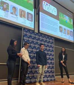
Emily Eastburn, a Ph.D. candidate in Bioengineering at Penn and a member of the Boerckel lab of the McKay Orthopaedic Research Laboratory, recently won the Ashton fellowship. The Ashton fellowship is an award for postdoctoral students in any field of engineering that are under the age of 25, third-generation American citizens, and residents of either Pennsylvania or New Jersey. A new member of the Boerckel lab, having joined earlier this fall, Eastburn will have the opportunity to conduct research throughout her Ph.D. program in the developmental mechanobiology and regeneration that the Boerckel lab focuses on.

Scholarship donors, students celebrate at ‘electric’ event

Nearly four years ago, when Angelica Du was a freshman, she recalled being completely “awestruck” upon walking into her first Scholarship Celebration.

“It’s just really warm,” the now-senior noted at this year’s event, which took place Wednesday, Nov. 20. “My donors have always been so warm with me.”

Seniors Angelica Du and Hayley Boote pose for a photo with Penn President Amy Gutmann at the Scholarship Celebration.

Du—with a smile that’s constant, as well as contagious—scanned the red-and-blue draped walls of the John R. Rockwell Gymnasium, completely transformed for the yearly event on campus, and eyed the appetizers being passed. She glanced at her proud mom, a few folks over. Hosted by the Undergraduate Named Scholarship Program, the Celebration is one that has grown to attract hundreds of scholarship donors and their recipients and families, for an evening of networking and good-old-fashioned catching up.

“[Angelica] tells me that she’s proud,” said Jerry Riesenbach, a Wharton School alumnus who helped support Du’s cost of education through the Class of 1960 scholarship fund. “And I said to her, she makes us proud. Being able to provide funds is one thing, but seeing the benefit that goes to these young people, who have such tremendous aspirations and are so grateful, is another.”

At Penn, Du, who will graduate with her bachelor’s in bioengineering in May and her master’s in December 2020, designs robots and conducts neurobiology research. She teaches thermodynamics and critical writing to her peers. She sings for a Disney-themed a cappella group, serves her community in a Christian union, celebrates her culture in the Penn Philippine Association, and advocates within several honor societies. This past summer, she worked at Thermo Fisher Scientific, running experiments for a next-generation sequencer that will take a patient’s DNA, sequence it, and diagnose it within 24 hours.

Read the full story at Penn Today.  Media contact Lauren Hertzler.

Student Spotlight: Katie Falcone

Master’s student Katie Falcone

Next up in our student spotlight series is graduate student Katie Falcone, a second-year Master’s student Bioengineering. Originally from the Philadelphia suburbs, Katie did her undergraduate degree at Drexel University’s Biomedical Engineering program and has been living in the University City area for almost nine years.


What drew you to the field of Bioengineering?

What originally drew me to this field was a “Women in Engineering Day” I attended at a local college while in high school. I had the opportunity to hear incredible women speak about their research regarding biomaterials and tissue engineering. This event showed me the impact this field can have on the world. This drove me to pursue an undergraduate degree in Biomedical Engineering, which only strengthened my passion. As I furthered my studies and began working full-time at a biotechnology company, I learned more about bioengineering. With encouragement from my coworkers and family, I decided to pursue my Master’s in Bioengineering and am delighted to have the opportunity to study at Penn.

What kind of research do you conduct, and what do you hope to focus on for your thesis?

I am actually a part-time student, who works full-time at a drug packaging and medical device company out in Exton, PA. Though I am not doing research on campus, my coursework has tied into previous research projects I have participated in at my job. My latest project entailed understanding different material properties used in container closure systems for mAb-based biologics and how they interact. This work was done to support an understanding of how to pick appropriate vial/syringe systems for various drug products in development.

What’s your favorite thing to do on Penn’s campus or in Philly?

My favorite thing to do is trying all the new restaurants and incredible foods this city has to offer. I think Philadelphia is so unique and has such rich cultural influences. With so many different neighborhoods and restaurant options you really can’t go wrong.

What did you study for your undergraduate degree, how does it pair with the work you’re doing now, and what advice would you give to your undergraduate self?

My undergraduate degree was in Biomedical Engineering. It has supported my graduate coursework very well and has given me a great opportunity to dive deeper into certain parts of my studies.

My advice to my younger self would be to take your time! It took me a little while to evaluate different graduate programs and choose which was right for me. Though it took some time, I ultimately decided what was best for me and couldn’t be happier with my choices.

What are you thinking about doing after graduate school?

Currently, I work full-time as an Associate Packaging Engineer at West Pharmaceutical Services in Exton, PA. I hope to take my degree to further my career and to help support my future aspirations at this company.

How to Build Your Own Makerspace for Under $1500

By Sophie Burkholder

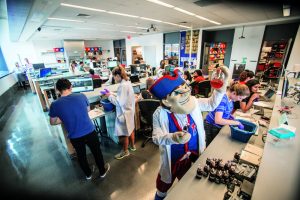
As technology and hands-on activities continue to become a larger part of education at all levels, a new movement of do-it-yourself projects is on the rise. Known as the “MakerSpace Movement,” the idea is that with the use of devices like 3-D printers, laser cutters, and simple circuitry materials, students, classes and communities can apply topics discussed in the classroom to real-life projects. Especially popular among STEM educators, the MakerSpace Movement is one that’s taken over labs in engineering schools around the country. Here at Penn, our own Stephenson Foundation Bioengineering Educational Lab and Bio-MakerSpace is equipped with all of the tools needed to bring student designs to fruition. In particular, the Stephenson Lab is the only lab on Penn’s campus that is open to all students and has both mechanical and electrical rapid prototyping equipment, as well as tools for biological and chemistry work.

Though Penn helps to fund the lab’s operation, many of the technologies and materials used in the Stephenson Lab and Bio-MakerSpace to help students throughout different class and independent projects are actually relatively affordable. Sevile Mannickarottu, Director of the Educational Laboratories, recently presented a paper describing the innovations and opportunities available to students through the MakerSpace attributes of the lab.

The Stephenson Lab mostly looks to support bioengineering majors, particularly in their lab courses and seniors design projects, but also encourages students of all disciplines to use the space for whatever MakerSpace-inspired ideas they might have, whether it be fixing a bike or measuring EMG signals for use in a mechanical engineering design.

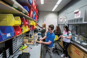
Believe it or not, however, some of the best parts of the Bio-MakerSpace can actually be purchased for a total of under $1500. Though that number is probably far beyond the individual budget of most students, it might be more affordable for a student club or dorm floor that receives additional funding from Penn. While the idea of building a MakerSpace from nothing might sound intimidating, the popularity of the movement actually helps to provide a wide range of technology and affordable options.

One of the hallmarks of the MakerSpace at the Stephenson Lab, and of any MakerSpace, is the 3-D printer. Certainly, the highest quality 3-D printers on the market are incredibly expensive, but the ones used in the Stephenson Lab are actually only $750 per printer. Even better, most spools of the PLA filaments used in printers like this one can be found online for under a price of $30 each. With access to free CAD-modeling services like OpenScad and SketchUp, all you need is a computer to start 3-D printing on your own.

But if you can’t afford a 3-D printer, or want to add more electric components to the plastic designs the printer can make, the Stephenson Lab also has NI myDAQ devices, external power sources, wires, resistors, voltage meters, Arduino kits, and other equipment that can all be purchased by students for less than $500.

The most expensive device is the NI myDAQ, which costs $200 for students, but $400 for everyone else. With access to software that includes a digital multimeter, oscilloscope, function generator, Bode analyzer, and several other applications, the myDAQ is essential to any project that involves data with electronic signals. But even without the myDAQ, components like breadboards, wire cutters, resistors, voltage regulators, and all of the other basic elements of circuitry can typically be found online for a total price of under $100.

The Stephenson Lab also provides students with Arduino Kits, which are a combination of hardware and software in circuitry and programming that can be purchased for under $100 from the Arduino website. With sensors, breadboards, and other essential circuit elements, the Arduino Kits also allow users to control their designs through a software code that corresponds to hands-on setup. Particularly for those new to understanding the relationship between codes and circuitry, an Arduino Kit can be a great place to start.

Using all of these items, you can easily start your own MakerSpace for under $1500, especially if you can take advantage of student pricing. At the heart of the MakerSpace movement is the notion that anyone, anywhere can bring their own ideas and innovations to reality with the right equipment. So if you have a project in mind, get started on building your own MakerSpace, with these tools or your own — it’s cheaper than you’d think!

Student Spotlight: Raveen Kariyawasam

Raveen Kariyawasam (BSE & BS ’21)

The first in our new student spotlight series is junior Raveen Kariyawasam. Raveen (BSE & BS ’21) is a dual degree student in the School of Engineering and Applied Science and Wharton, studying Bioengineering, Finance, and Management.


What drew you to the field of Bioengineering?

Growing up in Sri Lanka and being surrounded by relatives who were doctors, I have been fascinated by both modern and traditional medicine. However, during physician shadowing in high school, I came to the realization that I was far more fascinated with the technology doctors use rather than practicing medicine. Therefore, I made the decision to turn down studying medicine in the U.K. and come to Penn to study Bioengineering in the hopes of being more hands-on with medical technology.

Have you done research with a professor on campus? What did you like, and what didn’t you like about it?

I currently work in the Interventional Radiology Lab at the Hospital of the University of Pennsylvania (HUP) under Assistant Professor of Radiology Chamith Rajapakse. The best thing about research here is that I get to be hands-on with some of the most cutting edge technology in the world and help pioneer medical diagnostic techniques that aren’t traditionally being used anywhere else. The only downside is that the learning curve can be a little too steep.

What have been some of your favorite courses and/or projects in Bioengineering so far?

Without a doubt, my favorite BE class has to be BE 309 (Bioengineering Modeling, Analysis and Design Laboratory I) and especially the Computer-Cockroach Interface we have to develop for this lab.

What advice would you give to your freshman self?

There are way too many things happening at a given time at Penn. Take it easy and plan it out so you can do everything you want to! It’s totally possible. Who says you can’t work hard and play hard?!

What do you hope to pursue after obtaining your undergraduate degree?

My hope is to head my own health-tech startup and create technologies that will aid developing countries, starting out with my humble island of Sri Lanka first.

BE Senior Design Team Wins Berkman Prize

Senior Design Group MeVR

We would like to congratulate Penn Bioengineering Senior Design team MeVR on winning a Berkman Prize. MeVR consists of current BE seniors Nicole Chiou, Gabriel DeSantis, Ben Habermeyer, and Vera Lee. Awarded by the Penn Engineering Entrepreneurship Program, the Berkman Opportunity Fund provides grants to support students with innovative ideas that might turn into products and companies.

Bioengineering Seniors Ben Habermeyer (top left), Nicole Chiou (top right), Gabriel DeSantis (bottom right), and Vera Lee (bottom left)

MeVR is a bioresponsive virtual reality platform for administering biofeedback therapy. Biofeedback is the process of gaining greater awareness of involuntary physiological functions using sensors that provide information on the activity of those bodily systems, with the goal of gaining voluntary control over functions such as heart rate, muscle tension, and pain perception. This therapy is used to treat a variety of conditions such as chronic pain, stress, anxiety, and PTSD. These treatments cost on the order of hundreds to thousands of dollars, require the presence of a therapist to set up and deliver the therapy session, and are generally not interactive or immersive. MeVR is a platform to reduce these limitations of biofeedback therapy through an individualized, immersive, and portable device which guides users through biofeedback therapy using wearable sensors and a virtual reality environment which responds in real-time to biological feedback from the user’s body.

As part of the two-semester Senior Design course (BE 495 & BE 496), MeVR and the rest of the Bioengineering B.S.E. seniors will continue to develop their projects throughout the remainder of the academic year in George H. Stephenson Foundation Educational Laboratory & Bio-MakerSpace, culminating in their final presentations and the annual SEAS Senior Design Project Competition at the end of the spring 2020 semester.


Bioengineering Round-Up (October 2019)

by Sophie Burkholder

Innovations in Advancing a Cure for Diabetes

The blue circle is the global symbol for diabetes. Wikimedia Commons.

Diabetes is one of the more common diseases among Americans today, with the American Diabetes Association estimating that approximately 9.5 percent of the population battles the condition today. Though symptoms and causes may vary across types and patients, diabetes generally results from the body’s inability to produce enough insulin to keep blood sugar levels in check. A new experimental treatment from the lab of Sha Jin, Ph.D., a biomedical engineering professor at Binghamton University, aims to use about $1.2 million in recent federal grants to develop a method for pancreatic islet cell transplantation, as those are the cells responsible for producing insulin.

But the catch to this new approach is that relying on healthy donors of these islet cells won’t easily meet the vast need for them in diabetic patients. Sha Jin wants to use her grants to consider the molecular mechanisms that can lead pluripotent stem cells to become islet-like organoids. Because pluripotent stem cells have the capability to evolve into nearly any kind of cell in the human body, the key to Jin’s research is learning how to control their mechanisms and signaling pathways so that they only become islet cells. Jin also wants to improve the eventual culture of these islet cells into three-dimensional scaffolds by finding ways of circulating appropriate levels of oxygen to all parts of the scaffold, particularly those at the center, which are notoriously difficult to accommodate. If successful in her tissue engineering efforts, Jin will not only be able to help diabetic patients, but also open the door to new methods of evolving pluripotent stem cells into mini-organ models for clinical testing of other diseases as well.

A Treatment to Help Others See Better

Permanently crossed eyes, a medical condition called strabismus, affects almost 18 million people in the United States, and is particularly common among children. For a person with strabismus, the eyes don’t line up to look at the same place at the same time, which can cause blurriness, double vision, and eye strain, among other symptoms. Associate professor of bioengineering at George Mason University, Qi Wei, Ph.D., hopes to use almost $2 million in recent funding from the National Institute of Health to treat and diagnose strabismus with a data-driven computer model of the condition. Currently, the most common method of treating strabismus is through surgery on one of the extraocular muscles that contribute to it, but Wei wants her model to eventually offer a noninvasive approach. Using data from patient MRIs, current surgical procedures, and the outcomes of those procedures, Wei hopes to advance and innovate knowledge on treating strabismus.

A Newly Analyzed Brain Mechanism Could be the Key to Stopping Seizures

Among neurological disorders, epilepsy is one of the most common. An umbrella term for a lot of different seizure-inducing conditions, many versions of epilepsy can be treated pharmaceutically. Some, however, are resistant to the drugs used for treatment, and require surgical intervention. Bin He, Ph. D., the Head of the Department of Biomedical Engineering at Carnegie Mellon University, recently published a paper in collaboration with researchers at Mayo Clinic that describes the way that seizures originating at a single point in the brain can be regulated by what he calls “push-pull” dynamics within the brain. This means that the propagation of a seizure through the brain relies on the impact of surrounding tissue. The “pull” he refers to is of the surrounding tissue towards the seizure onset zone, while the “push” is what propagates from the seizure onset zone. Thus, the strength of the “pull” largely dictates whether or not a seizure will spread. He and his lab looked at different speeds of brain rhythms to perform analysis of functional networks for each rhythm band. They found that this “push-pull” mechanism dictated the propagation of seizures in the brain, and suggest future pathways of treatment options for epilepsy focused on this mechanism.

Hyperspectral Imaging Might Provide New Ways of Finding Cancer

A new method of imaging called hyperspectral imaging could help improve the prediction of cancerous cells in tissue specimens. A recent study from a University of Texas Dallas team of researchers led by professor of bioengineering Baowei Fei, Ph.D., found that a combination of hyperspectral imaging and artificial intelligence led to an 80% to 90% level of accuracy in identifying the presence of cancer cells in a sample of 293 tissue specimens from 102 patients. With a $1.6 million grant from the Cancer Prevention and Research Institute of Texas, Fei wants to develop a smart surgical microscope that will help surgeons better detect cancer during surgery.

Fei’s use of hyperspectral imaging allows him to see the unique cellular reflections and absorptions of light across the electromagnetic spectrum, giving each cell its own specific marker and mode of identification. When paired with artificial intelligence algorithms, the microscope Fei has in mind can be trained to specifically recognize cancerous cells based on their hyperspectral imaging patterns. If successful, Fei’s innovations will speed the process of diagnosis, and potentially improve cancer treatments.

People and Places

A group of Penn engineering seniors won the Pioneer Award at the Rothberg Catalyzer Makerthon led be Penn Health-Tech that took place from October 19-20, 2019. SchistoSpot is a senior design project created by students Vishal Tien (BE ‘20), Justin Swirbul (CIS ‘20), Alec Bayliff (BE ‘20), and Bram Bruno (CIS ‘20) in which the group will design a low-cost microscopy dianostic tool that uses computer vision capabilities to automate the diagnosis of schistosomiasis, which is a common parasitic disease. Read about all the winners here.

Virginia Tech University will launch a new Cancer Research Initiative with the hope of creating an intellectual community across engineers, veterinarians, biomedical researchers, and other relevant scientists. The initiative will focus not only on building better connections throughout departments at the university, but also in working with local hospitals like the Carilion Clinic and the Children’s National Hospital in Washington, D.C. Through these new connections, people from all different areas of science and engineering and come together to share ideas.

Associate Professor of Penn Bioengineering Dani Bassett, Ph.D., recently sat down with the Penn Integrates Knowledge University Professor Duncan Watts, Ph.D., for an interview published in Penn Engineering. Bassett discusses the origins of network science, her research in small-world brain networks, academic teamwork, and the pedagogy of science and engineering. You can read the full interview here.

An all-female group of researchers from Northern Illinois University developed a device for use by occupational therapists that can capture three-dimensional images of a patient’s hand, helping to more accurately measure the hand or wrist’s range of motion. The group presented the abstract for their design at this year’s meeting of the Biomedical Engineering Society here in Philadelphia, where Penn students and researchers presented as well.

Students’ Innovative Orthotic Device Wins Rothberg Catalyzer

NB: Penn Bioengineering would like to congratulate one of its current Senior Design teams (Alec Bayliff, Bram Bruno, Justin Swirbul, and Vishal Then) which took home the $500 Pioneer Award at this year’s Rothberg Catalyzer competition this past weekend! Keep reading for more information on the competition, awards, and winners.

Penn Health-Tech’s Rothberg Catalyzer is a two-day makerthon that challenges interdisciplinary student teams to prototype and pitch medical devices that aim to address an unmet clinical need.

The Catalyzer’s third competition was held last weekend and was won by MAR Designs, a team of Mechanical Engineering and Applied Mechanics graduate students: Rebecca Li, Ariella Mansfield and Michael Sobrepera.

MAR Designs took home the top prize of $10,000 for their project, an orthotic device that children with cerebral palsy can more comfortably wear as they sleep.

According to the team’s presentation, existing wrist orthoses “improve function and treat/prevent spasticity. However, patients report that these devices are uncomfortable which leads to lack of compliance and may also prevent patient’s eligibility for surgeries.” MAR Designs’ device initially allows full range of motion, but gradually straightens the wrist as the child is falling asleep.

In second place was Splash Throne. Team members Greg Chen, Nik Evitt, Jake Crawford and Meghan Lockwood proposed a toilet safety frame intended for elderly users. Embedded sensors track basic health information, like weight and heart-rate, as part of a preventative health routine.

Integrated Product Design students Jonah Arheim, Laura Ceccacci, Julia Lin and Alex Wan took third place with ONESCOPE, an untethered, hands-free laproscope designed to make minimally-invasive surgeries faster and safer.

Finally, SchistoSpot took home the Catalyzer’s Pioneer Award. Bioengineering and Computer and Information Science seniors Alec Bayliff, Bram Bruno, Justin Swirbul and Vishal Then designed a low-cost microscopy system that can aid in the diagnosis of the parasitic disease schistosomiasis by detecting eggs in urine samples, eliminating the need for a hospital visit.

The event was made possible by a three-year donation by scientist and entrepreneur Jonathan Rothberg, with the intent of inspiring the next generation of healthcare innovators.

Originally posted on the Penn Engineering Medium blog.

Penn Bioengineering at BMES 2019

The annual meeting of the Biomedical Engineering Society (BMES) will be held in our hometown of Philadelphia  October 16-19, 2019. The professional society for bioengineers and biomedical engineers will be taking over the city of Brotherly Love, and lots of faculty and students from Penn’s Bioengineering will be attending and presenting their research.

As previously mentioned here, Jason Burdick, Ph.D., the Robert D. Bent Professor of Bioengineering, is one of three chairs of the 2019 annual meeting. He shares this position with two other local faculty: Alisa Morss Clyne, Ph.D., Associate Professor of Mechanical Engineering and Mechanics at Drexel University; and Ruth Ochia, Ph.D., Associate Professor of Instruction in Bioengineering at Temple University. They have worked together since their appointment in 2017 to plan and chair the Philadelphia conference. Check out the video below with details of what to expect from BMES in Philly.

For those of you who have never been to BMES, the event is comprised of a mixture of academic and networking events, including keynote talks from top researchers, thousands of oral and poster presentations, participants from around the world, and social receptions. To plan your itinerary, click here for the program and agenda and here for the schedule at a glance. With the meeting being held locally this year, there are far too many presentations by Penn Bioengineering faculty and staff to list here, so check out BMES’s searchable scientific program or our searchable schedule of Penn faculty student activities at this year’s meeting (separated by day).

In addition to our academic participation, Penn Engineering and Bioengineering are also proud to sponsor this year’s meeting. Registered participants will have several venues to meet and mingle with Penn Engineering faculty, staff, and students and learn about its programs. Staff and volunteers will run a Penn Engineering booth (Booth #824) which will have literature on Penn departments and programs such as the Department of Bioengineering, the Center for Engineering MechanoBiology (CEMB), the Laboratory for Research on the Structure of Matter (LRSM), The Mahoney Institute for Neurosciences (MINS), and the Perelman School of Medicine’s Biomedical Graduate Studies group (BGS) and will be open 9:30am-5:00pm Thursday and Friday, and 9:30am-1:00pm during the conference.

For those interested in social events and networking, check out two back-to-back events on Friday night. From 6:30-8:30 pm, Penn’s Department of Bioengineering, CEMB, and LRSM will host a reception at the Philadelphia Marriott Downtown, Salon E. This will be followed by the meeting’s big BMES Dessert Bash at the Franklin Institute from 8:30-10:30 pm. (Please note: These events are open to registered conference participants only.) For those sticking around, there are no shortage of things to do in Philly, whether you are looking to site-see, shop, or dine.

We hope everyone has a wonderful time at the conference and enjoys Philadelphia! Let us know what activities you are enjoying most by tagging us on Twitter @pennbioeng or Instagram (pennbioengineering) and using the hashtag #pennbioengineering.

Blinking Eye-on-a-Chip is One of NSF’s ‘4 Awesome Discoveries’

Each week, the National Science Foundation highlights “4 Awesome Discoveries You Probably Didn’t Hear About” — a kid-friendly YouTube series that highlights particularly eye-popping NSF-supported research.

This week, one of those stories was literally about an eye, or rather, a synthetic model of one.

Dan Huh, associate professor in the Department of Bioengineering, and graduate student Jeongyun Seo, recently published a paper that outlined their new blinking eye-on-a-chip. Containing human cells and mechanical parts designed to mimic natural biological functions, including a motorized eyelid, the device was developed as platform for modeling dry eye disease and testing drugs to treat it.

See more of the series at the NSF’s Science360 site, and read more about Huh’s blinking-eye-on-a-chip research here.

Originally posted on the Penn Engineering Medium blog.