David F. Meaney, Solomon R. Pollack Professor of Bioengineering, has been named the Senior Associate Dean of Penn Engineering, effective January 1, 2020. This newly created leadership position will have oversight responsibilities in budget, space and infrastructure planning; facilities and research services; and will create and cultivate new interschool partnerships that will expand Penn Engineering’s footprint on campus.
Meaney is well known not only for his scholarship and innovation in neuroengineering and concussion science, but also for his leadership during his highly successful tenure as Chair of the Department of Bioengineering.
“Dave’s strong connections to the health schools will help strengthen Penn Engineering’s initiatives throughout campus,” says Vijay Kumar, Nemirovsky Family Dean of Penn Engineering. “He will have oversight of Penn Health-Tech, the Center for Engineering MechanoBiology and other efforts between engineering and the health schools, and Dave brings his unique creativity, energy and leadership experience to these collaborative efforts.”
Jason Burdick, Robert D. Bent Professor in the Department of Bioengineering, has been named a Fellow of the National Academy of Inventors (NAI), an award of high professional distinction accorded to academic inventors. Elected Fellows have demonstrated a prolific spirit of innovation in creating or facilitating outstanding inventions that have made a tangible impact on quality of life, economic development and the welfare of society.
Burdick’s research interests include developing degradable polymeric biomaterials that can be used for tissue engineering, drug delivery, and fundamental polymer studies. His lab focuses on developing polymeric materials for biomedical applications with specific emphasis on tissue regeneration and drug delivery. Burdick believes that advances in synthetic chemistry and materials processing could be the answer to organ and tissue shortages in medicine. The specific targets of his research include: scaffolding for cartilage regeneration, controlling stem cell differentiation through material signals, electrospinning and 3D printing for scaffold fabrication, and injectable hydrogels for therapies after a heart attack.
Danielle Bassett has been named the J. Peter Skirkanich Professor of Bioengineering.
Dr. Bassett is a Professor in the department of Bioengineering at the School of Engineering and Applied Science. She holds a Ph.D. in Physics from the University of Cambridge and completed her postdoctoral training at the University of California, Santa Barbara, before joining Penn in 2013.
Dr. Bassett has received numerous awards for her research, including an Alfred P Sloan Research Fellowship, a MacArthur Fellowship, an Office of Naval Research Young Investigator Award, a National Science Foundation CAREER Award and, most recently, an Erdos-Renyi Prize in Network Science to name but a few. She has authored over 190 peer-reviewed publications as well as numerous book chapters and teaching materials. She is the founding director of the Penn Network Visualization Program, a combined undergraduate art internship and K-12 outreach program bridging network science and the visual arts.
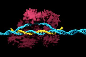
Positive results in first-in-U.S. trial of CRISPR-edited immune cells
3D render of the CRISPR-Cas9 genome editing system
Genetically editing a cancer patient’s immune cells using CRISPR/Cas9 technology, then infusing those cells back into the patient appears safe and feasible based on early data from the first-ever clinical trial to test the approach in humans in the United States. Researchers from the Abramson Cancer Center have infused three participants in the trial thus far—two with multiple myeloma and one with sarcoma—and have observed the edited T cells expand and bind to their tumor target with no serious side effects related to the investigational approach. Penn is conducting the ongoing study in cooperation with the Parker Institute for Cancer Immunotherapy and Tmunity Therapeutics.
“This trial is primarily concerned with three questions: Can we edit T cells in this specific way? Are the resulting T cells functional? And are these cells safe to infuse into a patient? This early data suggests that the answer to all three questions may be yes,” says the study’s principal investigator Edward A. Stadtmauer, section chief of Hematologic Malignancies at Penn. Stadtmauer will present the findings next month at the 61st American Society of Hematology Annual Meeting and Exposition.
Tulane researchers join NIH HEAL initiative for research into opioid crisis
A Tulane University professor and researcher of biomedical engineering will join fellow researchers from over 40 other institutions in the National Institute of Health’s Help to End Addiction Long-Term (HEAL) Initiative. Of the $945 million that make up the project, Michael J. Moore, Ph.D. will receive a share of $1.2 million to advance research in modeling human pain through computer chips, with the help of fellow Tulane researchers Jeffrey Tasker, Ph.D., and James Zadina, Ph.D., each with backgrounds in neuroscience.
Because of the opioid epidemic sweeping the nation, Moore notes that there’s a rapid search going on to develop non-addictive painkiller options. However, he also sees a gap in adequate models to test those new drugs before human clinical trials are allowed to take place. Here is where he hopes to step in and bring some innovation to the field, by integrating living human cells into a computer chip for modeling pain mechanisms. Through his research, Moore wants to better understand not only how some drugs can induce pain, but also how patients can grow tolerant to some drugs over time. If successful, Moore’s work will lead to a more rapid and less expensive screening option for experimental drug advancements.
New machine learning-assisted microscope yields improved diagnostics
Researchers at Duke University recently developed a microscope that uses machine learning to adapt its lighting angles, colors, and patterns for diagnostic tests as needed. Most microscopes have lighting tailored to human vision, with an equal distribution of light that’s optimized for human eyes. But by prioritizing the computer’s vision in this new microscope, researchers enable it to see aspects of samples that humans simply can’t, allowing for a more accurate and efficient diagnostic approach.
Led by Roarke W. Horstmeyer, Ph.D., the computer-assisted microscope will diffuse light through a bowl-shaped source, allowing for a much wider range of illumination angles than traditional microscopes. With the help of convolutional neural networks — a special kind of machine learning algorithm — Horstmeyer and his team were able to tailor the microscope to accurately diagnose malaria in red blood cell samples. Where human physicians typically perform similar diagnostics with a rate of 75 percent accuracy, this new microscope can do the same work with 90 percent accuracy, making the diagnostic process for many diseases much more efficient.
Case Western Reserve University researchers create first-ever holographic map of brain
A Case Western Reserve University team of researchers recently spearheaded a project in creating an interactive holographic mapping system of the human brain. The design, which is believed to be the first of its kind, involves the use of the Microsoft HoloLens mixed reality platform. Lead researcher Cameron McIntyre, Ph.D., sees this mapping system as a better way of creating holographic navigational routes for deep brain stimulation. Recent beta tests with the map by clinicians give McIntyre hope that the holographic representation will help them better understand some of the uncertainties behind targeted brain surgeries.
More than merely providing a useful tool, McIntyre’s project also brings together decades’ worth of neurological data that has not yet been seriously studied together in one system. The three-dimensional atlas, called “HoloDBS” by his lab, provides a way of finally seeing the way all of existing neuro-anatomical data relates to each other, allowing clinicians who use the tool to better understand the brain on both an analytical and visual basis.
Implantable cancer traps reduce biopsy incidence and improve diagnostic
Biopsies are one of the most common procedures used for cancer diagnostics, involving a painful and invasive surgery. Researchers at the University of Michigan are trying to change that. Lonnie Shea, Ph.D., a professor of biomedical engineering at the university, worked with his lab to develop implants with the ability to attract any cancer cells within the body. The implant can be inserted through a scaffold placed under the patient’s skin, making it a more ideal option than biopsy for inaccessible organs like lungs.
The lab’s latest work on the project, published in Cancer Research, details its ability to capture metastatic breast cancer cells in vivo. Instead of needing to take biopsies from areas deeper within the body, the implant allows for a much simpler surgical procedure, as biopsies can be taken from the implant itself. Beyond its initial diagnostic advantages, the implant also has the ability to attract immune cells with tumor cells. By studying both types of cells, the implant can give information about the current state of cancer in a patient’s body and about how it might progress. Finally, by attracting tumor and immune cells, the implant has the ability to draw them away from the area of concern, acting in some ways as a treatment for cancer itself.
People and Places
Cesar de la Fuente-Nunez, PhD
The Philadelphia Inquirer recently published an article detailing the research of Penn’s Presidential Assistant Professor in Psychiatry, Microbiology, and Bioengineering, Cesar de la Fuente, Ph.D. In response to a growing level of worldwide deaths due to antibiotic-resistant bacteria, de la Fuente and his lab use synthetic biology, computation, and artificial intelligence to test hundreds of millions of variations in bacteria-killing proteins in the same experiment. Through his research, de la Fuente opens the door to new ways of finding and testing future antibiotics that might be the only viable options in a world with an increasing level of drug-resistant bacteria
Emily Eastburn, a Ph.D. candidate in Bioengineering at Penn and a member of the Boerckel lab of the McKay Orthopaedic Research Laboratory, recently won the Ashton fellowship. The Ashton fellowship is an award for postdoctoral students in any field of engineering that are under the age of 25, third-generation American citizens, and residents of either Pennsylvania or New Jersey. A new member of the Boerckel lab, having joined earlier this fall, Eastburn will have the opportunity to conduct research throughout her Ph.D. program in the developmental mechanobiology and regeneration that the Boerckel lab focuses on.
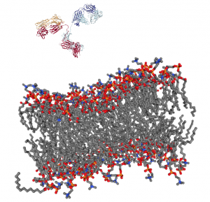
Getting a complex protein like an antibody through the membrane of a cell without damaging either is a long-standing challenge in the life sciences. Penn Engineers have found a plug-and-play solution that makes antibodies compatible with the delivery vehicles commonly used to ferry nucleic acids across that barrier.
For almost any conceivable protein, corresponding antibodies can be developed to block it from binding or changing shape, which ultimately prevents it from carrying out its normal function. As such, scientists have looked to antibodies as a way of shutting down proteins inside cells for decades, but there is still no consistent way to get them past the cell membrane in meaningful numbers.
Now, Penn Engineering researchers have figured out a way for antibodies to hitch a ride with transfection agents, positively charged bubbles of fat that biologists routinely use to transport DNA and RNA into cells. These delivery vehicles only accept cargo with a highly negative charge, a quality that nucleic acids have but antibodies lack. By designing a negatively charged amino acid chain that can be attached to any antibody without disrupting its function, they have made antibodies broadly compatible with common transfection agents.
Beyond the technique’s usefulness towards studying intracellular dynamics, the researchers conducted functional experiments with antibodies that highlight the technique’s potential for therapeutic applications. One antibody blocked a protein that decreases the efficacy of certain drugs by prematurely ejecting them from cells. Another blocked a protein involved in the transcription process, which could be an even more fundamental way of knocking out proteins with unwanted effects.
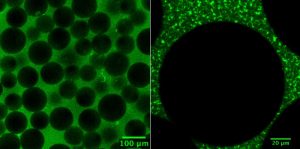
The researchers’ experiments involved making synthetic tissues with artificial “cells.” The fibrin network that surrounds these beads pull on them when compressed; by changing the number of beads in their experimental tissues, the researchers could suss out how cell-fiber interplay contributes to the tissue’s overall properties.
Tissue gets stiffer when it’s compressed. That property can become even more pronounced with injury or disease, which is why doctors palpate tissue as part of a diagnosis, such as when they check for lumps in a cancer screening. That stiffening response is a long-standing biomedical paradox, however: tissue consists of cells within a complex network of fibers, and common sense dictates that when you push the ends of a string together, it loosens tension, rather than increasing it.
Now, in a study published in Nature, University of Pennsylvania’s School of Engineering and Applied Science researchers have solved this mystery by better understanding the mechanical interplay between that fiber network and the cells it contains.
The researchers found that when tissue is compressed, the cells inside expand laterally, pulling on attached fibers and putting more overall tension on the network. Targeting the proteins that connect cells to the surrounding fiber network might therefore be the optimal way of reducing overall tissue stiffness, a goal in medical treatments for everything from cancer to obesity.
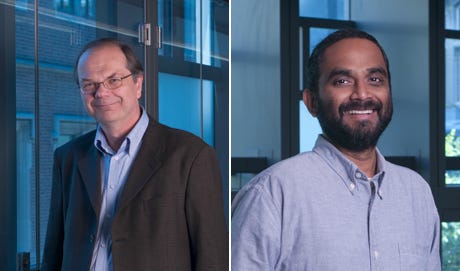
Paul Janmey and Vivek Shenoy
The study was led by Paul Janmey, Professor in the Perelman School of Medicine’s Department of Physiology and in Penn Engineering’s Department of Bioengineering, and Vivek Shenoy, Eduardo D. Glandt President’s Distinguished Professor in Penn Engineering’s Department of Materials Science and Engineering, Mechanical Engineering and Applied Mechanics, and Bioengineering, along with Anne van Oosten and Xingyu Chen, graduate students in Janmey’s and Shenoy’s labs. Van Oosten is now a postdoctoral fellow at Leiden University in The Netherlands.
Shenoy is Director of Penn’s Center for Engineering Mechanobiology, which studies how physical forces influence the behavior of biological systems; Janmey is the co-director of one of the Center’s working groups, organized around the question, “How do cells adapt to and change their mechanical environment?”
Together, they have been interested in solving the paradox surrounding tissue stiffness.
The blue circle is the global symbol for diabetes. Wikimedia Commons.
Diabetes is one of the more common diseases among Americans today, with the American Diabetes Association estimating that approximately 9.5 percent of the population battles the condition today. Though symptoms and causes may vary across types and patients, diabetes generally results from the body’s inability to produce enough insulin to keep blood sugar levels in check. A new experimental treatment from the lab of Sha Jin, Ph.D., a biomedical engineering professor at Binghamton University, aims to use about $1.2 million in recent federal grants to develop a method for pancreatic islet cell transplantation, as those are the cells responsible for producing insulin.
But the catch to this new approach is that relying on healthy donors of these islet cells won’t easily meet the vast need for them in diabetic patients. Sha Jin wants to use her grants to consider the molecular mechanisms that can lead pluripotent stem cells to become islet-like organoids. Because pluripotent stem cells have the capability to evolve into nearly any kind of cell in the human body, the key to Jin’s research is learning how to control their mechanisms and signaling pathways so that they only become islet cells. Jin also wants to improve the eventual culture of these islet cells into three-dimensional scaffolds by finding ways of circulating appropriate levels of oxygen to all parts of the scaffold, particularly those at the center, which are notoriously difficult to accommodate. If successful in her tissue engineering efforts, Jin will not only be able to help diabetic patients, but also open the door to new methods of evolving pluripotent stem cells into mini-organ models for clinical testing of other diseases as well.
A Treatment to Help Others See Better
Permanently crossed eyes, a medical condition called strabismus, affects almost 18 million people in the United States, and is particularly common among children. For a person with strabismus, the eyes don’t line up to look at the same place at the same time, which can cause blurriness, double vision, and eye strain, among other symptoms. Associate professor of bioengineering at George Mason University, Qi Wei, Ph.D., hopes to use almost $2 million in recent funding from the National Institute of Health to treat and diagnose strabismus with a data-driven computer model of the condition. Currently, the most common method of treating strabismus is through surgery on one of the extraocular muscles that contribute to it, but Wei wants her model to eventually offer a noninvasive approach. Using data from patient MRIs, current surgical procedures, and the outcomes of those procedures, Wei hopes to advance and innovate knowledge on treating strabismus.
A Newly Analyzed Brain Mechanism Could be the Key to Stopping Seizures
Among neurological disorders, epilepsy is one of the most common. An umbrella term for a lot of different seizure-inducing conditions, many versions of epilepsy can be treated pharmaceutically. Some, however, are resistant to the drugs used for treatment, and require surgical intervention. Bin He, Ph. D., the Head of the Department of Biomedical Engineering at Carnegie Mellon University, recently published a paper in collaboration with researchers at Mayo Clinic that describes the way that seizures originating at a single point in the brain can be regulated by what he calls “push-pull” dynamics within the brain. This means that the propagation of a seizure through the brain relies on the impact of surrounding tissue. The “pull” he refers to is of the surrounding tissue towards the seizure onset zone, while the “push” is what propagates from the seizure onset zone. Thus, the strength of the “pull” largely dictates whether or not a seizure will spread. He and his lab looked at different speeds of brain rhythms to perform analysis of functional networks for each rhythm band. They found that this “push-pull” mechanism dictated the propagation of seizures in the brain, and suggest future pathways of treatment options for epilepsy focused on this mechanism.
Hyperspectral Imaging Might Provide New Ways of Finding Cancer
A new method of imaging called hyperspectral imaging could help improve the prediction of cancerous cells in tissue specimens. A recent study from a University of Texas Dallas team of researchers led by professor of bioengineering Baowei Fei, Ph.D., found that a combination of hyperspectral imaging and artificial intelligence led to an 80% to 90% level of accuracy in identifying the presence of cancer cells in a sample of 293 tissue specimens from 102 patients. With a $1.6 million grant from the Cancer Prevention and Research Institute of Texas, Fei wants to develop a smart surgical microscope that will help surgeons better detect cancer during surgery.
Fei’s use of hyperspectral imaging allows him to see the unique cellular reflections and absorptions of light across the electromagnetic spectrum, giving each cell its own specific marker and mode of identification. When paired with artificial intelligence algorithms, the microscope Fei has in mind can be trained to specifically recognize cancerous cells based on their hyperspectral imaging patterns. If successful, Fei’s innovations will speed the process of diagnosis, and potentially improve cancer treatments.
People and Places
A group of Penn engineering seniors won the Pioneer Award at the Rothberg Catalyzer Makerthon led be Penn Health-Tech that took place from October 19-20, 2019. SchistoSpot is a senior design project created by students Vishal Tien (BE ‘20), Justin Swirbul (CIS ‘20), Alec Bayliff (BE ‘20), and Bram Bruno (CIS ‘20) in which the group will design a low-cost microscopy dianostic tool that uses computer vision capabilities to automate the diagnosis of schistosomiasis, which is a common parasitic disease. Read about all the winners here.
Virginia Tech University will launch a new Cancer Research Initiative with the hope of creating an intellectual community across engineers, veterinarians, biomedical researchers, and other relevant scientists. The initiative will focus not only on building better connections throughout departments at the university, but also in working with local hospitals like the Carilion Clinic and the Children’s National Hospital in Washington, D.C. Through these new connections, people from all different areas of science and engineering and come together to share ideas.
Associate Professor of Penn Bioengineering Dani Bassett, Ph.D., recently sat down with the Penn Integrates Knowledge University Professor Duncan Watts, Ph.D., for an interview published in Penn Engineering. Bassett discusses the origins of network science, her research in small-world brain networks, academic teamwork, and the pedagogy of science and engineering. You can read the full interview here.
An all-female group of researchers from Northern Illinois University developed a device for use by occupational therapists that can capture three-dimensional images of a patient’s hand, helping to more accurately measure the hand or wrist’s range of motion. The group presented the abstract for their design at this year’s meeting of the Biomedical Engineering Society here in Philadelphia, where Penn students and researchers presented as well.
We would like to congratulate Brit Shields, Ph.D., of the Penn Department of Bioengineering, on her recent promotion to Senior Lecturer. Shields got her start at Penn by completing her Ph.D. here in 2015 in History and Sociology of Science, with a dissertation on scientific diplomacy through the example of Richard Courant and New York University, where Shields completed an M.A. in Humanities and Social Thought: Science Studies. Following the conclusion of her doctorate, Shields immediately joined Penn as a lecturer in the Department of Bioengineering, teaching core undergraduate classes like the Senior Thesis course for B.A.S. degree candidates, and Engineering Ethics, one of the courses that fulfills the ethics requirement for all Penn engineering students. Furthermore, Shields has served as an advisor for undergraduate students on senior thesis in the History and Sociology of Science as well as Bioengineering.
In her new position, Shields will have the chance to further develop the engineering ethics curriculum for SEAS students. She will also take on a direct role with freshman bioengineering students as one of two bioengineering faculty members in charge of advising the incoming classes. Through these opportunities to better connect with students, Shields will be able to continue improving the ethics curriculum for all engineering majors, and increase its efficacy in imparting lessons that all engineers should take to the workforce with them. Beyond her roles in the classroom and as an advisor, Shields will also continue her research in the history and sociology of science and technology focusing on both scientific diplomacy and educational programs for engineers. She says that she “look[s] forward to collaborating with the school’s administration, faculty and students to further develop the engineering ethics curriculum. Being able to innovate in this field with such talented students is incredibly rewarding.”
We would like to congratulate Dr. Yale Cohen, Ph.D., on his recent appointment as the new Graduate Group Chair for Penn’s Department of Bioengineering. The Graduate Group is a group of faculty that graduate students in bioengineering can choose from to collaborate with on lab research. The Group includes members from nearly all of Penn’s schools, including Penn Engineering, Penn Dental, Penn Medicine, Penn Vet, and the School of Arts and Sciences.
Dr. Cohen specializes in otorhinolaryngology as his primary department, with research areas in computational and experimental neuroengineering. He will take over the role of Graduate Group Chair from Dr. Ravi Radhakrishnan, Ph.D, professor of bioengineering and chemical and biomolecular engineering, whose research specializes in cellular, molecular, and theoretical and computational bioengineering. During his tenure as Graduate Group Chair, Dr. Radhakrishnan says that “the most enjoyable part was the student talks during bioengineering seminars, and the talks at the bioengineering graduate student research symposium,” noting the way they made him realize the “depth and breadth of our graduate group, and how accomplished our students are.”
Also during his time as chair, Dr. Radhakrishnan says he was proud to expand the course BE 699 — the Bioengineering Department’s required seminar for all Ph.D. candidates — to include discussions of leadership and soft-skills, as well as instituting individualized development plans to help students track their work. In looking forward to Dr. Cohen’s appointment to the role, Dr. Radhakrishnan says that he is “a natural and accomplished scientist, educator, and amazing leader who connects so readily and well with our students and faculty.”
Dr. Cohen, looking forward to taking on his new role, says that he hopes to improve programs like the Graduate Association of Bioengineers (GABE) and the mentoring of graduate students so that they can access the wide range of wisdom that comprises the faculty, students, staff, and alumni associated with the Graduate Group. “I am thrilled to be the new chair of the BE Graduate Group and welcome your input and comments on how to improve an already outstanding program,” says Dr. Cohen.
Each week, the National Science Foundation highlights “4 Awesome Discoveries You Probably Didn’t Hear About” — a kid-friendly YouTube series that highlights particularly eye-popping NSF-supported research.
This week, one of those stories was literally about an eye, or rather, a synthetic model of one.
Dan Huh, associate professor in the Department of Bioengineering, and graduate student Jeongyun Seo, recently published a paper that outlined their new blinking eye-on-a-chip. Containing human cells and mechanical parts designed to mimic natural biological functions, including a motorized eyelid, the device was developed as platform for modeling dry eye disease and testing drugs to treat it.
See more of the series at the NSF’s Science360 site, and read more about Huh’s blinking-eye-on-a-chip research here.