In an era peppered by breathless discussions about artificial intelligence—pro and con—it makes sense to feel uncertain, or at least want to slow down and get a better grasp of where this is all headed. Trusting machines to do things typically reserved for humans is a little fantastical, historically reserved for science fiction rather than science.
Not so much for César de la Fuente, PhD, the Presidential Assistant Professor in Psychiatry, Microbiology, Chemical and Biomolecular Engineering, and Bioengineering in Penn’s Perelman School of Medicine and School of Engineering and Applied Science. Driven by his transdisciplinary background, de la Fuente leads the Machine Biology Group at Penn: aimed at harnessing machines to drive biological and medical advances.
“Biology is complexity, right? You need chemistry, you need mathematics, physics and computer science, and principles and concepts from all these different areas, to try to begin to understand the complexity of biology,” he said. “That’s how I became a scientist.”
Gabriella Daltoso, Sophie Ishiwari, Gabriela Cano, Caroline Amanda Magro, and Tifara Eliana Boyce
A team of recent Penn Bioengineering graduates have been included in list of prominent young Philadelphia innovators as chosen by The Philadelphia Business Journal and PHL Inno.
Gabriella Daltoso, Sophie Ishiwari, Gabriela Cano, Caroline Amanda Magro, and Tifara Eliana Boyce founded Sonura as their Senior Design Project in Bioengineering. The team, who all graduated in 2023, picked up a competitive President’s Innovation Prize for their beanie that promotes the cognitive and socioemotional development of newborns in the NICU by protecting them from the auditory hazards of their environments while fostering parental connection. Now, they have been included in the list of fourteen Inno Under 25 honorees for 2023.
“To determine this year’s list, the Philadelphia Business Journal and PHL Inno sought nominations from the public and considered candidates put forth by our editorial team. To be considered, nominees must be 25 years of age or younger and work for a company based in Greater Philadelphia and/or reside in the region.
Honorees span a wide range of industries, including consumer goods, biotechnology and environmental solutions. Many are products of the region’s colleges and universities, though some studied farther afield before setting up shop locally.”
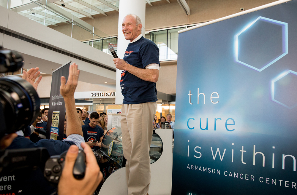
Carl June, at the flash mob celebration of the FDA approval of the CAR T cell therapy he developed, in August 2017. (Image: Courtesy of Penn Medicine Magazine)
For most of modern medicine, cancer drugs have been developed the same way: by designing molecules to treat diseased cells. With the advent of immunotherapy, that changed. For the first time, scientists engineered patients’ own immune systems to recognize and attack diseased cells.
One of the best examples of this pioneering type of medicine is CAR T cell therapy. Invented in the Perelman School of Medicine by Carl June, the Richard W. Vague Professor in Immunotherapy, CAR T cell therapy works by collecting T cells from a patient, modifying those cells in the lab so that they are designed to destroy cancerous cells, and reinfusing them into the patient. June’s research led to the first FDA approval for this type of therapy, in 2017. Six different CAR T cell therapies are now approved to treat various types of blood cancers. Carl June, at the flash mob celebration of the FDA approval of the CAR T cell therapy he developed, in August 2017. (Image: Courtesy of Penn Medicine Magazine)
CAR T cell therapy holds the potential to help millions more patients—if it can be successfully translated to other conditions. June and colleagues, including Daniel Baker, a fourth-year doctoral student in the Cell and Molecular Biology department, discuss this potential in a perspective published in Nature.
In the piece, June and Baker highlight other diseases that CAR T cell therapy could be effective.
“CAR T cell therapy has been remarkably successful for blood cancers like leukemias and lymphomas. There’s a lot of work happening here at Penn and elsewhere to push it to other blood cancers and to earlier stage disease, so patients don’t have to go through chemo first,” June says. “Another big priority is patients with solid tumors because they make up the vast majority of cancer patients. Beyond cancer, we’re seeing early signs that CAR T cell therapy could work in autoimmune diseases, like lupus.”
As for which diseases to pursue as for possible future treatment, June says, “essentially it boils down to two questions: Can we identify a population of cells that are bad? And can we target them specifically? Whether that’s asthma or chronic diseases or lupus, if you can find a bad population of cells and get rid of them, then CAR T cells could be therapeutic in that context.”
“What’s exciting is it’s not just theoretical at this point. There have been clinical reports in other autoimmune diseases, including myasthenia gravis and inflammatory myopathy,” Baker says. “But we are seeing early evidence that CAR T cell therapy will be successful beyond cancer. And it’s really opening the minds of people in the field to think about how else we could use CAR T. For example, there’s some pioneering work at Penn from the Epstein lab for heart failure. The idea is that you could use CAR T cells to get rid of fibrotic tissue after a cardiac injury, and potentially restore the damage following a heart attack.”
Baker adds, “there’s no question that over the last decade, CAR T cell therapy has revolutionized cancer. I’m hoping to play a role in bringing these next generation therapies to patients and make a real impact over the next decade. I think there’s potential for cell therapy to be a new pillar of medicine at large, and not just a new pillar of oncology.”
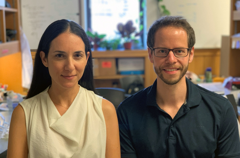
Perelman School of Medicine’s Maayan Levy, and Christoph Thaiss. (Image: Courtesy of Penn Medicine News)
When we hear about gut bacteria, we may think about probiotics and supplements marketed to help with digestion, about how taking antibiotics might affect our intestinal tract, or perhaps about trendy diets that aim to improve gut health.
But two researchers at Penn Medicine think that understanding the microbiome, the entirety of microbial organisms associated with the human body, might be the key to deciphering the fundamental mechanisms that make our bodies work. They think these microbes may work like a call center switchboard, making connections to help different organs, biological systems, and the brain communicate. Maayan Levy, and Christoph Thaiss, both assistant professors of microbiology at the Perelman School of Medicine, argue that the microbiome is instrumental to revealing how signals from the gastrointestinal tract are received by the rest of the body—which may hold the key to understanding inter-organ communication in general. Perelman School of Medicine’s Maayan Levy, and Christoph Thaiss. (Image: Courtesy of Penn Medicine News)
While the gut sends signals to all parts of the body to initiate various biological processes, the mechanisms underlying this communication—and communication between different organs involved in these processes—is relatively unknown.
“The more we learn about the role the microbiome plays in a wide range of diseases— from cancer to neurodegenerative diseases to inflammatory diseases—the more important it becomes to understand what exactly its role is,” says Thaiss. “And hopefully once we understand how it works, we can use the microbiome to treat these diseases.”
Levy and Thaiss joined the faculty at Penn Medicine after completing their graduate studies in 2018. Here, they continue to investigate the role of the microbiome in various biological processes.
In his lab, Thaiss focuses on the impact of the microbiome on the brain. He recently identified species of gut-dwelling bacteria that activate nerves in the gut to promote the desire to exercise. Most recently, Thaiss published a study that identified the cells that communicate psychological stress signals from the brain to the gastrointestinal tract, and cause symptoms of inflammatory bowel disease.
Meanwhile, in her lab, Levy examines how the microbiome influences the development of diseases, like cancer, and other conditions throughout the body.
A recent publication authored by Levy suggested that the ketogenic diet (high fat, low carbohydrate) causes the production of a metabolite called beta-hydroxybutyrate (BHB), that suppresses colorectal cancer in small animal models.
Now, Levy is collaborating with Bryson Katona, an assistant professor of Medicine in the division of gastroenterology who specializes in gastrointestinal cancers, to investigate whether BHB has the same effect in patients with Lynch syndrome, which causes individuals to have a genetic predisposition to many different kinds of cancer, including colon cancer. These efforts are part of a growing emphasis at Penn on finding methods to intercept cancer in its earliest stages.
“It’s remarkable that we were able to quickly take the findings from our animal models and rapidly design a clinical trial,” Levy says. “One of the most exciting aspects of our work is not only making discoveries about how our bodies work on a biological level, but then being able to work with the world’s leading clinical experts to translate these discoveries into therapies for patients.”
Further, studies led by Levy and Thaiss often utilize human samples and data from the Penn Medicine BioBank, to validate animal model findings in the tissue of human patients suffering from the diseases which they are investigating.
While Levy and Thaiss pursue different research interests with their labs, they also collaborate often, building on their previous research into what the microbiome does, and its role in the biological processes that keep us healthy. Their long-term goal is to learn about the mechanisms by which the gastrointestinal tract influences disease processes in other organs to treat various diseases of the body using the gastrointestinal tract as a noninvasive entry point to the body.
“Some of the most common and devastating diseases in humans—like cancer or neurodegeneration—are difficult to treat because they are no existing therapies that can reach the brain,” says Thaiss. “If we can understand how the gastrointestinal tract interacts with other organs in the body, including the brain, we might be able to develop treatments that ‘send messages’ to these organs through the body’s natural communication pathways.”
“Obviously there is a lot more basic biology to be uncovered before we get there,” adds Levy. “Most importantly, we want to map all the different routes by which the gastrointestinal tract interacts with the body, and how that communication happens.”
Christopher Thaiss is Assistant Professor in Microbiology in the Perelman School of Medicine. He is a member of the Penn Bioengineering Graduate Group.
Genetic diseases that involve the central nervous system (CNS) often impact children before birth, meaning that once a child is born, irreversible damage has already been done. Given that many of these conditions result from a mutation in a single gene, there has been growing interest in using gene editing tools to correct these mutations before birth.
However, identifying the appropriate vehicle to deliver these gene editing tools to the CNS and brain has been a challenge. Viral vectors used to deliver gene therapies have some potential drawbacks, including pre-existing viral immunity and vector-related adverse events, and other options like lipid nanoparticles (LNPs) have not been investigated extensively in the perinatal brain.
Now, researchers in the Center for Fetal Research at Children’s Hospital of Philadelphia (CHOP) and Penn Engineering have identified an ionizable LNP that can deliver mRNA base editing tools to the brain and have shown it can mitigate CNS disease in perinatal mouse models. The findings, published in ACS Nano, open the door to mRNA therapies that could be delivered pre- or postnatally to treat genetic CNS diseases.
The research team began by screening a library of ionizable LNPs – microscopic fat bubbles that have a positive charge at low pH but neutral charge at physiological conditions in the body. After identifying which LNPs were best able to penetrate the blood-brain barrier in fetal and newborn mice, they optimized their top-performing LNP to be able to deliver base editing tools. The LNPs were then used to deliver mRNA for an adenine base editor, which would correct a disease-causing mutation in the lysosomal storage disease, MPSI, by changing the errant adenine to guanine.
The researchers showed that their LNP was able to improve the symptoms of the lysosomal storage disease in the neonatal mouse brain, as well as deliver mRNA base editing tools to the brain of other animal models. They also showed the LNP was stable in human cerebrospinal fluid and could deliver mRNA base editing tools to patient-derived brain tissue.
“This proof-of-concept study – co-led by Rohan Palanki, an MD/PhD student in my lab, and Michael Mitchell’s lab at Penn Bioengineering – supports the safety and efficacy of LNPs for the delivery of mRNA-based therapies to the central nervous system,” said co-senior author William H. Peranteau, MD, an attending surgeon in the Division of General, Thoracic and Fetal Surgery at CHOP and the Adzick-McCausland Distinguished Chair in Fetal and Pediatric Surgery. “Taken together, these experiments provide the foundation for additional translational studies and demonstrate base editing facilitated by a nonviral delivery carrier in the NHP fetal brain and primary human brain tissue.”
This story was written by Dana Bate. It originally appeared on CHOP’s website.
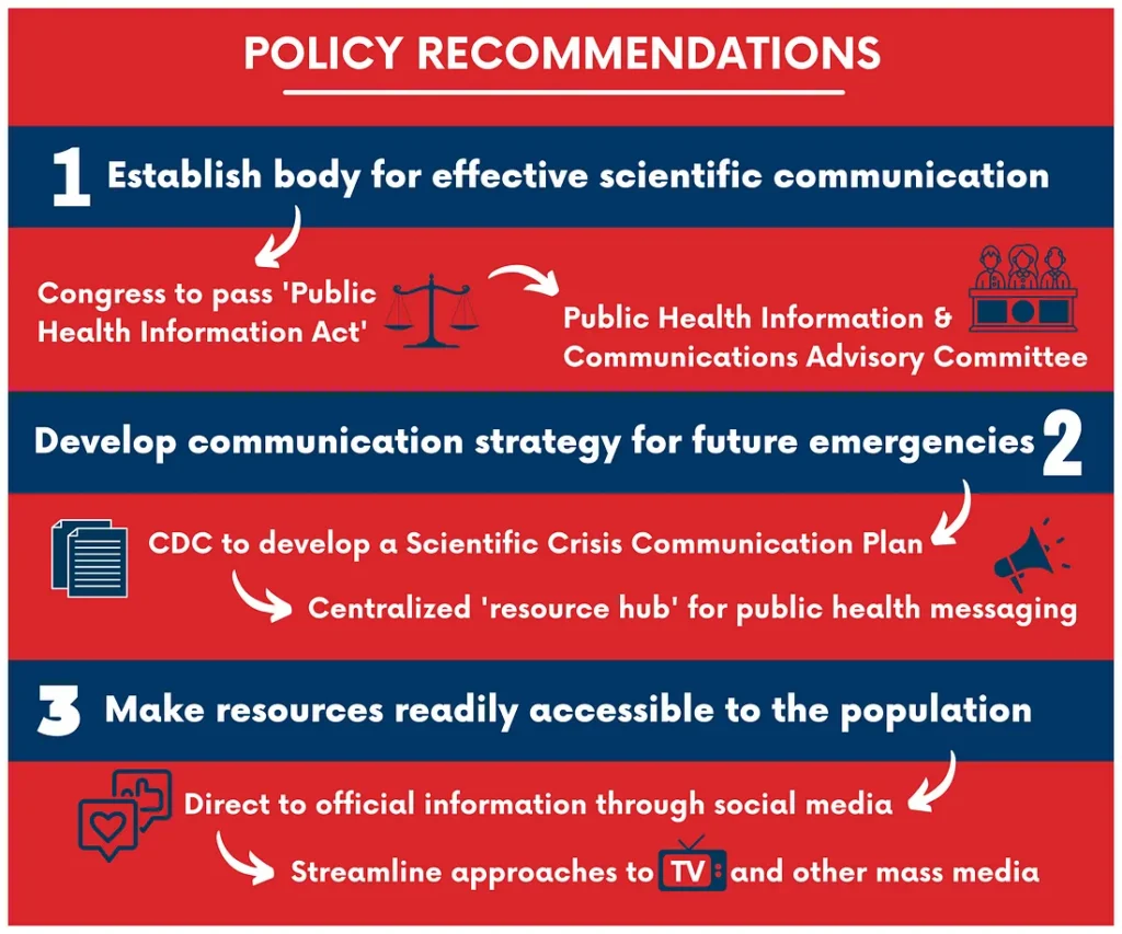
Three graduate students in Bioengineering have collaborated to craft a list of recommendations to improve science communication during national health emergencies.
Doctoral students Miles J. Arnett, Dimitris Boufidis, and Melanie Hilman are part of the Penn Science Policy and Diplomacy Group (PSPDG), student organization which creates opportunities for students to get hands-on experience in Science Policy, Diplomacy, and Communication.
Their brief reviews the public health response to the COVID-19 pandemic and recommends specific improvements to science policy and communication by national scientific institutions:
“The public health response to the pandemic was dramatically weakened by an uncoordinated communication strategy, inconsistent messaging, and fractured media environments. These shortcomings had a real human cost, with an estimated hundreds of thousands of Americans dying as a consequence of high rates of vaccine hesitancy. Now, in the aftermath of the pandemic, we have a chance to learn from this crisis and develop a more robust science communication infrastructure for future health emergencies.“
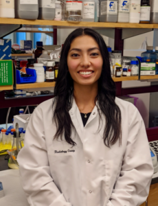
Cosette Tomita, a master’s student in Bioengineering, spoke with Penn Engineering Graduate Admissions about her research in cellular therapy and her path to Penn Engineering.
“What were you doing before you came to Penn Engineering?
After college I wanted to get some industry experience before going to graduate school, so I spent a year working for a pharmaceutical company in New Jersey. I learned a lot—but mostly I learned that I wanted to go back into academia. So I was looking for a more research-oriented position to boost my graduate school applications, and I found a position at Penn’s cyclotron facility. Shortly after that, I applied to the master’s program. I’m still working at the cyclotron, so I’m doing the program part time.
How has your experience in the program been so far?
I love the research I’m doing here. I love the collaboration we have and the fact that I’m able to work with whoever I want to. And I can only say good things about my PI, Robert Mach. He’s a very busy man, but he makes time for his people. And he recognizes when somebody has a lot on their plate and he will go to bat for that person.
What’s your research all about?
The focus of my PI’s lab is on neurodegenerative diseases and opiate use, so we’re looking to make imaging agents and antagonists that can help with the opioid crisis.
For my project, I wanted to look at treating neurodegenerative disease from the perspective of cellular therapy. My PI doesn’t have that expertise, so when I came to him with this idea, he said I should talk to Mark Sellmyer in the bioengineering department. He does a lot of cellular therapies, cell engineering, protein engineering and things of that nature. So his lab is more biological.
I don’t have a grant for my research, so my advisors are supporting it out of their own pockets. They could have said, no, you need to work on this project that’s already going on in the lab. But they gave me the intellectual freedom to do what I wanted to do.”
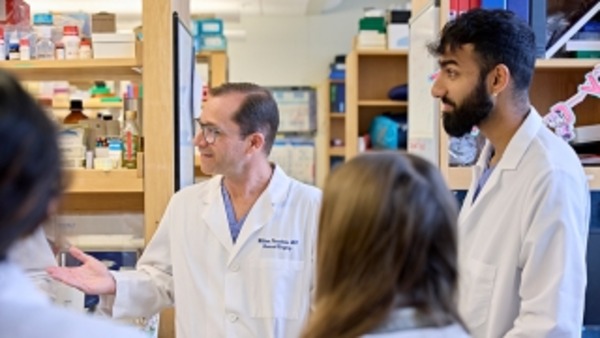
Matthew Aronson (left), Ph.D. student in Bioengineering, and Riccardo Gottardi, Assistant Proessor in Bioengineering and Pediatrics.
Riccardo Gottardi, Assistant Professor in Pediatrics in the Perelman School of Medicine and in Bioengineering in the School of Engineering and Applied Science, has been named a “Young Innovator of Cellular and Molecular Bioengineering” by Cellular and Molecular Bioengineering, the official journal of the Biomedical Engineering Society (BMES). Gottardi is Chief Scientist in the Pediatric Airway Frontier Program at the Children’s Hospital of Philadelphia (CHOP). He leads the Bioengineering and Biomaterials (Bio2) Lab, and was recognized here for his research to prevent subglottic stenosis in children.
Gottardi’s work in subglottic stensosis, a severe narrowing of the airway in response to intubation, was recently profiled in CHOP’s Cornerstone Blog. CHOP’s award press release describes Gottardi’s innovative treatment:
“Prior studies by Dr. Gottardi’s lab used in vitro models to demonstrate that incorporating AMPs into polymer-coated tubes can inhibit bacterial growth and modulate the upper-airway microbiome. In a recent study in Cellular and Molecular Engineering, led by [Bioengineering] PhD student Matthew Aronson of the Gottardi Lab, the researchers went a step further and used both ex vivo and in vivo models to show how their patent-pending antimicrobial peptide-eluting endotracheal tube (AMP-ET) effectively targeted the local airway microbiota, reducing inflammation and resolving stenosis.
‘I am honored to be recognized by Cellular and Molecular Engineering for this exciting and notable award,” Dr. Gottardi said. “We are hopeful that our airway innovation will show similar success in human trials, so that we can improve outcomes for intubated pediatric patients.’”
A research team led by engineers at the University of Pennsylvania and Northwestern University scientists has created a new synthetic biology approach, or a “QR code for cancer cells,” to follow tumor cells over time, finding there are meaningful differences in why a cancer cell dies or survives in response to anti-cancer therapies.
Remarkably, what fate cancer cells choose after months of therapy is “entirely predictable” based on seemingly small, yet important, differences that appear even before treatment begins. The researchers also discovered the reason is not genetics, contrary to beliefs held in the field.
The study outlined the team’s new technology platform that developed a QR code for each of the millions of cells for scientists to find and use later — much like tagging swans in a pond. The QR code directs researchers to a genome-wide molecular makeup of these cells and provides information about how they’ve reacted to cancer treatment.
“We think this work stands to really change how we think about therapy resistance,” said Arjun Raj, co-senior author and Professor in Bioengineering in the School of Engineering and Applied Science at the University of Pennsylvania. “Rather than drug-resistant cells coming in just one flavor, we show that even in highly controlled conditions, different ‘flavors’ can emerge, raising the possibility that each of these flavors may need to be treated individually.”
In the study, the lab and collaborators sought to apply synthetic biology tools to answer a key question in cancer research: What makes certain tumors come back a few months or years after therapy? In other words, could the lab understand what causes some rare cells to develop therapeutic resistance to a drug?
“There are many ways cells become different from each other,” said Yogesh Goyal, the co-senior author at Northwestern University. “Our lab asks, how do individual cells make decisions? Understanding this in the context of cancer is all the more exciting because there’s a clinically relevant dichotomy: A cell dies or becomes resistant when faced with therapies.”
Using the interdisciplinary team, the scientists put the before-and-after cloned cells through a whole genome sequencing pipeline to compare the populations and found no systematic underlying genetic mutations to investigate the hypothesis. Raj and Goyal helped develop the QR code framework, FateMap, that could identify each unique cell that seemed to develop resistance to drug therapy. “Fate” refers to whether a cell dies or survives (and if so, how), and the scientists “map” the cells across their lifespan, prior to and following anti-cancer therapy. FateMap is the result of work from several research institutions, and it applies an amalgamation of concepts spanning several disciplines, including synthetic biology, genome engineering, bioinformatics, machine learning and thermodynamics.
“Some are different by chance — just as not all leaves on a tree look the same — but we wanted to determine if that matters,” Goyal said. “The cell biology field has a hard time defining if differences have meaning.”
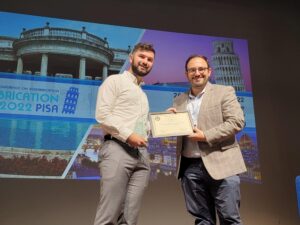
Paul Gehret (left) and Riccardo Gottardi, PhD, at Biofabrication 2022, the International Conference on Biofabrication.
Bioengineering researchers at Children’s Hospital of Philadelphia are developing a less invasive and quicker method to create cartilage implants as an alternative to the current treatment for severe subglottic stenosis, which occurs in 10 percent of premature infants in the U.S.
Subglottic stenosis is a narrowing of the airway, in response to intubation. Severe cases require laryngotracheal reconstruction that involves grafting cartilage from the rib cage with an invasive surgery. With grant support from the National Institutes of Health, Riccardo Gottardi, PhD, who leads the Bioengineering and Biomaterials (Bio2) Lab at CHOP, is refining a technology called Meniscal Decellularized scaffold (MEND). Working with a porcine model meniscus, the researchers remove blood vessels and elastin fibers to create pathways that allow for recellularization. Dr. Gottardi and his team then harvest ear cartilage progenitor cells (CPCs) with a minimally invasive biopsy, combine them with MEND, and create cartilage implants that could be a substitute for the standard laryngotracheal reconstruction.
While laryngotracheal reconstruction in the adult population has a success rate of up to 96%, success rates in children range from 75% to 85%, and children often require revision surgery due to a high incidence of restenosis. The procedure also involves major surgery to remove cartilage from the rib cage, which is more difficult for childrens’ smaller bodies.
“Luckily not many children suffer from severe subglottic stenosis, but for those who do, it is really serious,” said Dr. Gottardi, who also is assistant professor in the Department of Pediatrics and Department of Bioengineering at CHOP and the University of Pennsylvania. “With our procedure, we have an easily accessible source for the cartilage and the cells, providing a straightforward and noninvasive treatment option with much potential.”
Riccardo Gottardi is an Assistant Professor in the Department of Pediatrics, Division of Pulmonary Medicine in the Perelman School of Medicine and in the Department of Bioengineering in the School of Engineering and Applied Science. He also holds an appointment in the Children’s Hospital of Philadelphia (CHOP).
Paul Gehret is a Ph.D. student in Bioengineering, an Ashton Fellow and a NSF Fellow. His research focuses on leveraging decellularized cartilage scaffolds and novel cell sources to reconstruct the pediatric airway.

 Cosette Tomita, a master’s student in Bioengineering, spoke with Penn Engineering Graduate Admissions about her research in cellular therapy and her path to Penn Engineering.
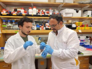
 A research team led by engineers at the