Dan Huh’s Space-based Organ-on-a-Chip Experiments Featured in WIRED

By Lauren Salig

SpaceX launched its 17th resupply mission to the International Space Station on May 4, with bioengineering professor Dan Huh’s organ-on-a-chip experiments in tow.

Dan Huh, the Wilf Family Term Assistant Professor in the Department of Bioengineering, researches human organs and the diseases that infect them by engineering devices made of living cells that act as stand-ins for organs. Huh’s lab has developed imitations of many organs, including the placenta and the eye, but it’s his lung-on-a-chip and his bone-marrow-on-a-chip that are reaching unprecedented heights as part of a new experiment taking place at the International Space Station (ISS).

On May 4, SpaceX launched a ISS-bound cargo capsule carrying Huh’s organ-on-a-chip experiments, which will remain in space for a month. Once back on Earth, the chips that spent time in space will be compared to control chips from Huh’s lab that are being monitored in parallel. Huh’s team is looking to see how being in space affects bacterial infections in lungs and white blood cell behavior in bone marrow. The researchers’ hope is that their studies will reveal important information about how human organs function both in space and on Earth.

Daniel Oberhaus of WIRED wrote an article describing the multiple organs-on-a-chip experiments being conducted at the ISS, including the two experiments headed by Huh:

Dan Huh is a bioengineer at the University of Pennsylvania and the lead researcher on the lung tissue chip headed to the ISS. This lung chip models a human airway and will be infected with Pseudomonas aeruginosa, a species of bacteria that had previously been found on the ISS. On Earth this bacteria is usually associated with respiratory infections, which are one of the leading types of illness on long-duration missions to the ISS.

Huh says scientists still know very little about why astronauts’ immune response seems to become suppressed in orbit, and the tissue chips are aimed at building a better understanding of the phenomenon.


Originally posted at the Penn Engineering Medium Blog.

Read the entire article at WIRED.

Penn Engineers: Cells Require Gene Expression Feedback to Keep Moving

By Lauren Salig

When cells move throughout the body, they do so by dragging themselves, using molecular “arms” to pull themselves closer to where they need to be while unlatching themselves from the area they’re moving away from. In a recent study, Penn Engineers looked at a few mechanobiological factors that help regulate cells’ migration towards their destination, providing new insight into the gene expression feedback loops that keep them from getting stuck.

Joel Boerckel and Devon Mason

The research was led by Joel Boerckel, Assistant Professor of Orthopaedic Surgery in the Perelman School of Medicine and in Bioengineering in Penn Engineering, and bioengineering graduate student Devon Mason. Co-authors include bioengineering graduate student Joseph Collins and researchers from the University of Notre Dame, Indiana University and Purdue University.

The study was published in the Journal of Cell Biology.

Read the full story at the Penn Engineering Medium Blog.

BE’s Danielle Bassett Profiled in Science Magazine

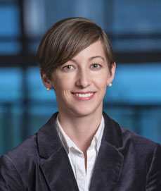
Danielle Bassett, PhD

Danielle Bassett, Eduardo D. Glandt Faculty Fellow and Associate Professor in theDepartment of Bioengineering, grew up in central Pennsylvania where she and her 10 siblings were homeschooled. Back then, Bassett had aspirations to become a professional pianist, a dream shattered by stress fractures in her arm at age 16.

Now, Bassett is a renowned physicist and MacArthur fellow who has pushed the field of network science, which studies connections and interactions between parts of a whole, to new realms. Bassett’s research focuses on brain function, including work on how brains of people with schizophrenia are organized, how brain communication changes with learning, and how the brain is able to switch between tasks.

Kelly Servick of Science sat down with Bassett to talk through her incredible journey from child pianist to leading network scientist:

““By 17, discouraged by her parents from attending college and disheartened at her loss of skill while away from the keys, she expected that responsibilities as a housewife and mother would soon eclipse any hopes of a career. ‘I wasn’t happy with that plan,’ she says.

Instead, Bassett catapulted herself into a life of research in a largely uncharted scientific field now known as network neuroscience. A Ph.D. physicist and a MacArthur fellow by age 32, she has pioneered the use of concepts from physics and math to describe the dynamic connections in the human brain. ‘She’s now the doyenne of network science,’ says theoretical neuroscientist Karl Friston of University College London. ‘She came from a formal physics background but was … confronted with some of the deepest questions in neuroscience.’”

Continue reading about Bassett’s career path and evolving research interests at Science.

Reposted from the Penn Engineering Medium blog. Media contact Evan Lerner.

Week in BioE: April 19, 2019

by Sophie Burkholder

New Vascularized Patches Could Help Patient Recovery from Heart Attacks

Heart attacks are the result of a stoppage of blood flow to the heart – an interruption to normal function that can result in severe tissue damage, or even tissue death. This loss of healthy tissue function is one of the biggest challenges in treating patients that undergo heart attacks, as the damaged tissue increases their risk of having future attacks. One of the main solutions to this issue right now is the creation of cardiac tissue scaffolds using stem cells to create a platform for new and healthy tissue to grow in vivo. A group of biomedical engineers at Michigan Technological University hopes to expand on this basis by focusing not just on cellular alignment in the scaffold but on that of microvessels too. Led by Feng Zhao, Ph.D., Associate Professor of Biomedical Engineering, the team hopes that this new attention on microvessel organization will improve the vasculature of the scaffolds, and thus improve the success of the scaffolds in vivo, allowing for a better recovery from heart attacks.

Some Stem Cells May Be More Fit Than Others

Stem cells are one of the hottest research areas in the field of bioengineering today. Widely known as the cells in the human embryo that have the ability to eventually transform into specific cells for the brain, lung, and every other organ, stem cells are also of recent interest because researchers found ways to reverse this process, transforming organ-specific cells back to the pluripotent stem cell level. This achievement however, is mostly applicable to individual stem cells, and doesn’t fully encapsulate the way this process might work on a larger population level. So Peter Zandstra, Ph. D., a bioengineering faculty member at the University of British Columbia, decided to research just that.

Using mouse embryonic fibroblasts (MEFs), Zandstra and his lab attempted to track the cells throughout their reprogramming, to more clearly trace each back to its respective parent population. Surprisingly, they found that after only one week of reprogramming, nearly 80% of the original cell population had been removed, meaning that most of the parent generation was not “fit” enough to undergo the process of reprogramming, indicating that perhaps some stem cells will have a better chance of survival in this process than others. This research may suggest that not all cells have the capacity to undergo reprogramming, as many researchers originally thought.

A New Microdevice Will Help Model Bronchial Spasms

The difficulty in breathing associated with asthma is the result of bronchial spasms, which are a kind of muscle contraction in the airways. But little was known about just how these spasms occurred in patients, so Andre Levchenko, Ph.D., Professor of Biomedical Engineering at Johns Hopkins, and his lab created a microdevice to model them. Calling the device a “bronchi on a chip,” Levchenko and his team used a microphysiological model to look at some of the biochemical and mechanical signals associated with these kinds of muscle contractions. They found that the contractions operate in a positive feedback system, so that those caused by disturbance from allergens will subsequently cause even more contractions to occur. But surprisingly, they also found that a second contraction, if triggered at the right time during the initial contraction, could actually stop the process and allow the muscles to relax. Because asthma is a notoriously difficult disease to translate from animal to human models, this new device opens the door to understanding different mechanisms of asthma before taking research to clinical trials.

New CHOP Research Center to Focus Research on Pediatric Airway Disorders

A new bioengineering lab at the Children’s Hospital of Philadelphia called the Center for Pediatric Airway Disorders will specialize in a variety of airway procedures for pediatric patients such as tracheal reconstruction and recurrent laryngeal nerve reinnervation. This new lab will be one of the first to give a unique focus to the application of bioengineering to pediatric laryngology. The interdisciplinary center brings together students and researchers from all different fields, including materials science and microbiology, to find new ways of repairing tissue and regenerating organs related to respiratory disorders. Specific areas of research will involve the modeling of children’s vocal cords, understanding the mechanisms of fibrosis, and improving surgical procedures.

Deeper Understanding of Sickle Cell Anemia Could Lead to New Treatments

Though sickle cell anemia is a common and well-known disease, a new study of its causes at the nanoscale level might reveal previously unknown information about the assembly of hemoglobin fibers. Using microscopes with the ability to visualize these molecules at such a small level, researchers at the University of Minnesota found that the beginning organizations that lead to sickle cell anemia are much less ordered than originally thought. Led by Associate Professor of Biomedical Engineering David Wood, Ph.D., the team of researchers used this higher level of microscopy to find that hemoglobin self-assembly process, which was originally thought to be 96% efficient, is actually only 4% efficient. Wood hopes that this new knowledge will help allow for the development of new and better treatments for patients with sickle cell anemia, as there are currently only two FDA-approved ones on the market.

People & Places

Penn Today asked five Penn researchers about the women in STEM who have been a source of inspiration and encouragement throughout their own careers. Their responses include active researchers who have paved the way for better inclusion in STEM and famous female scientists from the past who broke boundaries as they made strides with their research.

Dr. Danielle Bassett, the Eduardo D. Glandt Faculty Fellow and associate professor of bioengineering and electrical and systems engineering in the School of Engineering and Applied Science, has two heroes: “Ingrid Daubechies for her work on wavelets, or “little waves,” which are beautiful mathematical objects that can be used to extract hidden structure in complex data. “Also, Maryam Mirzakhani for inspiring a child to believe that mathematics is simply painting. Would that we all could see the world just that bit differently.”

Read the full story on Penn Today.

Joel Boerckel, Ph.D, Assistant Professor of Orthopaedic Surgery and Bioengineering

This week, we want to congratulate Joel Boerckel, Ph.D., Assistant Professor of Orthopaedic Surgery and Bioengineering, and his lab on receiving a second R01 Grant from the National Institute of Arthritis and and Musculoskeletal Skin Diseases for their work on defining the roles of YAP and TAZ in embryonic bone morphogenesis and mechanoregulation of fracture repair. Dr. Boerckel is a member of the McKay Orthopaedic Research Laboratory.

We would also like to congratulate Christopher Yip, Ph. D., on being appointed as the new dean of the University of Toronto’s Faculty of Applied Science and Engineering. A professor in both the Department of Chemical Engineering and Applied Chemistry the Institute of Biomaterials and Biomedical Engineering, Dr. Yip’s research involves the use of molecular imaging to understand the self-assembly of proteins.

BE’s Jason Burdick Receives the 2019 Acta Biomaterialia Silver Medal

by Sophie Burkholder

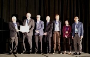
Dr. Burdick (second from the left) receives his award at the Annual Meeting of the U.S. Society for Biomaterials, April 2019

The Department of Bioengineering would like to congratulate our very own Jason A. Burdick, Ph. D., on being awarded the Acta Biomaterialia Silver Medal. Dr. Burdick is the Robert D. Bent Professor and a member of both the Laboratory for Research on the Structure of Matter (LRSM) and Center for Engineering Mechanobiology (CEMB) here at Penn.

The Acta Biomaterialia Silver Medal is an award from the monthly peer-reviewed scientific journal Acta Biomaterialia that recognizes leaders in academia, industry, and the public sector for mid-career leadership in and significant contribution to the field of biomaterials engineering. Dr. Burdick is the third recipient of the award so far, which includes a silver medal, an inscribed certificate, and reward of $5000. As the principal investigator of the Polymeric Biomaterials Laboratory in Penn’s Department of Bioengineering, Dr. Burdick leads research with a focus in polymer design, musculoskeletal tissue engineering, the control of stem cells with material cues, and the control of molecule delivery with polymers.

The Silver and Gold Medalists (Dr. Burdick and Dr. Antonios G. Mikos respectively) were presented with their own brand of wine in celebration of their achievement.

Specifically, Dr. Burdick’s innovation in the application of hydrogels to the musculoskeletal and cardiovascular systems brought him recognition for this award. His recent publications in Acta Biomaterialia include a study of bioactive factors for cartilage repair and regenesis in collaboration with fellow Penn Professor of Bioengineering Robert Mauck, Ph. D, and a study of adhesive biolinks that mimic the behavior of the extracellular matrix. The Acta Biomaterialia Silver Medal is only the most recent of several awards that Dr. Burdick has received, including both the George H. Heilmeier Faculty Award for Excellence in Research and the Clemson Award for Basic Research, and we can’t wait to see where his continued innovation in biomaterial engineering will take him next.

Organs-on-a-Chip Hurtle Toward the Final Frontier

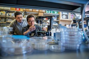
Graduate student Andrei Georgescu and Assistant Professor Dan Huh in Huh’s lab. Adapting the organ-on-a-chip technology for a trip to the International Space Station presented Huh’s team with a number of engineering challenges. (Photo: Kevin Monko)

Throughout the 60-year history of the U.S. space program—from the Mercury capsules of the 1960s to today’s International Space Station—astronauts have been getting sick. Researchers know being in orbit seems to suppress their immune systems, creating a more fertile ground for infections to grow. But nobody really understands why.

Early on the morning of April 26, a SpaceX Falcon 9 rocket will launch a cargo mission to the ISS from Cape Canaveral Air Force Station. Along with fresh water, food, and other necessities for the crew, the craft will be carrying two experiments designed by Penn scientists that could help shed light on why bugs have bedeviled space travelers.

For more than a decade, Dan Huh, the Wilf Family Term Assistant Professor of Bioengineering in the School of Engineering and Applied Science, has been developing super-small devices that use living cells to stand in for larger organs. These organs-on-a-chip hold great promise for all kinds of research, from diagnosing disease to curing them. They’re also a way to test things, including drugs and cosmetics, in a way that mimics real life without relying on animal subjects.

Read the full story at Penn TodayMedia contact Gwyneth K. Shaw

Bioengineering Chair and Students Honored at the 2019 SEAS Awards

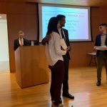
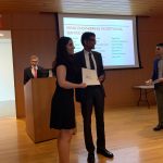
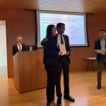
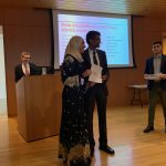
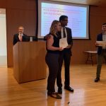
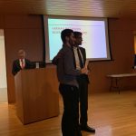
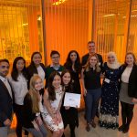
Each spring, the School of Engineering and Applied Science at the University of Pennsylvania hosts an awards recognition dinner to honor exceptional work in the school: The Faculty honor students for outstanding service and academics, while the students choose faculty members for their commitment to teaching and advising. This year, the Department of Bioengineering won big with honors for both our Department Chair and our undergraduates. Read about each of the award winners and see photos from the awards ceremony below. Congratulations to all the winners!

David F. Meaney, Ph.D.

Dr. David F. Meaney, Solomon R. Pollack Professor and Chair of Bioengineering, was awarded with the Ford Motor Company Award for Faculty Advising, which recognizes “dedication to helping students realize their educational, career and personal goals.” Dr. Meaney is beloved by the students in BE for his engaging teaching style, his commitment to student wellness and advancement, as well as his weekly Penn Bioengineering spin classes, and so we are delighted to see him recognized in this way by the wider student body  Read more about the award here and Dr. Meaney here.

Eshwar Inapuri (BAS 2019), a graduating senior completing his Bachelor of Applied Science degree in BE with minors in Biophysics and Chemistry, was awarded the Ben and Bertha Gomberg Kirsch Prize. This competitive award is decided by the SEAS faculty from among the Engineering undergraduate body and distinguishes a member of the B.A.S. senior class in  who “in applying the flexibility of the program, has created a personal academic experience involving the most creative use of the resources of the University.”

The Hugo Otto Wolf Memorial Prize, awarded to one or more members of each department’s senior class, distinguishes students who meet with great approval of the professors at large through “thoroughness and originality” in their work. This year, BE chose to share the award between Ethan Zhao (BSE 2019) and Shelly Teng (BSE 2019).

The Herman P. Schwan Award is decided by the Bioengineering Department and honors a graduating senior who demonstrates the “highest standards of scholarship and academic achievement.” The 2019 recipient of the Schwan Award is Joseph Maggiore (BSE 2019).

Every year, four BE students are recognized with Exceptional Service Awards for their outstanding service to the University and their larger communities. Our winners this year are Dana Abulez (BSE 2019), Daphne Cheung (BSE 2019), Lamis Elsawah (BSE 2019), and Kayla Prezelski (BSE 2019). All four of these recipients are also currently in the Accelerated Master’s program in BE.

And finally, BE also awards a single lab group (four students) with the Albert Giandomenico Award which reflects their “teamwork, leadership, creativity, and knowledge applied to discovery-based learning in the laboratory.” This year’s group consists of Caroline Atkinson (BSE 2019), Shuting (Sarah) Cai (BSE 2019), Rebecca Kellner (BSE 2019), and Harrison Troche (BSE 2019).

A full list of SEAS award descriptions and recipients can be found here.

Week in BioE: April 5, 2019

by Sophie Burkholder

Tulane Researchers Use Cancer Imaging Technique to Help Detect Preeclampsia

Preeclampsia is potentially life-threatening pregnancy disorder that typically occurs in about 200,000 expectant mothers every year. With symptoms of high blood pressure, swelling of the hands and feet, and protein presence in urine, preeclampsia is usually treatable if diagnosed early enough. However, current methods for diagnosis involve invasive procedures like cordocentesis, a procedure which takes a sample of fetal blood.

Researchers at Tulane School of Medicine led by assistant professor of bioengineering Carolyn Bayer, Ph.D., hope to improve diagnostics for preeclampsia with the use of spectral photoacoustic imaging. Using this technique, Bayer’s team noticed a nearly 12 percent decrease in placental oxygenation in rats with induced preeclampsia when compared to normal pregnant rats after only two days. If success in using this imaging technology continues at the clinical level, Bayer plans to find more applications of it in the detection and diagnosis of breast and ovarian cancers as well.

New CRISPR-powered device detects genetic mutations in minutes 

Two groups of researchers from the University of California, Berkeley and the Keck Graduate Institute of the Claremont Colleges recently collaborated to design what they call a “CRISPR-Chip” –  a combination of the CRISPR-Cas9 System with a graphene transistor to sequence DNA for the purpose of genetic mutation diagnosis. While companies like 23andMe made genetic testing and analysis more common and accessible for the general public in recent years, the CRISPR-Chip looks to streamline the technology even more.

This new chip eliminates the long and expensive amplification process involved in the typical polymerase chain reaction (PCR) used to read DNA sequences. In doing so, the CRISPR-Chip is much more of a point-of-care diagnostic, having the ability to quickly detect a given mutation or sequence when given a pure DNA sample. Led by Kiana Aran, Ph.D., the research team behind the CRISPR-Chip hopes that this new combination of nanoelectronics and modern biology will allow for a new world of possibilities in personalized medicine.

New Method of Brain Stimulation Might Alleviate Symptoms of Depression

Depression is one of the most common mental health disorders in the United States, with nearly 3 million cases every year. For most patients suffering from depression, treatment involves prolonged psychotherapy, antidepressant medication, or even electroconvulsive therapy in extreme cases. Now, scientists at the University of North Carolina School of Medicine study the use of transcranial alternating current stimulation (tACS) to alleviate symptoms of depression.

Led by Flavio Frohlich, Ph.D., who has an adjunct appointment in biomedical engineering, this team of researchers based this new solution on information from each patient’s specific alpha oscillations, which are a kind of wave that can be detected by an electroencephalogram (EEG). Those who suffer from depression tend to have imbalanced alpha oscillations, so Frohlich and his team sought to use tACS to restore this balance in those patients. After seeing positive results from data collected two weeks after patients in a clinical trial receives the tACS treatment, Frohlich hopes that future applications will include treatment for even more mental health disorders and psychiatric illnesses.

University of Utah Researchers Receive Grant to Improve Hearing Devices for Deaf Patients

Engineers at the University of Utah are part of team that recently received a $9.7 million grant from the National Institute of Health (NIH) to design new implantable hearing devices for deaf patients, with the hope to improve beyond the sound quality of existing devices. The work will build upon a previous project at the University of Utah called the Utah Electrode Array, a brain-computer interface originally developed by Richard Normann, Ph.D., that can send and receive neural impulses to and from the brain. This new device will differ from a typical cochlear implant because the Utah Electrode Array assembly will be attached directly to the auditory nerve instead of the cochlea, providing the patient with a much higher resolution of sound.

People & Places

Vivek Shenoy, Eduardo D. Glandt President’s Distinguished Scholar in the Department of Materials Science and Engineering and Secondary Faculty in Bioengineering, has been named the recipient of the 2018–19 George H. Heilmeier Faculty Award for Excellence in Research for “for pioneering multi-scale models of nanomaterials and biological systems.”

The Heilmeier Award honors a Penn Engineering faculty member whose work is scientifically meritorious and has high technological impact and visibility. It is named for George H. Heilmeier, a Penn Engineering alumnus and advisor whose technological contributions include the development of liquid crystal displays and whose honors include the National Medal of Science and Kyoto Prize.

Read the rest of the story on Penn Engineering’s Medium blog.

We would also like to congratulate Jay Goldberg, Ph.D., from Marquette University on his election as a fellow to the National Academy of Inventors. Nominated largely for his six patents involving medical devices, Goldberg also brings this innovation to his courses. One in particular called Clinical Issues in Biomedical Engineering Design allows junior and senior undergraduates to observe the use of technology in clinical settings like the operating room, in an effort to get students thinking about how to improve the use of medical devices in these areas.


Penn Bioengineers: Cells Control Their Own Fate by Manipulating Their Environment

by Lauren Salig

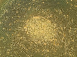
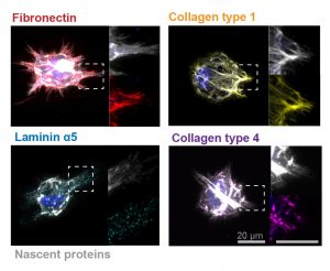
In these images, the researchers labeled new proteins white, and antibodies against other proteins in different colors. The co-localization of new proteins and antibodies show how cells can impact their local environments.

As different as muscle, blood, brain and skin cells are from one another, they all share the same DNA. Stem cells’ transformation into these specialized cells — a process called cell fate determination — is controlled through various signals from their surroundings.

A recent Penn Engineering study suggests that cells may have more control over their fate than previously thought.

Jason Burdick, Robert D. Bent Professor of Bioengineering, and Claudia Loebel, a postdoctoral researcher in his lab, led the study. Robert Mauck, Mary Black Ralston Professor for Education and Research in Orthopaedic Surgery at Penn’s Perelman School of Medicine, also contributed to the research.

Their study was published in Nature Materials.

Read the full story in the Penn Engineering Medium Blog. Media contact Evan Lerner.

Week in BioE (March 22, 2019)

by Sophie Burkholder

A New Microscopy Technique Could Reduce the Risk of LASIK Surgery
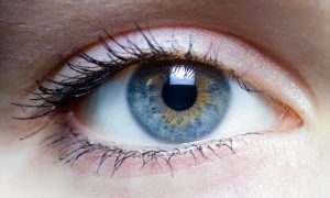
Though over ten million Americans have undergone LASIK vision corrective surgery since the option became available about 20 years ago, the procedure still poses some risk to patients. In addition to the usual risks of any surgery however, LASIK has even more due to the lack of a precise way to measure the refractive properties of the eye, which forces surgeons to make approximations in their measurements during the procedure. In an effort to eliminate this risk, a University of Maryland team of researchers in the Optics Biotech Laboratory led by Giuliano Scarcelli, Ph. D., designed a microscopy technique that would allow for precise measurements of these properties.

Using a form of light-scattering technology called Brillouin spectroscopy, Scarcelli and his lab found a way to directly determine a patient’s refractive index – the quantity surgeons need to know to be able to measure and adjust the way light travels through the eye. Often used as a way to sense mechanical properties of tissues and cells, this technology holds promise for taking three-dimensional spatial observations of these structures around the eye. Scarcelli hopes to keep improving the resolution of the new technique, to further understanding of the eye, and reduce even more of the risks involved with LASIK surgery.

Taking Tissue Models to the Final Frontier

Space flight is likely to cause deleterious changes to the composition of bacterial flora, leading to an increased risk of infection. The environment may also affect the susceptibility of microorganisms within the spacecraft to antibiotics, key components of flown medical kits, and may modify the virulence of bacteria and other microorganisms that contaminate the fabric of the International Space Station and other flight platforms.

“It has been known since the early days of human space flight that astronauts are more prone to infection,” says Dongeun (Dan) Huh, Wilf Family Term Assistant Professor in Bioengineering at Penn Engineering. “Infections can potentially be a serious threat to astronauts, but we don’t have a good fundamental understanding of how the microgravity environment changes the way our immune system reacts to pathogens.”

In collaboration with G. Scott Worthen, a physician-scientist in neonatology at the Children’s Hospital of Philadelphia, Huh will attempt to answer this question by sending tissues-on-chips to space. Last June, the Center for the Advancement of Science in Space (CASIS) and the National Center for Advancing Translational Sciences (NCATS), part of the National Institutes of Health (NIH), announced that the duo had received funding to study lung host defense in microgravity at the International Space Station.

Huh and Worthen aim to model respiratory infection, which accounts for more than 30 percent of all infections reported in astronauts. The project’s goals are to test engineered systems that model the airway and bone marrow, a critical organ in the immune system responsible for generating white blood cells, and to combine the models to emulate and understand the integrated immune responses of the human respiratory system in microgravity.

Read the rest of the article on Penn Engineering’s Medium Blog. Media contacts Evan Lerner and Janelle Weaver.

Sappi Limited Teams Up with the University of Maine to Develop Paper Microfluidics

At the Westbrook Technology Center of Sappi, a global pulp and paper company, researchers found ways to apply innovations in paper texture for medical use. So far, these include endeavors in medical test devices and patches for patient diagnostics. In collaboration with the Caitlin Howell, Ph.D., Assistant Professor of Chemical and Biomedical Engineering at the University of Maine, Sappi hopes to continue advances in these unconventional uses of their paper, especially as the business in paper for publishing purposes declines.

Sappi’s projects with the university focus on the development of paper microfluidics devices as what’s now becoming a widespread solution for obstacles in point-of-care diagnostics. One project in particular, called Sharklet, uses a paper that mimics shark skin as a way to impede unwanted microbial growth on the device – a key characteristic needed for its transition into commercial use. Beyond this example, Sappi’s work in developing paper microfluidics underscores the benefits of these devices in their mass producibility and adaptability.

New Observations of the WNT Pathway Deepen the Understanding of Protein Signaling in Cellular Development

Scientists at Rice University recently found that a protein signaling pathway called WNT, typically associated with its role in early organism development, can both listen for signals from a large amount of triggers and influence cell types throughout embryonic development. These new findings, published in PNAS, add to the already known functions of WNT, deepening our understanding of it and opening the doors to new potential applications of it in stem cell research.

Led by Aryeh Warmflash, Ph. D., researchers discovered that the WNT pathway is different between stem cells and differentiated cells, contrary to prior belief that it was the same for both. Using CRISPR-Cas9 gene editing technology, the Warmflash lab observed that the WNT signaling pathway is actually context-dependent throughout the process of cellular development. This research brings a whole new understanding to the way the WNT pathway operates, and could open the doors to new forms of gene therapy and treatments for diseases like cancers that involve genetic pathway mutations.

People and Places

In a recent article from Technical.ly Philly, named Group K Diagnostics on a list of ten promising startups in Philadelphia. Group K Diagnostics founder Brianna Wronko graduated with a B.S.E. from Penn’s Department of Bioengineering in 2017, and her point-of-care diagnostics company raised over $2 million in funding last year. Congratulations Brianna!

We would also like to congratulate Pamela K. Woodward, M.D., on her being named as the inaugural Hugh Monroe Wilson Professor of Radiology at the Washington University School of Medicine in St. Louis. Also a Professor of Biomedical Engineering at the university, Dr. Woodward leads a research lab with a focus on cardiovascular imaging, including work on new standards for diagnosis of pulmonary blood clots and on an atherosclerosis imaging agent.

Lastly, we would like to congratulate all of the following researchers on their election to the National Academy of Engineering:

  • David Bishop, Ph. D., a professor at the College of Engineering at Boston University whose current research involves the development of personalized heart tissue as an all-encompassing treatment for patients with heart disease.
  • Joanna Aizenberg, Ph. D., a professor of chemistry and chemical biology at Harvard University who leads research in the synthesis of biomimetic inorganic materials
  • Gilda Barabino, Ph. D., the dean of the City College of New York’s Grove School for Engineering whose lab focuses on cartilage tissue engineering and treatments for sickle cell disease.
  • Karl Deisseroth, M.D., Ph. D., a professor of bioengineering at Stanford University whose research involves the re-engineering of brain circuits through novel electromagnetic brain stimulation techniques.
  • Rosalind Picard, Ph.D., the founder and director of the Affective Computing Research Group at the Massachusetts Institute of Technology’s Media Lab whose research focuses on the development of technology that can measure and understand human emotion.
  • And finally, Molly Stevens, Ph. D., the Research Director for Biomedical Material Sciences at the Imperial College of London with research in understanding biomaterial interfaces for biosensing and regenerative medicine.