Week in BioE (January 24, 2018)

Using AI to Better Understand Cancer Immunology
ImmunoMap
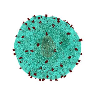
A T cell with receptors highlighted in red

T lymphocytes in the immune system play a vital role in the body to recognize invasion by an outside element. When foreign bacteria enter the body, receptors on the T cell surface detect antigens associated with the bacteria and send a signal deploying phagocytes to attack and defeat the invading bacteria. While evolution and vaccination make the immune system very efficient, the inability of T cell receptors (TCRs) to detect cancer makes normal T cells relatively ineffective in resisting cancer. One of the ways to overcome this limitation of the immune system is to better understand how the TCRs respond to antigens. Analyses of the proteins involved in TSR responses are useful but limited by several factors, including the dizzying amount of data involved. Data analysis techniques have been helpful but have offered little information about the general reactions of TSRs, rather than how they react to specific antigens.  

A possible solution to this obstacle is ImmunoMap, developed by scientists collaborating between Johns Hopkins University and Memorial Sloan Kettering Cancer Center. In a study recently published in Cancer Immunology Research, the authors, led by Jonathan P. Schneck, M.D., Ph.D., a professor of pathology at Johns Hopkins associated with the university’s Institute for Cell Engineering and Institute for Nanobiotechnology, describe their creation and deployment of ImmunoMap, a group of artificial intelligence algorithms that use machine learning to process large amounts of sequencing data and compare data from different antigens with each other.

The authors trained ImmunoMap initially using data from a mouse model of melanoma, in which the algorithm demonstrated significantly better performance than traditional methods. Subsequently, ImmunoMap was applied to patient response data from a melanoma clinical trial of the chemotherapy agent nivolumab. The algorithm discovered a new group of patients that would respond positively to nivolumab treatment — a finding missed by popular past methods. More testing of ImmunoMap is necessary, but the technology could make significant contributions to the monitoring of cancer patients receiving chemotherapy. In addition, it could to help to better predict response in patients before they begin specific chemotherapy regimes.

Wearables Improving Health

Among the most troubling health disparities related to global wealth inequality is the higher rate of mortality among children suffering from cancer. Fever is a common symptom of children undergoing cancer treatment, and this symptom may indicate more serious health issues that require the attention of a doctor. However, continuously monitoring skin temperature in children from low resource settings is difficult. Seeking to help remedy this problem, undergraduate engineering students at Harvard collaborated with the Dana-Farber/Boston Children’s Cancer & Blood Disorders Center’s Global Health Initiative to develop tools for earlier fever detection and treatment.

In a course taught by David Mooney, Ph.D., Robert P. Pinkas Family Professor of Bioengineering at Harvard, students developed a wearable device that sounds an alarm when the wearer needs medical help. The app can send patients’ recorded messages to their doctors, who can then review the temperature data and messages from the children before responding. Fashioned like a wristwatch, the extra-durable and waterproof device will next move into pilot testing among a larger patient population.

Meanwhile, at Northwestern, John A. Rogers, Ph.D., the Louis Simpson and Kimberly Querrey Professor of Materials Science and Engineering, Biomedical Engineering, and Neurological Surgery in Northwestern’s McCormick School of Engineering, has partnered with cosmetics giant L’Oréal to create the world’s smallest wearable. The device, which is smaller than an adult fingernail, measures UV sun exposure for the wearer and can tell when they should go back inside instead of risking overexposure. Unsurprisingly, it’s solar powered, and it was demonstrated a couple of weeks ago at a consumer electronics show in Las Vegas.


Growing Hydrogels Like Human Tissue

Scientists at Carnegie Mellon University and Nanyang Technological University in Singapore have collaborated in a process to create polyacrylamide gels that grow in a manner resembling natural tissue. K. Jimmy Hsia, Ph.D., Professor of Biomedical Engineering at Carnegie Mellon, is co-lead author of a new study in PNAS describing this new growth mode.

In the study, Dr. Hsia and his coauthors report that, in the same way that growth factors secreted by a living organism affect the generation of new tissue, oxygen can be modulated to control how hydrogels grow. Moreover, while growth is under way, the process could be continued to efficiently manage the mass transfer of nutrients from cell to cell. Finally, the authors detail the mechanical processes that help to shape the final product. With this new process, the ability to design and create materials for applications such as robotics and tissue engineering comes a step closer to resembling living tissue as closely as possible.

People and Places

Engineers at Virginia Tech have been awarded a $1.1 million grant from the Virginia Research Investment Committee to develop a device that uses low-energy electric fields for the treatment of brain tumors. Rafael Davalos, Ph.D., L. Preston Wade Professor of Biomedical Engineering and Mechanics, is the chief investigator on the grant.

The Department of Biomedical Engineering has announced the appointment of Kam W. Leong, Ph.D., as the Samuel Y. Sheng Professor of Biomedical Engineering. Dr. Leong earned his Ph.D. in chemical engineering from the University of Pennsylvania and taught at Duke and Johns Hopkins before arriving at Columbia in 2006. He was previously the James B. Duke Professor of Biomedical Engineering at Duke. Congratulations to Dr. Leong!

A Call to Understand Brain Network Mechanisms of Mental Disorders

The sheer complexity of the human brain means that, despite the tremendous advances made in neuroscience, there is still much we don’t know about what goes on inside our heads and how it goes awry in mental disorders. Even with the most advanced techniques, much of what we’ve learned about the brain is descriptive — telling that something is different between health and unhealthy function — but not why that something is different or how we could change it.

mental disorders
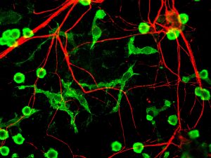
Rat microglia and neurons stained for different proteins

Among the approaches that have provided important insights into these questions is network science, which seeks to understand the brain as a complex system of multiple interacting components. Now, in a review published recently in Neuron, Danielle Bassett, Ph.D., Eduardo D. Glandt Faculty Fellow and Associate Professor of Bioengineering, and Richard Betzel, Ph.D., a postdoc in Dr. Bassett’s lab, have collaborated with scientists from the University of Heidelberg in Germany. The review covers a broad range of discoveries and innovations, moving from earlier, two-dimensional approaches to understanding the brain, such as graph theory, to newer approaches including multilayer networks, generative network models, and network control theory.

“Stating what is different in brain networks of individuals with disorders of mental health is not the same as identifying why” says Bassett. “Here we propose that emerging tools from network science can be used to identify true mechanisms of mental health disorders, and bridge molecular and genetic mechanisms through brain physiology, thus informing interventions in the form of pharmacological manipulations and brain stimulation.”

Brain Network Control Emerges over Childhood and Adolescence

network control


The developing human brain contains a cacophony of electrical and chemical signals from which emerge the powerful adult capacities for decision-making, strategizing, and critical thinking. These signals support the trafficking of information across brain regions, in patterns that share many similarities with traffic patterns in railway and airline transportation systems. Yet while air traffic is guided by airport control towers, and railway routes are guided by signal control rooms, it remains a mystery how the information traffic in the brain is guided and how that guidance changes as kids grow.

In part, this mystery has been complicated by the fact that, unlike transportation systems, the brain is not hooked up to external controllers. Control must happen internally. The problem becomes even more complicated when we think about the sheer number of routes that must exist in the brain to support the full range of human cognitive capabilities. Thus, the controllers would need to produce a large set of control signals or use different control strategies. Where internal controllers might be, how they produce large variations in routing, and whether those controllers and their function change with age are important open questions.

A recent paper published in Nature Communications – a product of collaboration among the Departments of Bioengineering and Electrical & Systems Engineering at the University of Pennsylvania and the Department of Psychiatry of Penn’s Perelman School of Medicine – offers some interesting answers. In their article, Danielle Bassett, Ph.D., Eduardo D. Glandt Faculty Fellow and Associate Professor in the Penn BE Department, Theodore D. Satterthwaite, M.D., Assistant Professor in the Penn Psychiatry Department, postdoctoral fellow Evelyn Tang, and their colleagues suggest that control in the human brain works in a similar way to control in man-made robotic and other mechanical systems. Specifically, controllers exist inside each human brain, each region of the brain can perform multiple types of control, and this control grows as children grow.

As part of this study, the authors applied network control theory — an emerging area of systems engineering – to explain how the pattern of connections (or network) between brain areas directly informs the brain’s control functions. For example, hubs of the brain’s information trafficking system (like Grand Central Station in New York City) show quite different capacities for and sensitivities to control than non-hubs (like Newton Station, Kansas). Applying these ideas to a large set of brain imaging data from 882 youths in the Philadelphia area between the ages of 8 and 22 years old, the authors found that the brain’s predicted capacity for control increases over development. Older youths have a greater predicted capacity to push their brains into nearby mental states, as well as into distant mental states, indicating a greater potential for diversity of mental operations than in younger youths.

The investigators then asked whether the principles of network control could explain the specific manner in which connections in the brain change as youths age. They used tools from evolutionary game theory – traditionally used to study Darwinian competition and evolving populations in biology – to ‘evolve’ brain networks in silico from their 8-year old state to their 22-year-old state. The results demonstrated that the optimization of network control is a principle that explains the observed changes in brain connectivity as youths develop over childhood and adolescence. “One of the observations that I think is particularly striking about this study,” Bassett says, “is that the principles of network controllability are sufficient to explain the observed evolution in development, suggesting that we have identified a quintessential rule of developmental rewiring.”

This research informs many possible future directions in scientific research. “Showing that network control properties evolve during adolescence also suggests that abnormalities of this developmental process could be related to cognitive deficits that are present in many neuropsychiatric disorders,” says Satterthwaite. The discovery that the brain optimizes certain network control functions over time could have important implications for better understanding of neuroplasticity, skill acquisition, and developmental psychopathology.

Second Roundtable With Undergrads

Second roundtableLate last semester, Penn Bioengineering Department chair David Meaney and senior lecturer LeAnn Dourte held a second roundtable with BE undergrads Eric Helfgott, Joseph Maggiore, Kayla Prezelski, and Margaret Schroeder. They picked up on topics from the last roundtable, extending the topics to balancing an engineering workload and other commitments.


Tissue Folding Processes Further Unraveled

tissue folding
Alex Hughes, Ph.D.

One of the key processes in embryonic development and growth through childhood and adolescence is that of how tissue folds into the specific shapes required for them to function in the body.  For instance, mesenchymal stem cells, which form a variety of tissues including bones, muscles, and fat, are required to “know” what shapes to take on as they form organ systems and other structures. Therefore, a big concern in tissue engineering is determining how to control these processes of tissue folding.

Just in time for his arrival at Penn Bioengineering, Dr. Alex Hughes, a new assistant professor in the department, is the lead author on a new paper in Developmental Cell that explores this concern. The study was coauthored with Dr. Zev Gartner of the University of California, Berkeley, where Dr. Hughes just finished a postdoctoral fellowship. In the paper, the authors used three-dimensional cell-patterning techniques, embryonic tissue explants, and finite element modeling to determine that the folding process involves the interaction of a protein called myosin II with the extracellular matrix, itself the molecular material that provides a structural framework for developing tissues. With the knowledge gained in the initial experiments, the authors were then able to reproduce the tissue folding process in the lab.

“Bioengineers are currently thinking about building tissues,” Dr. Hughes says, “not just at the level of organoids, but at the level of organs in the body. One of my interests at Penn is to harness developmental principles that link these length scales, allowing us to design medically relevant scaffolds and machines.”

Equipped with the knowledge gleaned from this research, future studies could contribute further to the ability to generate tissues and even organ systems in laboratories. Ultimately, this knowledge could revolutionize transplant medicine, as well as variety of other fields.