Among the myriad medical complications caused by diabetes, pressure sores of the feet are among the most troubling. Because of the common complication of peripheral neuropathy, people with diabetes are often unable to determine how much pressure is being exerted on their feet. As a result, they cause foot ulcers, which can become infected, leading in the worst cases to amputation.
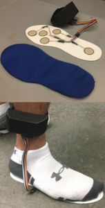
The Flysole combines an insole with five sensors (top) and an ankle band (bottom) to house the electrical components, including the circuit for the pressure sensors as well as the microcontroller and SD card to log the pressure data.
One of the senior design teams from the Department of Bioengineering at the University of Pennsylvania developed a project to address this problem. Their solution was Flysole (right), a prognostic implant that diabetic patients can wear to collect data on foot pressure so that the doctor can prescribe an optimal orthotic to prevent sores from developing. The team was named one of the three winners of this year’s competition.
The team, which consisted of Parag Bapna, Karthik Ramesh, Jane Shmushkis, and Amey Vrudhula, designed the Flysole as a lightweight insole with ankle band paired with software that generates a profile of the pressure on the sole of the patient’s foot. The insole has five sensors to collect these data. The cost is approximately $75 per pair.
In addition, the team made the Flysole to be reusable by including a polyurethane laminate sleeve for the individual patient. Future improvements envisioned by the students include improving the software to include recommendations for orthotics and alternate arrangements for the sole sensors.
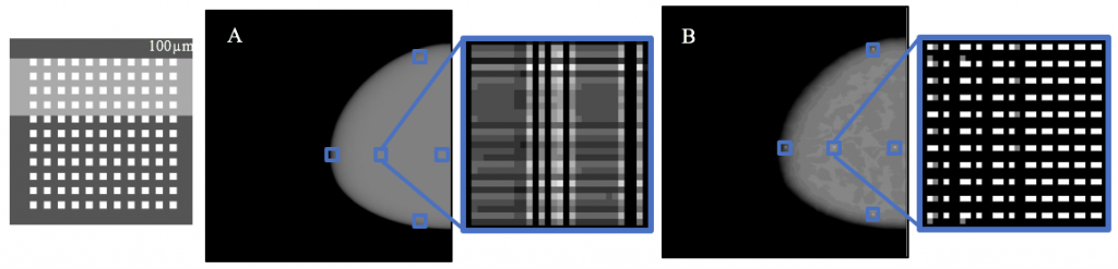
An embedded 11×11 cluster of 100-micron objects (which models a cluster of microcalcifications — one of the earliest indicators of breast cancer). (A) shows the results from the current standard of imaging only along the chest wall. (B) shows the results of our method that considers the patient’s unique breast geometry using a Custom V imaging pattern. (B) resolves the embedded cluster as a distinct cluster of objects while (A) appears to blur the final image.
Breast cancer continues to affect more than 10% of all women — and a small percentage of men — despite significant advances in diagnosis and treatment. While a majority cases today can be successfully treated, early detection is essential to beginning treatment before it’s too late.
Among the more recent innovations in screening has been three-dimensional mammography. However, this modality has lacked the ability to personalize the scan to the individual patient’s breast, instead only acquiring several two-dimensional images along the chest wall, resulting in a lack of individualization for the patient.
A senior design project team at the University of Pennsylvania’s Department of Bioengineering, however, has helped to develop a more personalized 3D imaging technology, which acquires a series of images but instead following the contour of the breast itself. With their efforts, the team earned one of the three awards given to student teams yearly.
The four-student team, consisting of Lucy Chai, Elizabeth Kobe, Margaret Nolan, and Sushmitha Yarrabothula, picked up a project begun last year (a common practice with senior design projects) and demonstrated with their work that the imaging technique could be applied using a digital phantom (a computerized breast model) with great clarity, including successful resolution of a simulated mass just one-tenth of a millimeter in size.
Now, the four seniors will hand off the project to another team, continuing this multi-year research. Ultimately, before it can be applied in actual patients, the modality will need to be tested against the current standard of care in terms of its ability to detect small masses in the breast. Nevertheless, this year’s team moved the ball downfield significantly.
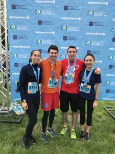
Four students from the Bioengineering Department at the University of Pennsylvania participated in this year’s Blue Cross Broad Street Run, which was held on Sunday, May 7, in Philadelphia.
(left to right) Melissa Schweizer, Mike Patterson, Kyle O’Neil, Margaret Schroeder
The four students (right) ran the annual event, which begins at Broad Street and W. Fisher Avenue, in the Logan section of North Philadelphia and runs almost the entire length of Broad to the Navy Yard in South Philadelphia. Broad Street is one of Philadelphia’s main thoroughfares and runs 11 miles along the city’s north-south axis. This year was the 38th year that the Broad Street Run has taken place.
“The Broad Street Run is one of the greatest Philadelphia running traditions,” department chair David Meaney, PhD, said, “and it is remarkable that our students would take time from their finals for an ‘easy’ ten-mile run — remarkable but not surprising.”
In January 1951, Henrietta Lacks, a 30-year-old African-American woman from Baltimore, was diagnosed with cervical cancer at the Johns Hopkins Medical Center. She was treated with radium brachytherapy, the standard of care at the time, but her condition worsened. In August, a week after she turned 31, she was admitted to the hospital with severe abdominal pain. Less than three months later, she died. An autopsy showed widespread metastasis of the original cancer.
Henrietta Lacks had died, but strangely, her cancer cells have lived on. Unbeknownst to her and her family, Henrietta’s doctors had sampled her cancer cells for research — a common practice at the time, particularly from patients treated in wards. Those cells were given to George Gey, a JHU biologist who had been trying for years to establish a cancer cell line that could be grown outside the body. Henrietta Lacks’s cells ended up being the first cell line so established.
The cell line was named “HeLa” by Gey’s laboratory assistant, who coded cell samples using the first two letters of the donor’s first and last names. With Henrietta Lacks’s cells, Gey was able to establish an immortal cell line, i.e., a line of cells that would continue to divide indefinitely. The ability of these cells to divide like this lent itself to the line being used in numerous scientific studies since the 1950s, including Jonas Salk’s development of the polio vaccine.
Notwithstanding the tremendous accomplishments achieved using the HeLa cell line, the case nevertheless evokes serious ethical issues regarding the consent of patients to having their tissue used for research. In recent years, the case has attracted significant attention, with a book, The Immortal Life of Henrietta Lacks, published by Rebecca Skloot in 2010 and now an HBO feature film of the same title produced by and starring Oprah Winfrey as Lacks’s daughter. The film debuted on April 22.
Brittany Shields, PhD, a senior lecturer in the Department of Bioengineering at the University of Pennsylvania, discussed some of the issue raised by Lacks’s story. “Henrietta Lacks’s story has brought public attention to a number of ethical issues in biomedical research, including the role of informed consent, privacy, and commercialization in the collection, use and dissemination of biospecimens,” Dr. Shields says.
“In the United States, biomedical research at federally-funded institutions must follow the policy set by the Department of Health and Human Services. The current policy, known as the ‘Common Rule,’ calls for informed consent and oversight through Institutional Review Boards for research conducted with human beings,” she explains.
However, she continues, “these regulations may or may not apply in different situations related to biospecimens. If an anonymous biospecimen had already been collected for another purpose, informed consent is generally not required.”
In the case of Henrietta Lacks, or more precisely her descendants, an agreement was reached between the family and the National Institute of Health stipulating that the family must give consent when certain genetic information gleaned from the cell line is used. However, controversy between the family and the medical research community has persisted.
When someone talks about using “your network” to find a job or answer a question, most people understand that to mean the interconnected web of your friends, family, and acquaintances. But we all have another key network that shapes our life in powerful ways: our brains.
In the brain, impulses whiz from one brain region to another, helping you formulate all of your thoughts and decisions. As science continues to unlock the complexities of the brain, a group of researchers has found evidence that brain networks and social networks actually influence and inform one another.
It found that people who show greater changes in connectivity in their mentalizing system during social exclusion compared to inclusion tend to have a less tightly knit social network — that is, their friends tend not to be friends with one another. By contrast, people with more close-knit social networks, in which many people in the network tend to know one another, showed less change in connectivity in their mentalizing regions.
Cataracts are a leading cause of vision loss worldwide. In the world’s more developed countries, laser is commonly used for cataract removal. However, in much of the developing world, lasers are expensive or difficult to acquire. In these countries, cataract surgeries are still largely performed freehand, with all of the attendant risks that such procedures involve.
One of this year’s senior design projects in the Department of Bioengineering at Penn was a surgical tool for ophthalmic surgeons to perform capsulorhexis, the fancy term for the circular incision necessary to remove a cataract. The team, which included seniors Akshatha Bhat, Nimay Kulkarni, Steven Polomski, and Ananya Sureshkumar, collaborated with the Aravind Eye Hospital in Puducherry, India, created a surgical device called the Rhex to create this round incision
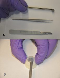
A) The Rhex (top), compared to forceps (center) and a scalpel (bottom); B) Rhex inserted into a model eye.
The Rhex (right) consists of a stem with a ring at the end, into which a blade is fitted. It can be inserted into a scleral incision, pressed, and rotated to perform the capsulorhexis. Initial testing indicated the Rhex could create a circulation incision approximately 6.7 mm in diameter, with eccentricity (indicating deviation from a perfect circle) of 0.254±0.08, which was within the acceptable range determined by the team.
The students designed the Rhex to be autoclavable and to use disposable blades. The next step will be to decrease the size of the instrument further and perhaps to use translucent or even transparent material to produce newer prototypes, which could be particularly useful, since cataract surgeries are open performed using backlighting.
With increasing age in the population, Parkinson’s disease has become increasingly common. One of the most frustrating effects of the disease is freezing of gait (FOG), in which a patient will suddenly stop while walking and find it difficult to begin again. Falls are a common consequence.
Despite intensive research, FOG is poorly understood. However, studies have shown that certain external stimuli, including metronomes and devices that provide visual cues, can be helpful. With this knowledge, a team of bioengineering students set to tackle this issue with their senior design project.
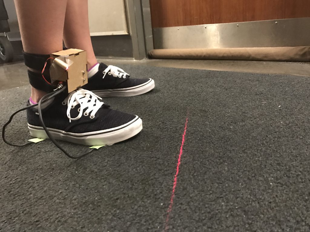
The team —whose members were Priyanka Ghosh, Fiona La, Laurel Leavitt, and Lia Lombardi — came up with ShuffleAssist, a wearable device that uses force sensors and an internal measurement unit to detect FOG and automatically provide a cue for the patient. The patient can choose a metronome beat or visual laser cue that can be provided either as determined by the device or continually, for patients who so choose.
ShuffleAssist tested well among normal subjects, detecting FOG correctly 98% of the time within approximate one second. In addition, the students were able to create their prototype for a cost of $107 per unit, compared to similarly intended products already on the market costing more than twice that much.
The next step for the team is to test the device in actual patients with Parkinson’s. The students have left the device with a faculty member in the Perelman School of Medicine who treats patients with motor disorders. This faculty member will offer the device to patients for testing.
See below for a video demonstration of ShuffleAssist.
The Department of Bioengineering at the University of Pennsylvania is proud to announce that Konrad Kording, PhD, currently professor of physical medicine and rehabilitation, physiology, and applied mathematics at Northwestern University, will join the BE faculty in the fall.
Dr. Kording, a neuroscientist with advanced degrees in experimental physics and computational neuroscience, is a native of Germany. After earning his PhD in 2001 at the Swiss Federal Institute of Technology in Zurich, he held fellowships at University College, London, and MIT before arriving at Northwestern in 2006.
Kording’s groundbreaking interdisciplinary research uses data science to understand brain function, improve personalized medicine, collaborate with clinicians to diagnose diseases based on mobile phone data, and even understand the careers of professors. Across many areas of biomedical research, his group analyzes large datasets to test new models and thus get closer to an understanding of complex problems in bioengineering, neuroscience, and beyond.
Dr. Kording’s appointment will be shared between the BE Department and the Department of Neuroscience in the Perelman School of Medicine.
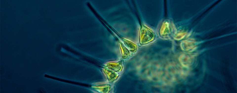
The Scripps Institution of Oceanography at the University of California, San Diego, announced last week that one of its faculty members, Andrew Barton, PhD, received a Simons Foundation Early Career Award to study phytoplankton — a type of algae that requires sunlight to survive and that serves as the basis for much of the marine food chain.
Dr. Barton’s research will use the Scripps Plankton Camera System, which provides real-time photographic images to monitor these phytoplankton. While not exactly offering the excitement or cuteness factor of the Golden Retriever Puppy Cam, this sort of technology is incredibly important to better understanding certain aspects of marine biology.
“This is an interesting project that brings cutting edge image-processing technology to the natural habitat to study complex organismal dynamics in the real-world setting,” says Brian Chow, PhD, assistant professor of bioengineering at the University of Pennsylvania. “Establishing the critical interplay between an organism’s form and function and the forces of its local and global environments are important problems in physical biology in general. Diatoms have long been studied by bioengineers interested in self-assembly, programmed assembly, biomineralization, and biomimicry, so the work may lead to some novel insights for our field.”
Congratulations to Dr. Barton on receiving this prestigious award.
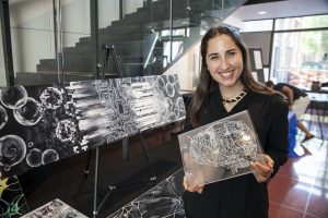
In high school, Rebecca Kellner (right) always had a dual love of art and science. When she entered the University of Pennsylvania as a freshman, she thought that her interest in art would always be separate from her pursuit of science. “I’ve always loved art and science and I wondered how I would integrate my passions into one area of study,” Rebecca says. “Then I heard about the Network Visualization Program run by Dr. Danielle Bassett . In this program, the intersection of art and science is celebrated, and this intersection is a place where I feel right at home.”
The Penn Network Visualization Program, begun in 2014, had long been a dream of Dr. Bassett. She wanted a forum where young artists and research scientists could interact with each other. “Science and art are often perceived to be at odds with each other, two fundamentally different ways of understanding the world. As a scientist, I’ve learned that the visual impact of the information I present is crucially important. Networks are visually intuitive,” says Bassett, “and represent an opportunity to foster a common language between scientists and artists.”
In this six-week summer program, young artists spend time with scientists at Penn who are performing cutting-edge research in network science as applied to social systems, human biology, and physical materials, with the underlying goal of advancing bioengineering. Faculty from the Warren Center for Network and Data Science who have volunteered their time and creativity to the project include Eleni Katifori, Erol Akcay, and Randy Kamien of the School of Arts and Sciences; Robert Ghrist and Victor Preciado of the School of Engineering and Applied Sciences; Sandra Gonzalez-Bailon of the Annenberg School of Communications; and Francis Diebold of the Wharton School of Business. During the course of the internship, the artists produce works of art interpreting and capturing the intricacies of these networks in novel ways. Artistic supervision and project advice are provided by local artists affiliated with the program. The goal of the internship is to provide scientists with new conceptualizations of their research and to provide the intern with new knowledge in scientific art applications.
Rebecca was thrilled when she was accepted into the program. During her internship she worked with a variety of scientists. Her final artwork focused on the research of Dr. Ann Hermundstad (Janelia), the postdoctoral researcher in the Physics of Living Matter Group, University of Pennsylvania Department of Physics and Astronomy. Dr. Hermundstad’s research focuses on what and how the brain sees. Fascinated by these networks, Rebecca created a painting and a laser-etched acrylic book.
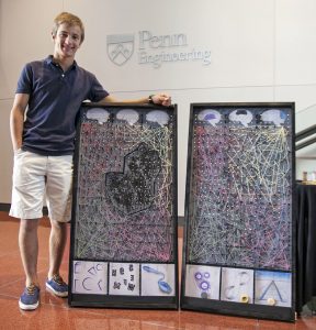
Nicholas Hanchak
The program also invites six high school students who have exhibited creativity and academic achievement. Nicholas Hanchak (right) from Westtown School participated during the summer of 2016. “I love art, science and baseball and I am thinking about architecture as a possible career,” Nicholas says. “The Penn program challenged me to find new ways to combine these interests.” For his final project, Nicholas created a Plinko Game Board showing the difference between the networks in a healthy brain and in a brain damaged by stroke.
“Artists and scientists are kindred spirits because they both are interested in observing what is in front of them,” says Dr. Bassett. “The Network Visualization program offers an opportunity for scientists and artists to inform each other in very tangible ways.”
The program runs every other summer. During the fall, several of the artists’ pieces are showcased in Philadelphia-area middle and high schools, particularly in disadvantaged areas. These efforts are enabled by ongoing collaborations with the Netter Center for Community Partnerships and Penn’s Center for Curiosity, and they are partially funded by the National Science Foundation. Bassett hopes this outreach effort will encourage children to explore intersections between the arts and sciences, while instilling a growing appreciation of their networked world.