More Cancers May be Treated with Drugs than Previously Believed

by Alex Gardner

3D illustration of cancer cells
nucleus and membrane of pathogen micro organisms in blue background

Up to 50 percent of cancer-signaling proteins once believed to be immune to drug treatments due to a lack of targetable protein regions may actually be treatable, according to a new study from the Perelman School of Medicine at the University of Pennsylvania. The findings, published this month in Nature Communications, suggest there may be new opportunities to treat cancer with new or existing drugs.

Researchers, clinicians, and pharmacologists looking to identify new ways to treat medical conditions—from cancer to autoimmune diseases—often focus on protein pockets, areas within protein structures to which certain proteins or molecules can bind. While some pockets are easily identifiable within a protein structure, others are not. Those hidden pockets, referred to as cryptic pockets, can provide new opportunities for drugs to bind to. The more pockets scientists and clinicians have to target with drugs, the more opportunities they have to control disease.

The research team identified new pockets using a Penn-designed neural network, called PocketMiner, which is artificial intelligence that predicts where cryptic pockets are likely to form from a single protein structure and learns from itself. Using PocketMiner—which was trained on simulations run on the world’s largest super computer—researchers simulated single protein structures and successfully predicted the locations of cryptic pockets in 35 cancer-related protein structures in thousands of areas of the body. These once-hidden targets, now identified, open up new approaches for potentially treating existing cancer.

What’s more, while successfully predicting the cryptic pockets, the method scientists used in this study was much faster than previous simulation or machine-learning methods. The network allows researchers to nearly instantaneously decide if a protein is likely to have cryptic pockets before investing in more expensive simulations or experiments to pursue a predicted pocket further.

“More than half of human proteins are considered undruggable due to an apparent lack of binding proteins in the snapshots we have,” said Gregory R. Bowman, PhD, a professor of Biochemistry and  Biophysics and Bioengineering at Penn and the lead author of the study. “This PocketMiner research and other research like it not only predict druggable pockets in critical protein structures related to cancer but suggest most human proteins likely have druggable pockets, too. It’s a finding that offers hope to those with currently untreatable diseases.”

Read the full story in Penn Medicine News.

A Potential Strategy to Improve T Cell Therapy in Solid Tumors

A new Penn Medicine preclinical study demonstrates a simultaneous ‘knockout’ of two inflammatory regulators boosts T cell expansion to attack solid tumors.

by Meagan Raeke

Image: Courtesy of Penn Medicine News

A new approach that delivers a “one-two punch” to help T cells attack solid tumors is the focus of a preclinical study by researchers from the Perelman School of Medicine. The findings, published in the Proceedings of the National Academy of Sciences, show that targeting two regulators that control gene functions related to inflammation led to at least 10 times greater T cell expansion in models, resulting in increased anti-tumor immune activity and durability.

CAR T cell therapy was pioneered at Penn Medicine by Carl H. June, the Richard W. Vague Professor in Immunotherapy at Penn and director of the Center for Cellular Immunotherapies (CCI) at Abramson Cancer Center, whose work led to the first approved CAR T cell therapy for B-cell acute lymphoblastic leukemia in 2017. Since then, personalized cellular therapies have revolutionized blood cancer treatment, but remained stubbornly ineffective against solid tumors, such as lung cancer and breast cancer.

“We want to unlock CAR T cell therapy for patients with solid tumors, which include the most commonly diagnosed cancer types,” says June, the new study’s senior author. “Our study shows that immune inflammatory regulator targeting is worth additional investigation to enhance T cell potency.”

One of the challenges for CAR T cell therapy in solid tumors is a phenomenon known as T cell exhaustion, where the persistent antigen exposure from the solid mass of tumor cells wears out the T cells to the point that they aren’t able to mount an anti-tumor response. Engineering already exhausted T cells from patients for CAR T cell therapy results in a less effective product because the T cells don’t multiply enough or remember their task as well.

Previous observational studies hinted at the inflammatory regulator Regnase-1 as a potential target to indirectly overcome the effects of T cell exhaustion because it can cause hyperinflammation when disrupted in T cells—reviving them to produce an anti-tumor response. The research team, including lead author David Mai, a bioengineering graduate student in the School of Engineering and Applied Science, and co-corresponding author Neil Sheppard, head of the CCI T Cell Engineering Lab, hypothesized that targeting the related, but independent Roquin-1 regulator at the same time could boost responses further.

“Each of these two regulatory genes has been implicated in restricting T cell inflammatory responses, but we found that disrupting them together produced much greater anti-cancer effects than disrupting them individually,” Mai says. “By building on previous research, we are starting to get closer to strategies that seem to be promising in the solid tumor context.”

Read the full story in Penn Medicine News.

June is a member of the Penn Bioengineering Graduate Group. Read more stories featuring June’s research here.

Student Summer Research Spotlight: Dahin Song

Dahin Song
Dahin Song (BE 2024)

Dahin Song, a third year undergraduate student in Bioengineering, penned a guest blog post for Penn Career Services as part of their ongoing series of posts by recipients of the 2022 Career Services Summer Funding Grant. In this post, Song talks about her opportunity to conduct research in the SMART Lab of Daeyeon Lee, Professor and Evan C. Thompson Term Chair for Excellence in Teaching in the Department of Chemical and Biomolecular Engineering and member of the Penn Bioengineering Graduate Group. During her summer research, Song worked on increasing the stability of the monolayer in microbubbles, gas particles which have been put to therapeutic use. She writes:

“My project was on increasing the stability of the monolayer using cholesterol; theoretically, this would decrease the permeability while maintaining the fluidity of the monolayer. Being given my own project at the get-go was initially intimidating; initial learning curve was overwhelming – along with new wet lab techniques and protocols, I learned a whole new topic well enough to ask meaningful questions. But in retrospect, throwing myself headlong into a project was the best method to immerse me in the research environment, especially as a first-time researcher. I learned how to read papers efficiently, troubleshoot research problems, navigate in a laboratory environment, and be comfortable with working independently but more importantly, with others.”

Read “The Itsy Bitsy Bubble” in the Career Services blog.

New Insights into the Mechanisms of Tumor Growth

by

3d render of cells secreting exosomes
A team of researchers led by the School of Arts & Science’s Wei Guo offers new insights into a mechanism that promotes tumor growth. “This information could be used to help clinicians diagnose cancers earlier in the future,” says Guo.

In many instances, the physical manifestation of cancers and the ways they are subsequently diagnosed is via a tumor, tissue masses of mutated cells and structures that grow excessively. One of the major mysteries in understanding what goes awry in cancers relates to the environments within which these structures grow, commonly known as the tumor microenvironment.

These microenvironments play a role in facilitating tumor survival, growth, and spread. Tumors can help generate their own infrastructure in the form of vasculature, immune cells, signaling molecules, and extracellular matrices (ECMs), three-dimensional networks of collagen-rich support scaffolding for a cell. ECMs also help regulate cellular communications, and in the tumor microenvironment ECMs can be a key promoter of tumor growth by providing structural support for cancerous cells and in modulating signaling pathways that promote growth.

Now, new research led by the School of Arts & Science’s Wei Guo and published in the journal Nature Cell Biology has bridged the complex structural interactions within the tumor microenvironment to the signals that trigger tumor growth. The researchers studied cancerous liver cells grown on ECMs of varying stiffness and discovered that the stiffening associated with tumor growth can initiate a cascade that increases the production of small lipid-encapsulated vesicles known as exosomes.

“Think of these exosomes as packages that each cell couriers out, and, depending on the address, they get directed to other cells,” says Ravi Radhakrishnan, professor of bioengineering in the School of Engineering and Applied Science and a co-author of the paper.

“By recording the number of packages sent, the addresses on these packages, their contents, and most importantly, how they’re regulated and generated, we can better understand the relationship between a patient’s tumor microenvironment and their unique molecular signaling signatures, hinting at more robust personalized cancer therapies,” Radhakrishnan says.

While studying exosomes in relation to tumor growth and metastasis has been well-documented in recent years, researchers have mostly focused on cataloging their characteristics rather than investigating the many processes that govern the creation and shuttling of exosomes between cells. As members of Penn’s Physical Sciences Oncology Center (PSOC), Guo and Radhakrishnan have long collaborated on projects concerning tissue stiffness. For this paper, they sought to elucidate how stiffening promotes exosome trafficking in cancerous intracellular signaling.

“Our lab previously found that high stiffness promotes the secretion of exosomes,” says Di-Ao Liu, co-first author of the paper and a graduate student in the Guo Lab. “Now, we were able to model the stiffening processes through experiments and identify molecular pathways and protein networks that cause this, which better links ECM stiffening to cancerous signaling.”

Read the full story in Penn Today.

Targeted Prenatal Therapy for Mothers and Their Babies Addresses Longstanding Gap in Health Equity

by

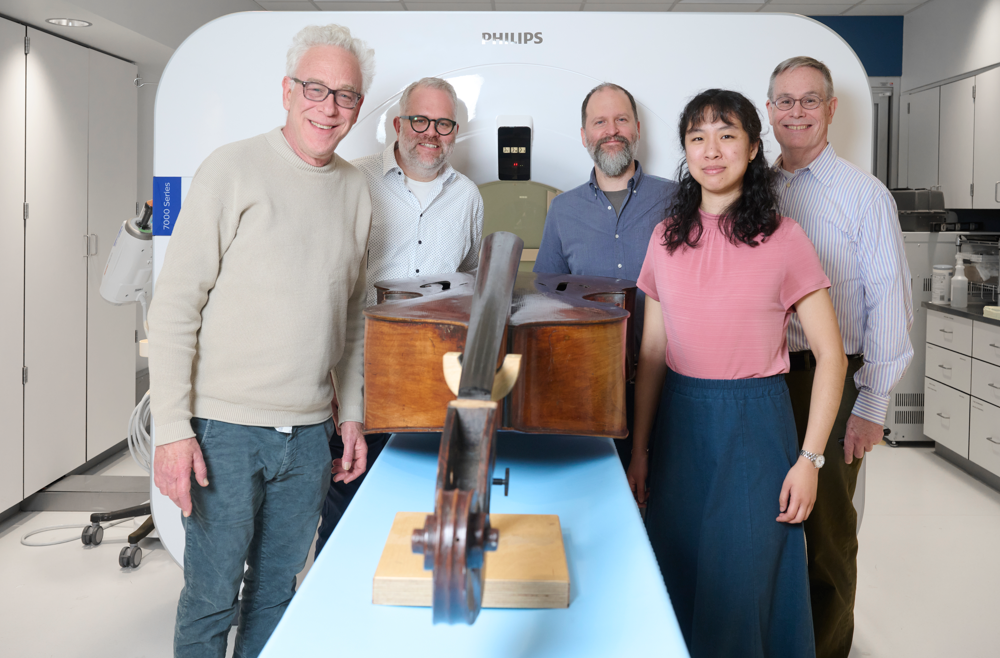
The research team from left to right includes Kelsey Swingle, Hannah Safford, Alex Hamilton, Ajay Thatte, Hannah Geisler, and Mike Mitchell.

New research on reproductive health demonstrates the first successful delivery of mRNA to placental cells to treat pre-eclampsia at its root.

Pre-eclampsia is a leading cause of stillbirths and prematurity worldwide, occurring in 3 – 8 % of pregnancies. A disorder characterized by high maternal blood pressure, it results from insufficient vasodilation in the placenta, restricting blood flow from the mother to the fetus.

Currently, a health-care plan for someone with pre-eclampsia involves diet and movement changes, frequent monitoring, blood pressure management, and sometimes early delivery of the baby. These standards of care address symptoms of the condition, not the root cause, and further perpetuate health inequity.

Now, Penn engineers are addressing this longstanding gap in reproductive health care with targeted RNA therapy.

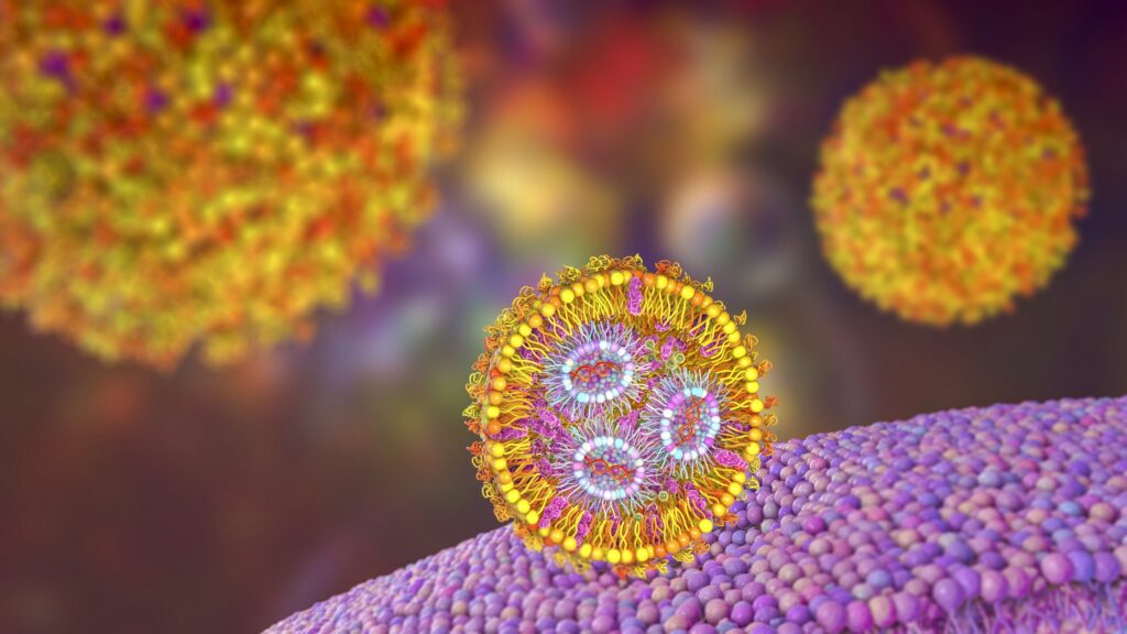
The COVID vaccines demonstrated how lipid nanoparticles (LNPs) efficiently deliver mRNA to target cells. The success of LNPs is opening doors for a variety of RNA therapies aiming to treat the root causes of illness and disease. However, drug development and health care have consistently neglected a portion of the population in need of targeted care the most – pregnant people and their babies.

Targeted Treatment for Pre-eclampsia. Current treatment: Early delivery. Results in high maternal blood pressure, restricted blood flow to the fetus. New treatment: Targeted RNA therapy and blood pressure monitoring. Strategically designed Lipid Nanoparticles deliver mRNA to placental cells. Vascular endothelial growth factor expands blood vessels, restores blood flow.In one of the first studies of its kind, published in the Journal of the American Chemical Society, Michael Mitchell, J. Peter and Geri Skirkanich Assistant Professor of Innovation in Bioengineering, and Kelsey Swingle, Ph.D. student in the Mitchell Lab and lead author, describe their development of an LNP with the ability to target and deliver mRNA to trophoblasts, endothelial cells, and immune cells in the placenta.

Once these cells receive the mRNA, they create vascular endothelial growth factor (VEGF), a protein that helps expand the blood vessels in the placenta to reduce the mother’s blood pressure and restore adequate circulation to the fetus. The researchers’ successful trials in mice may lead to promising treatments for pre-eclampsia in humans.

Read the full story in Penn Engineering Today.

Penn Bioengineering Student is a Hertz Fellowship Finalist

Savan Patel (Class of 2023)

Savan Patel, a fourth year Penn Bioengineering student, is one of 42 finalists competing for a 2023 Hertz Fellowship in applied science, mathematics, and engineering, one of the most prestigious Ph.D. fellowships in the United States. Chosen annually, the Hertz Fellowship is awarded to the nation’s most promising graduate students in science and technology.

From the Hertz Foundation website:

“Since 1963, the Hertz Foundation has granted fellowships empowering the nation’s most promising young minds in science and technology. Hertz Fellows receive five years of funding valued at up to $250,000, which offers flexibility from the traditional constraints of graduate training and the independence needed to pursue research that best advances our security and economic vitality […]

Over the foundation’s 60-year history of awarding fellowships, more than 1200 Hertz Fellows have established a remarkable track record of accomplishments. Their ranks include two Nobel laureates; recipients of 10 Breakthrough Prizes and three MacArthur Foundation “genius awards”; and winners of the Turing Award, the Fields Medal, the National Medal of Technology, and the National Medal of Science. In addition, 50 are members of the National Academies of Sciences, Engineering and Medicine, and 34 are fellows of the American Association for the Advancement of Science. Hertz Fellows hold over 3,000 patents, have founded more than 375 companies and have created hundreds of thousands of science and technology jobs.”

Patel is studying Bioengineering and Finance in the Jerome Fisher Program in Management and Technology (M&T), an interdisciplinary dual degree program coordinated by Penn Engineering and the Wharton School of Business. He is currently a member of the lab of Michael J. Mitchell, J. Peter and Geri Skirkanich Assistant Professor of Innovation in Bioengineering. Patel’s research interests lie at the interface of drug delivery and immunoengineering. His current project involves the use of modified cholesterol molecules to induce shifts in the biodistribution of ionizable lipid nanoparticles (LNPs). Following graduation, he intends to pursue a Ph.D. in bioengineering in which hopes to develop translatable immunotherapies and drug delivery platforms.

If chosen, the Hertz Fellowship will fund Patel’s graduate studies. Selected from over 750 applicants, Patel is one of fifteen undergraduates and one of two bioengineering students to make the final round of interviews. After a culminating round of interviews, the 2023 Class of Hertz Fellows will be announced in May.

Learn more about the Hertz Fellowship and read the full list of finalists here.

Carl June and Avery Posey Lead the Way in CAR T Cell Therapy

Perelman School of Medicine (PSOM) professors and Penn Bioengineering Graduate Group members Carl June and Avery Posey are leading the charge in T cell therapy and the fight against cancer.

Avery Posey, PhD
Carl June, MD

Advances in genome editing through processes such as CRISPR, and the ability to rewire cells through synthetic biology, have led to increasingly elaborate approaches for modifying and supercharging T cells for therapy. Avery Posey,  Assistant Professor of Pharmacology, and Carl June, the Richard W. Vague Professor in Immunotherapy, explain how new techniques are providing tools to counter some of the limitations of current CAR T cell therapies in a recent Nature feature.

The pair were also part of a team of researchers from PSOM, the Children’s Hospital of Philadelphia (CHOP), and the Corporal Michael J. Crescenz VA Medical Center to receive an inaugural $8 million Therapy ACceleration To Intercept CAncer Lethality (TACTICAL) Award from the Prostate Cancer Foundation. Their project will develop new clinic-ready CAR T cell therapies for Metastatic Castrate-Resistant Prostate Cancer (mCRPC).

Read “The race to supercharge cancer-fighting T cells” in Nature.

Read about the TACTICAL Award in the December 2022 Awards & Accolades section of Penn Medicine News.

Inside the Mitchell Lab: Crossing Biological Barriers

by

Black and white photo of Mike Mitchell working in the lab.
Mike Mitchell, Ph.D.

Engineers in the Center for Precision Engineering for Health (CPE4H) are focusing on innovations in diagnostics and delivery, cellular and tissue engineering, and the development of new devices that integrate novel materials with human tissues. Below is an excerpt from “Going Small to Win Big: Engineering Personalized Medicine,” featuring the research from the laboratory of Michael Mitchell, J. Peter and Geri Skirkanich Assistant Professor of Innovation in Bioengineering.

The Challenge

Solid tumors evade the immune system’s ability to attack them in part due to the tumors’ tough, fibrous biological barriers that circulating immune cells can’t cross. Researchers need to identify ways to deliver individualized treatments that can better target these tumors without causing damage to healthy tissues or affecting overall quality of life.

The Status Quo

Current cancer treatments typically involve surgery, radiation or chemo- therapy to eliminate solid tumors. These treatments are invasive and can cause numerous negative downstream effects. Newer treatments involve engineering a patient’s immune system to recognize and fight cancerous cells, but are so far only effective against certain “liquid” cancers, where the mutated cells circulate freely in the blood and bone marrow and are small enough to be picked off by the patient’s upgraded T cells. Additionally, existing methods can also require that the cell engineering take place in a lab rather than directly inside the body.

The Mitchell Lab’s Fix

Members of the lab of Michael Mitchell, J. Peter and Geri Skirkanich Assistant Professor of Innovation in Bioengineering, are looking to utilize nanoparticle delivery technology developed by their lab to engineer a different type of immune cell, the macrophage, in order to fight solid- tumor cancers from the inside.

The Mitchell lab is using lipid nanoparticles (LNPs) to carry mRNA and DNA sequences inside of macrophages, a type of immune cell that can consume tumor cells if engineered correctly. In theory, a patient would receive an injection carrying the LNP payload, and the macrophages, whose name literally means “big eaters,” would take up the genetic sequence, alter their function and be able to recognize a patient’s own unique tumor cells in the body.

Because of the way macrophages operate, they could cross the tumor’s biological barrier and attack the cells, destroying the tumor from the inside. An added benefit of the Mitchell Lab’s technology is that the destroyed tumor cells would then also allow other immune cells to present their antigens to circulating T cells, which could then learn to fight those same cancer cells in the future.

“One of the longstanding challenges that we face in the context of cancer and immunotherapies is that every tumor has unique antigens that are specific to patients,” says Mitchell. “This is why we’ve had a lot of trouble developing targeted therapies. Personalizing an approach by harnessing an individual’s immune system gives each patient a greater chance of a positive outcome.”

Read the full story in Penn Engineering magazine.

Inside the Jiang Lab: An Inventory of Immunity

by

Black and white photo of Jenny Jiang working in her lab on a laptop.
Jenny Jiang, Ph.D.

Engineers in the Center for Precision Engineering for Health (CPE4H) are focusing on innovations in diagnostics and delivery, cellular and tissue engineering, and the development of new devices that integrate novel materials with human tissues. Below is an excerpt from “Going Small to Win Big: Engineering Personalized Medicine,” featuring the research from the laboratory of Jenny Jiang, J. Peter and Geri Skirkanich Associate Professor of Innovation in Bioengineering.

The Challenge

In order to create personalized immune therapies, researchers need to untangle what is happening between an individual patient’s immune cells and the antigens that they interact with on a molecular level. Immune cell-antigen interactions need to be understood in four different areas in order to create a full picture: the unique genetic sequence of the T cell’s antigen receptors, the antigen specificity of that cell, and both the gene and protein expression of the same cell.

The Status Quo

Prior methods of understanding interactions between T cells and antigens could only get a picture of one or two of these four elements because of technology constraints. Other roadblocks included that cells cultured or engineered in a laboratory setting are not in a natural environment so they won’t express genes or proteins in the way T cells would in the body, and technologies that assess the antigen specificity of T cells were not cost-effective for looking at large numbers of antigens.

The Jiang Lab’s Fix

The lab of Jenny Jiang, J. Peter and Geri Skirkanich Associate Professor of Innovation in Bioengineering, developed a technology called TetTCR-SeqHD, which solves these problems. Using this technology, scientists can now simultaneously profile samples of large numbers of single T cells in the four dimensions using high- throughput screening.

The Jiang Lab’s technology is essentially a method for getting a “full-body scan” of an individual’s T cells and creates a catalog of the different types of T cells and the antigens they respond (or don’t respond) to, paving the way for the ability to better target immune therapies to an individual patient.

“Individual T cells are unique, and that’s the challenge of using one treatment to fit all,” says Jiang. “Identifying antigen specificity and creating therapies that target that specificity in an individual’s T cells will be key to truly personalizing immune therapies in the future.”

Read the full story in Penn Engineering magazine.

New Single Cell Analysis Tool

by Nathi Magubane

Researchers at Penn and colleagues have developed a tool to analyze single cells that assesses both the patterns of gene activation within a cell and which sibling cells shared a common progenitor.

3D illustration of a cell held by a pipet and a needle
Arjun Raj of the School of Engineering and Applied Science and the Perelman School of Medicine, former postdoc Lee Richman, now of Brigham and Women’s Hospital, and colleagues have developed a new analysis tool that combines a cell’s unique gene expression data with information about the cell’s origins. The method can be applied to identify new cell subsets throughout development and better understand drug resistance.

Recent advances in analyzing data at the single-cell level have helped biologists make great strides in uncovering new information about cells and their behaviors. One commonly used approach, known as clustering, allows scientists to group cells based on characteristics such as the unique patterns of active or inactive genes or by the progeny of duplicating cells, known as clones, over several generations.

Although single-cell clustering has led to many significant findings, for example, new cancer cell subsets or the way immature stem cells mature into “specialized” cells, researchers to this point had not been able to marry what they knew about gene-activation patterns with what they knew about clone lineages.

Now, research published in Cell Genomics led by University of Pennsylvania professor of bioengineering Arjun Raj has resulted in the development of ClonoCluster, an open-source tool that combines unique patterns of gene activation with clonal information. This produces hybrid cluster data that can quickly identify new cellular traits; that can then be used to better understand resistance to some cancer therapies.

“Before, these were independent modalities, where you would cluster the cells that express the same genes in one lot and cluster the others that share a common ancestor in another,” says Lee Richman, first paper author and a former postdoc in the Raj lab who is now at Brigham and Women’s Hospital in Boston. “What’s exciting is that this tool allows you to draw new lines around your clusters and explore their properties, which could help us identify new cell types, functions, and molecular pathways.”

Researchers in the Raj Lab use a technique known as barcoding to assign labels to cells they are interested in studying, particularly useful for tracking cells, clustering data based on cells’ offspring, and following lineages over time. Believing they could parse more valuable information out of this data by incorporating the cell’s unique patterns of gene activation, the researchers applied ClonoCluster to six experimental datasets that used barcoding to track dividing cells’ offspring. Specifically, they looked at the development of chemotherapy resistance and of stem cells into specialized tissue types.

Read the full story in Penn Today.