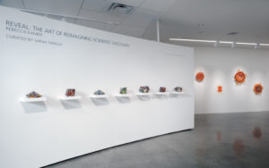
Rebecca Kamen, Penn artist-in-residence and visiting scholar, has a new exhibition titled “Reveal: The Art of Reimagining Scientific Discovery” at American University Museum at the Katzen Arts Center that explores curiosity and the creative process across art and science. (Image: Greg Staley)
Rebecca Kamen, Penn artist-in-residence and visiting scholar, has long been interested in science and the natural world. As a Philadelphia native and an artist with a 40-plus-year career, her intersectional work sheds light on the process of scientific discovery and its connections to art, with previous exhibitions that celebrate Apollo 11’s “spirit of exploration and discovery” to new representations of the periodic table of elements.
Now, in her latest exhibition, Kamen has created a series of pieces that highlight how the creative processes in art and science are interconnected. In “Reveal: The Art of Reimagining Scientific Discovery,” Kamen chronicles her own artistic process while providing a space for self-reflection that enables viewers to see the relationship between science, art, and their own creativity.
The exhibit, on display at the Katzen Art Center at American University, was inspired by the work of Penn professor Dani Bassett and American University professor Perry Zurn, the exhibit’s faculty sponsor. The culmination of three years of work, “Reveal” features collaborations with a wide range of scientists, including philosophers at American University, microscopists at the National Institutes of Health studying SARS-CoV-2 , and researchers in Penn’s Complex Systems Lab and the Addiction, Health, and Adolescence (AHA!) Lab.
“Reveal: The Art of Reimagining Scientific Discovery,” presented by the Alper Initiative for Washington Art and curated by Sarah Tanguy, is on display at the American University Museum in Washington, D.C., until Dec. 12.
The exhbition catalog, which includes an essay on “Radicle Curiosity” by Perry Zurn and Dani S. Bassett, can be viewed online.
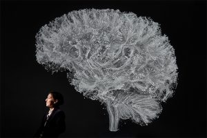
Among shots of a towering thunderstorm reaching into the stratosphere, the moon Daphnis peeking through Saturn’s rings, and an extremely close-up of a highly-endangered pangolin in Mozambique, one of Science’s favorite photos of 2019 was taken in Penn Engineering’s Raisler Lounge.
There, Danielle Bassett, J. Peter Skirkanich Professor in the Department of Bioengineering, poses underneath a giant visualization of the brain’s structural connections, projected on the wall behind her. Bassett’s research combines elements of physics, mathematics, engineering and neuroscience to provide a new look at how brain function arises from these networks of neurons.
Kelly Servick of Science profiled Bassett last year, revealing how a child whose parents discouraged her from attending college went on to become a pioneer in a highly interdisciplinary way of understanding the brain.
Read Bassett’s profile in Science here, and see the rest of the journal’s favorite photos of the year here.
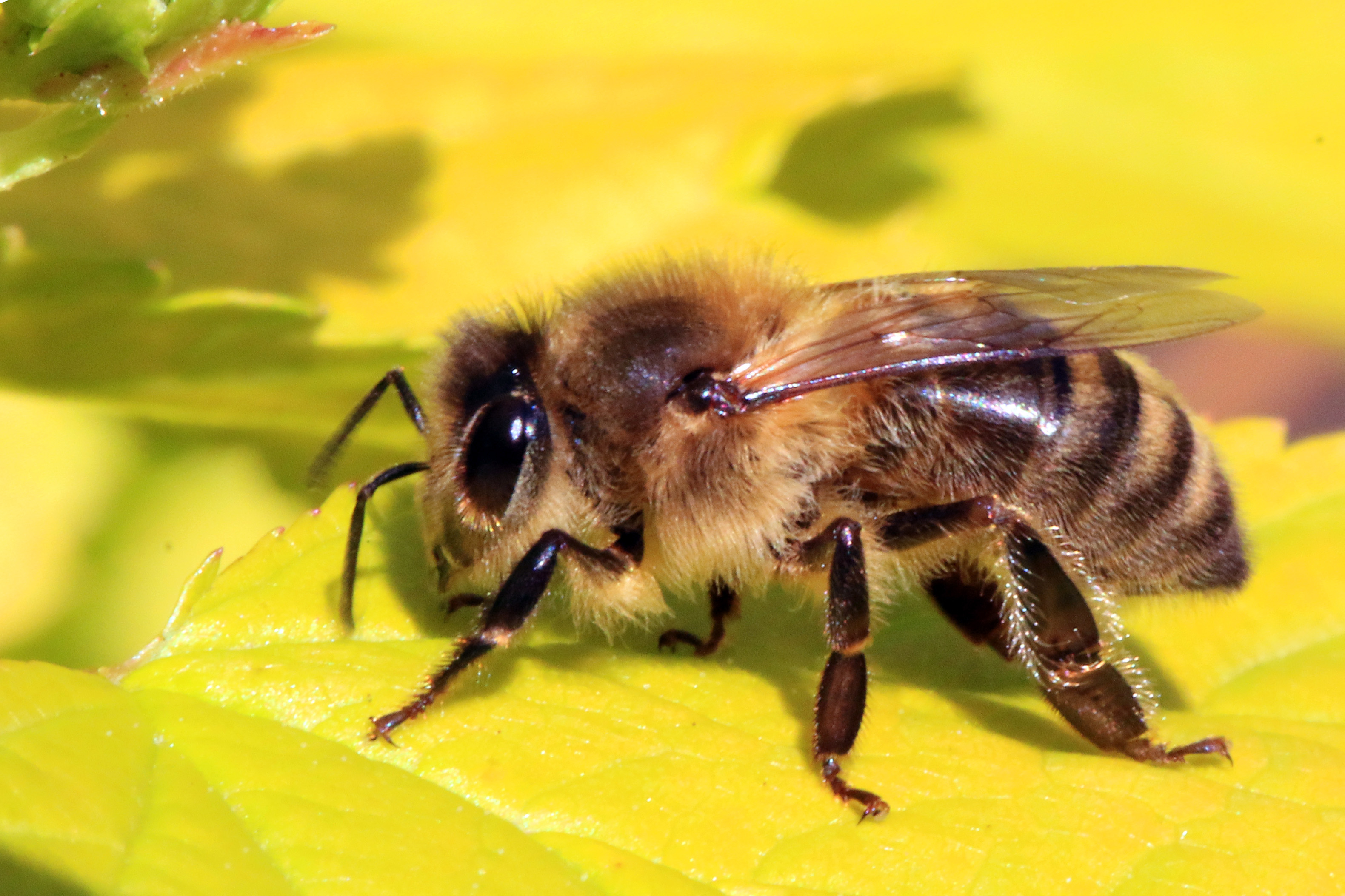
You might have heard reporting over the last few years that honeybees are dying at faster-than-usual rates. Over the last decade, colony collapse rates increased significantly, causing precipitous losses in the overall bee population. The consequences could be grave: in addition to providing honey, bee pollination is an important factor in agriculture, affecting major crops such as melons. squashes, and several kinds of nuts. Loss of this factor could substantially increase prices or even result in shortages.
To address this crisis, scientists at Washington State University focused on the role played by pesticides in colony collapse disorder. These poisons are particularly toxic to bees in tiny amounts, with the problem compounded by the ability of these toxins to build up in the bees’ bodies. A group of students led by Waled Suliman, PhD, a postdoctoral research associate in WSU’s Department of Biological Systems Engineering, developed a powder that acts like a magnet to draw pesticide out of the insects’ bodies. The bees then excrete the pesticide-laden particles like any other kind of waste.
The initiative, called Gaminus, has already tested its material in bees and found that the design works as planned. In coming months, they intend to continue their research by measuring toxin levels in the excreted particles.
Advances in Visualization
An important field within bioengineering is visualization, or the ability to use technology to enable scientists to see biological processes not normally visible to the naked eye. If you’ve seen a fetal ultrasound, for instance, then you’ve seen how one part of this area has advanced enormously in recent years. However, integrating visualization technologies with surgery remains a major challenge, particularly for minimally invasive surgeries. One key obstacle is that surgeons must rely on video screens during surgery, rather than being able to look down and feel the tissue with their hands.
A startup at the Cleveland Clinic is attempting to integrate perioperative visualization with HoloLens, a brand of smart glasses developed by Microsoft, to produce “mixed reality,” i.e., a combination of actual vision and virtual reality. With a grant from the National Heart, Lung, and Blood Institute awarded to Centerline Biomedical, the Cleveland Clinic startup, and to Karl West, Director of Medical Device Solutions at Cleveland Clinic and a staff member in the Lerner Research Institute’s Department of Biomedical Engineering, the integrated visualization device will be tested in a preclinical model of cardiac stent placement.
Elsewhere in the Midwest, Nathan Gianneschi, PhD, Professor of Chemistry, Biomedical Engineering and Materials Science and Engineering at Northwestern University, has been leading an effort to augment transmission electron microscopy (TEM). In its common form, TEM provides highly detailed images of submicroscopic organisms and structures and can provide visualization of nanomaterials as they grow. Gianneschi’s new approach, called liquid cell TEM (LCTEM), uses an irradiated region of a liquid cell to facilitate real-time visualization. The work is detailed in a recent article in ACS Central Science. You can see video posted online at the journal website.
Turning Red
Ultraviolet and infrared light appear beyond either end of the visible light spectrum. Past work using either ultraviolet or infrared light to activate fluorescent proteins can help visualize biochemistry in vivo, but it can also damage cells because of the activating light or the chemicals produced by illuminating the proteins. Recently, Young L. Kim, PhD, Associate Professor of Biomedical Engineering at Purdue, led a team of scientists who produced red fluorescent silk to kill harmful bacteria when the protein is activated by external green light. Dr. Kim and his colleagues report their findings in Advanced Science. The silk requires further testing, but if ultimately proved successful, it could overcome a current limitation of the use light-activated fluorescent biomaterials in controlling pathogens, which is that the light itself, often in the ultraviolet part of the spectrum, comes with its own potentially negative effects on health.
Absorbable Stents for Cardiac Care
Vascular stents to reopen blocked coronary arteries are usually the treatments used for patients with mild coronary artery disease. These simple devices are a small tube, sometimes coated with a drug to prevent clotting, inserted into the artery to restore flow. Stents can fail over time, requiring reimplantation, and the stents may also narrow over time and reduce blood flow to the surrounding tissue. To overcome this problem, Donghui Zhu, PhD, Associate Professor in the Department of Biomedical Engineering at the University of North Texas, developed a stent that is fully biodegradable and disappears over time as the damaged tissue heals. Dr. Zhu recently won a $2 million grant from the National Institutes of Health to test the stent in a series of trials.
People and Places
Penn State University has won a research grant from the American Heart Association, which will be used to support its 10-week Penn State Summer Translational Cardiovascular Science Institute (STCSI). Led by Keefe Manning, PhD, Professor of Biomedical Engineering at Penn State, the STCSI will provide $4,000 stipends for undergraduate students to conduct summer research on cardiovascular disease.
Finally, here at Penn Bioengineering, we are immensely proud to announce that our PhD student Jina Ko was named one of 14 PhD candidates in the inaugural class of Schmidt Science Fellows. Schmidt Fellows are each awarded a $100,000 stipend to cover the cost of living while conducting postdoctoral research. Congratulations, Jina!
Spina bifida is a fairly common type of birth defect caused by incomplete closure of the backbone and tissue surrounding the spinal cord. Fetal surgery can repair the defect before delivery, but this invasive surgery can lead to high risk preterm delivery.
A new material may dramatically reduce the invasiveness of surgery needed to correct spina bifida. In a new article in Macromolecular Bioscience, surgeons and bioengineers from the University of Colorado report on one of these alternatives. One of the lead authors was Daewon Park, Ph.D., assistant professor of bioengineering. Dr. Park and his colleagues developed a reverse thermal gel, which is an injectable liquid that forms a gel at higher temperatures when injected into the body. Ultimately a gel like this one could be injected at or near the spine, where it would cover the defect in a spina bifida patient, harden into a gel, and ultimately repair the defect by deploying stem cells or engineered tissue.
The research team’s most recent study indicates that their gel retained its stability in amniotic fluid and was compatible with neural tube cells. They also tested the gel in two animal models, with successful results. The gel is still far from being used in actual fetal surgery cases, but the authors will continue to test the gel under conditions increasingly similar to the human amniotic sac.
Building Better Brains
UCLA scientists have developed an improved system for generating brain structures from stem cells. The team of scientists, led by Bennett G. Novitch, Ph.D., professor of neurobiology at UCLA, report their findings in Cell Reports. Importantly, the methods used by Dr. Novitch and his colleagues fine-tuned and simplified earlier efforts in this area, developing a method that did not require any specific reactors to generate the tissue. They were also able to generate tissue resembling the basal ganglia for the first time, indicating promise for using these tissues to model diseases affecting that part of the brain, including Parkinson’s disease.
Next, the authors demonstrated the usefulness of these “organoids” in modeling damage due to Zika virus. After exposing the generated organoids to Zika, the authors measured the cellular responses of the tissue, demonstrating the ability to use these tissues to model the disease. Given the recent epidemic of Zika virus in the Western Hemisphere, which focused attention on the virus’s effects on the human brain, in addition to microcephaly and other birth defects when the disease is transmitted from pregnant mothers to their children, understanding how Zika affects the developing brain is key to determining how to prevent the damage it causes and possibly repairing it. Reliable models of brain development are necessary, and the UCLA team’s findings seem to indicate that they’ve found one.
Rebuilding Brain Circuits After Injury
Among the issues in the prevention and treatment of head injury is that we still lack complete information about the mechanism underlying these injuries. However, a key piece of basic research recently published by a team at the University of North Carolina, Chapel Hill, demonstrates that a key aspect of this mechanism occurs in the axons, which are the stalks that grow from neurons to signal to each other. In an article published in Nature Communications, the team, led by Anne Marion Taylor, Ph.D., assistant professor of biomedical engineering at UNC, reports on using microfluidics technology to determine how neurons react when axons are severed. The authors found that damage to axons causes a compensatory loss of collateral connections to neighboring neurons. This loss in connectivity could be reversed by adding a protein, netrin-1, into the solution surrounding the neurons. Although netrin-1 was already known for its importance in rebuilding damaged axons, this work showed that netrin-1 has more widespread effects in rebuilding neural circuits after trauma.
Seeing Inside the Body
One of the key problems with minimally invasive surgical procedures is difficulty in shining sufficient light in the target region to see and manipulate tissue during surgery. Glass filaments are currently used, but they pose a health risk because they can break off in the body. A new citrate polymer fiber invented by scientists at Penn State University represents a much safer alternative to glass filaments. Reporting in Biomaterials, the scientists, led by Jian Yiang, Ph.D., associate professor of biomedical engineering at Penn State, describe how they developed the fiber and show how much less likely this polymer filament is to break when manipulated. If biocompatibility tests show that this polymer does not affect tissue health, it could eventually appear in surgical microscopes and make glass filaments a thing of the past.
Creating better illumination tools is one way of seeing inside the body. An elegant device developed by a team including MIT biomedical engineer Giovanni Traverso, Ph.D., consists of a proof-of-concept ingestable sensor that attaches to the stomach lining and can provide upper gastrointestinal system information for two days. The team reports on the device in a recent issue of Nature Biomedical Engineering. Unlike earlier ingestable diagnostic devices, the sensor developed by Dr. Traverso and his colleagues uses the contraction of the gut to power the device. It also surpassed pill-sized ingestable cameras by providing data from a longer time period.
Implants That Grow With Chilren
Using implants to treat medical problems in children is difficult for one simple reason: children can quickly outgrow their devices. The problem has been particularly acute among pediatric cardiac surgeons, for whom implants are commonly used devices. Now, thanks to collaboration between surgeons and a team including Jeffrey Karp, Ph.D., a Harvard biomedical engineer, valve implants that grow with patients could be here soon. Inspired by the clever design of a Chinese finger trap, the collaborators developed an implant that grows longer but thinner over time. They report on their device in Nature Biomedical Engineering, including its proof-of-principle testing in growing piglets. Based on these data, the study authors will continue to adapt and develop the device.
Say What?
Humans are especially good at listening to many voices at once or focusing on one voice in a crowded room. However, we really don’t know how we do this so well. Scientists at Imperial College London (ICL) solved part of this mystery. Reporting in eLife, the ICL authors developed a mathematical method to measure how the response to speech in a person’s brainstem changed when that person’s attention moved from one person to another. Perceptible changes in brainstem activity occurred when a person was intently listening to someone, and it disappeared when the person ignored speech. In addition to supplementing what we already know about how the brain stem participates in sensory tasks, the ICL team’s findings mean that damage to the brainstem – common to several neurological disorders – can easily affect how we process speech and interact with each other.