Project Builds on Breast Cancer Screening Tech

breast cancer
An embedded 11×11 cluster of 100-micron objects (which models a cluster of microcalcifications — one of the earliest indicators of breast cancer). (A) shows the results from the current standard of imaging only along the chest wall. (B) shows the results of our method that considers the patient’s unique breast geometry using a Custom V imaging pattern. (B) resolves the embedded cluster as a distinct cluster of objects while (A) appears to blur the final image.

Breast cancer continues to affect more than 10% of all women — and a small percentage of men — despite significant advances in diagnosis and treatment. While a majority cases today can be successfully treated, early detection is essential to beginning treatment before it’s too late.

Among the more recent innovations in screening has been three-dimensional mammography. However, this modality has lacked the ability to personalize the scan to the individual patient’s breast, instead only acquiring several two-dimensional images along the chest wall, resulting in a lack of individualization for the patient.

A senior design project team at the University of Pennsylvania’s Department of Bioengineering, however, has helped to develop a more personalized 3D imaging technology, which acquires a series of images but instead following the contour of the breast itself. With their efforts, the team earned one of the three awards given to student teams yearly.

The four-student team, consisting of Lucy Chai, Elizabeth Kobe, Margaret Nolan, and Sushmitha Yarrabothula, picked up a project begun last year (a common practice with senior design projects) and demonstrated with their work that the imaging technique could be applied using a digital phantom (a computerized breast model) with great clarity, including successful resolution of a simulated mass just one-tenth of a millimeter in size.

Now, the four seniors will hand off the project to another team, continuing this multi-year research. Ultimately, before it can be applied in actual patients, the modality will need to be tested against the current standard of care in terms of its ability to detect small masses in the breast. Nevertheless, this year’s team moved the ball downfield significantly.

Cataract Surgery Instrument With an Eye on India

Cataracts are a leading cause of vision loss worldwide. In the world’s more developed countries, laser is commonly used for cataract removal. However, in much of the developing world, lasers are expensive or difficult to acquire. In these countries, cataract surgeries are still largely performed freehand, with all of the attendant risks that such procedures involve.

One of this year’s senior design projects in the Department of Bioengineering at Penn was a surgical tool for ophthalmic surgeons to perform capsulorhexis, the fancy term for the circular incision necessary to remove a cataract. The team, which included seniors Akshatha Bhat, Nimay Kulkarni, Steven Polomski, and Ananya Sureshkumar, collaborated with the Aravind Eye Hospital in Puducherry, India, created a surgical device called the Rhex to create this round incision

cataract-rhex
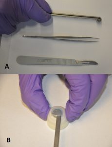
A) The Rhex (top), compared to forceps (center) and a scalpel (bottom); B) Rhex inserted into a model eye.

The Rhex (right) consists of a stem with a ring at the end, into which a blade is fitted. It can be inserted into a scleral incision, pressed, and rotated to perform the capsulorhexis. Initial testing indicated the Rhex could create a circulation incision approximately 6.7 mm in diameter, with eccentricity (indicating deviation from a perfect circle) of 0.254±0.08, which was within the acceptable range determined by the team.

The students designed the Rhex to be autoclavable and to use disposable blades. The next step will be to decrease the size of the instrument further and perhaps to use translucent or even transparent material to produce newer prototypes, which could be particularly useful, since cataract surgeries are open performed using backlighting.


Design Project Offers Help for Parkinson’s Patients

parkinson's
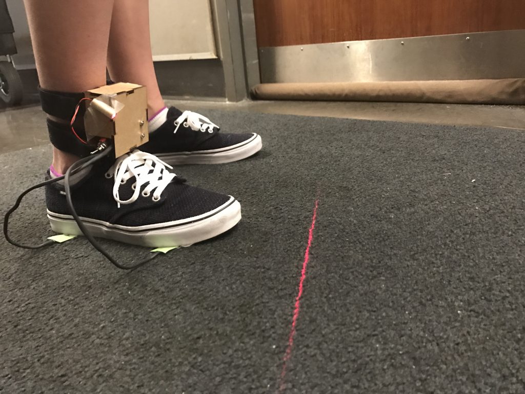
ShuffleAssist demonstrated by a student

With increasing age in the population, Parkinson’s disease has become increasingly common. One of the most frustrating effects of the disease is freezing of gait (FOG), in which a patient will suddenly stop while walking and find it difficult to begin again. Falls are a common consequence.

Despite intensive research, FOG is poorly understood. However, studies have shown that certain external stimuli, including metronomes and devices that provide visual cues, can be helpful. With this knowledge, a team of bioengineering students set to tackle this issue with their senior design project.

The team whose members were Priyanka Ghosh, Fiona La, Laurel Leavitt, and Lia Lombardi — came up with ShuffleAssist, a wearable device that uses force sensors and an internal measurement unit to detect FOG and automatically provide a cue for the patient. The patient can choose a metronome beat or visual laser cue that can be provided either as determined by the device or continually, for patients who so choose.

ShuffleAssist tested well among normal subjects, detecting FOG correctly 98% of the time within approximate one second. In addition, the students were able to create their prototype for a cost of $107 per unit, compared to similarly intended products already on the market costing more than twice that much.

The next step for the team is to test the device in actual patients with Parkinson’s. The students have left the device with a faculty member in the Perelman School of Medicine who treats patients with motor disorders. This faculty member will offer the device to patients for testing.

See below for a video demonstration of ShuffleAssist.