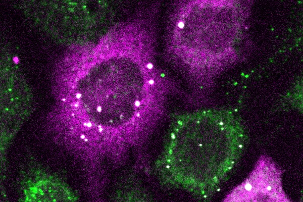
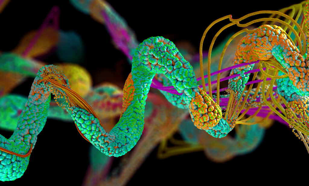
The bright white spots represent tiny clusters of proteins detected by CluMPS. (Photo by: Thomas Mumford)
Penn Engineers have pioneered a new way to visualize the smallest protein clusters, skirting the physical limitations of light-powered microscopes and opening new avenues for detecting the proteins implicated in diseases like Alzheimer’s and testing new treatments.
In a paper in Cell Systems, Lukasz Bugaj, Assistant Professor in Bioengineering, describes the creation of CluMPS, or Clusters Magnified by Phase Separation, a molecular tool that activates by forming conspicuous blobs in the presence of target protein clusters as small as just a few nanometers. In essence, CluMPS functions like an on/off switch that responds to the presence of clusters of the protein it is programmed to detect.
Normally, says Bugaj, detecting such clusters requires laborious techniques. “With CluMPS, you don’t need anything beyond the standard lab microscope.” The tool fuses with the target protein to form condensates orders of magnitude larger than the protein clusters themselves that resemble the colorful blobs in a lava lamp. “We think the simplicity of the approach is one of its main benefits,” says Bugaj. “You don’t need specialized skills or equipment to quickly see whether there are small clusters in your cells.”
In the human body, the lungs and their vasculature can be likened to a building with an intricate plumbing system. The lungs’ blood vessels are the pipes essential for transporting blood and nutrients for oxygen delivery and carbon dioxide removal. Much like how pipes can get rusty or clogged, disrupting normal water flow, damage from respiratory viruses, like SARS-CoV-2 or influenza, can interfere with this “plumbing system.”
In a recent study, researchers looked at the critical role of vascular endothelial cells in lung repair. Their work, published in Science Translational Medicine, was led by Andrew Vaughan of the University of Pennsylvania’s School of Veterinary Medicine and shows that, by using techniques that deliver vascular endothelial growth factor alpha (VEGFA) via lipid nanoparticles (LNPs), that they were able to greatly enhance modes of repair for these damaged blood vessels, much like how plumbers patch sections of broken pipes and add new ones.
“While our lab and others have previously shown that endothelial cells are among the unsung heroes in repairing the lungs after viral infections like the flu, this tells us more about the story and sheds light on the molecular mechanisms at play,” says Vaughan, assistant professor of biomedical sciences at Penn Vet. “Here we’ve identified and isolated pathways involved in repairing this tissue, delivered mRNA to endothelial cells, and consequently observed enhanced recovery of the damaged tissue. These findings hint at a more efficient way to promote lung recovery after diseases like COVID-19.”
They found VEGFA’s involvement in this recovery, while building on work in which they used single cell RNA sequencing to identify transforming growth factor beta receptor 2 (TGFBR2) as a major signaling pathway. The researchers saw that when TGFBR2 was missing it stopped the activation of VEGFA. This lack of signal made the blood vessel cells less able to multiply and renew themselves, which is vital for the exchange of oxygen and carbon dioxide in the tiny air sacs of the lungs.
“We’d known there was a link between these two pathways, but this motivated us to see if delivering VEGFA mRNA into endothelial cells could improve lung recovery after disease-related injury,” says first author Gan Zhao, a postdoctoral researcher in the Vaughan Lab.
“LNPs have been great for vaccine delivery and have proven incredibly effective delivery vehicles for genetic information. But the challenge here was to get the LNPs into the bloodstream without them heading to the liver, which is where they tend to congregate as its porous structure lends favor to substances passing from the blood into hepatic cells for filtration,” says Mitchell, an associate professor of bioengineering at Penn Engineering and a coauthor of the paper. “So, we had to devise a way to specifically target the endothelial cells in the lungs.”
Lulu Xue, a postdoctoral researcher in the Mitchell Lab and a co-first author of the paper, explains that they engineered the LNP to have an affinity for lung endothelial cells, this is known as extra hepatic delivery, going beyond the liver.
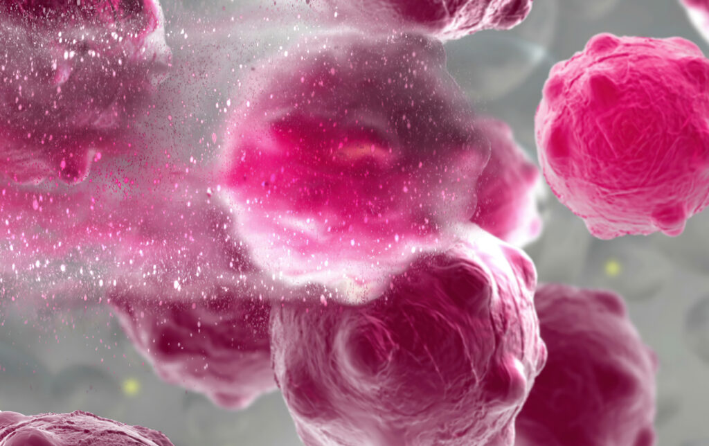
3d illustration of a damaged and disintegrating cancer cell. (Image: iStock/vitanovski)
The development of any type of second cancer following CAR T cell therapy is a rare occurrence, as found in an analysis of more than 400 patients treated at Penn Medicine, researchers from the Perelman School of Medicine at the University of Pennsylvania reported today in Nature Medicine. The team also described a single case of an incidental T cell lymphoma that did not express the CAR gene and was found in the lymph node of a patient who developed a secondary lung tumor following CAR T cell therapy.
CAR T cell therapy, a personalized form of immunotherapy in which each patient’s T cells are modified to target and kill their cancer cells, was pioneered at Penn. More than 30,000 patients with blood cancers in the United States—many of whom had few, if any, remaining treatment options available—have been treated with CAR T cell therapy since the first such therapy was approved in 2017. Some of the earliest patients treated in clinical trials have gone on to experience long-lasting remissions of a decade or more.
Secondary cancers, including T cell lymphomas, are a known, rare risk of several types of cancer treatment, including chemotherapy, radiation, and stem cell transplant. CAR T cell therapy is currently only approved to treat blood cancers that have relapsed or stopped responding to treatment, so patients who receive CAR T cell therapies have already received multiple other types of treatment and are facing dire prognoses.
In November 2023, the FDA announced an investigation into several reported cases of secondary T cell malignancies, including CAR-positive lymphoma, in patients who previously received CAR T cell therapy products. In January 2024, the FDA began requiring drugmakers to add a safety label warning to CAR T cell products. While the FDA review is still ongoing, it remains unclear whether the secondary T cell malignancies were caused by CAR T cell therapy.
As a leader in CAR T cell therapy, Penn has longstanding, clearly established protocols to monitor each patient both during and after treatment – including follow-up for 15 years after infusion – and participates in national reporting requirements and databases that track outcomes data from all cell therapy and bone marrow transplants.
Marco Ruella, M.D.
“When this case was identified, we did a detailed analysis and concluded the T cell lymphoma was not related to the CAR T cell therapy. As the news of other cases came to light, we knew we should go deeper, to comb through our own data to better understand and help define the risk of any type of secondary cancer in patients who have received CAR T cell products,” said senior author Marco Ruella, MD, an assistant professor of Hematology-Oncology and Scientific Director of the Lymphoma Program. “What we found was very encouraging and reinforces the overall safety profile for this type of personalized cell therapy.”
More than 34 million Americans suffer from pulmonary diseases like asthma, emphysema and chronic bronchitis. While medical treatments can keep these ailments in check, there are currently no cures. Part of the reason, notes Dan Huh, is that it’s incredibly hard to study how these diseases actually work. While researchers can grow cells taken from human lungs in a dish, they cannot expect them to act like they would in the body. In order to mimic the real deal, it’s necessary to recreate the complex, 3D environment of the lung — right down to its tiny air sacs and blood vessels — and to gently stretch and release the tissue to simulate breathing.
Huh, Associate Professor in Bioengineering, is the cofounder of Vivodyne, a Penn Engineering biotech spinoff that is creating tissues like these in the lab. Vivodyne uses a bioengineering technology that Huh has been developing for more than a decade. While a postdoctoral fellow at Harvard’s Wyss Institute, he played a central role in creating a novel device called an “organ on a chip,” which, as the name implies, assembles multiple cell types on a tiny piece of engineered plastic to create an approximation of an organ.
“While those chips represented a major innovation,” says Huh, “they still weren’t truly lifelike. They lacked many of the essential features of their counterparts in the human body, such as the network of blood vessels running between different kinds of tissue, which are essential for transporting oxygen, nutrients, waste products and various biochemical signals.”
Noor Momin, Stephenson Foundation Term Assistant Professor of Innovation
While growing up, Noor Momin, who joined the Department of Bioengineering in January as the Stephenson Foundation Term Assistant Professor of Innovation, imagined becoming a physician. Becoming a doctor seemed like a tangible way for someone interested in science to make a difference. Not until college did she realize the impact she could have as a bioengineer instead.
“I was taping microscope slides together,” Momin recalls of her initial experience as an undergraduate researcher at the University of Texas at Austin. “I didn’t even know what a Ph.D. was.”
It wasn’t until co-authoring her first paper, which explores how lipids, the water-repelling molecules that make up cell membranes (and also fats and oils), can switch between more fluid and less fluid arrangements, that Momin understood the degree to which bioengineering can influence medicine. “Someone could potentially use that paper for drug design,” Momin says.
As Momin sees it, the conventional wisdom of treating the heart like a mechanical pump, whose pipes can be replaced or whose throughput can be treated to prevent clogging in the first place, overshadows the immune system’s critical role in the development of heart disease.
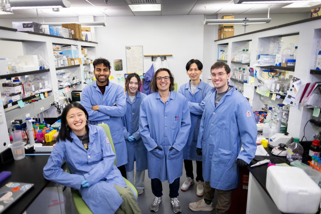
From left: Emily Han, Rohan Palanki, Jacqueline Li, Michael Mitchell, Dongyoon Kim, and Marshall Padilla of Penn Engineering.
Imagine the brain as an air traffic control tower, overseeing the crucial and complex operations of the body’s ‘airport.’ This tower, essential for coordinating the ceaseless flow of neurological signals, is guarded by a formidable layer that functions like the airport’s security team, diligently screening everything and everyone, ensuring no unwanted intruders disrupt the vital workings inside.
However, this security, while vital, comes with a significant drawback: sometimes, a ‘mechanic’—in the form of critical medication needed for treating neurological disorders—is needed inside the control tower to fix arising issues. But if the security is too stringent, denying even these essential agents entry, the very operations they’re meant to protect could be jeopardized.
Now, researchers led by Michael Mitchell of the University of Pennsylvania are broaching this long-standing boundary in biology, known as the blood-brain barrier, by developing a method akin to providing this mechanic with a special keycard to bypass security. Their findings, published in the journal Nano Letters, present a model that uses lipid nanoparticles (LNPs) to deliver mRNA, offering new hope for treating conditions like Alzheimer’s disease and seizures—not unlike fixing the control tower’s glitches without compromising its security.
“Our model performed better at crossing the blood-brain barrier than others and helped us identify organ-specific particles that we later validated in future models,” says Mitchell, associate professor of bioengineering at Penn’s School of Engineering and Applied Science, and senior author on the study. “It’s an exciting proof of concept that will no doubt inform novel approaches to treating conditions like traumatic brain injury, stroke, and Alzheimer’s.”
A pair of proteins, YAP and TAZ, has been identified as conductors of bone development in the womb and could provide insight into genetic diseases such as osteogenesis imperfecta, known commonly as “brittle bone disease.” This research, published in Developmental Cell and led by members of the McKay Orthopaedic Research Laboratory of the Perelman School of Medicine, adds understanding to the field of mechanobiology, which studies how mechanical forces influence biology.
“Despite more than a century of study on the mechanobiology of bone development, the cellular and molecular basis largely has remained a mystery,” says the study’s senior author, Joel Boerckel, an associate professor of orthopaedic surgery. “Here, we identify a new population of cells that are key to turning the body’s early cartilage template into bone, guided by the force-activated gene regulating proteins, YAP and TAZ.”
Kaitlin Mrksich, an undergraduate student in Penn Bioengineering, was honored with the Student Award for Outstanding Research (Undergraduate) by the Society for Biomaterials (SFB). This prestigious award recognizes undergraduate students who have shown outstanding achievement in biomaterials research.
Mrksich is a third-year student from Hinsdale, Illinois. She is interested in developing drug delivery systems that can serve as novel therapeutics for a variety of diseases. She works in the lab of Michael Mitchell, Associate Professor in Bioengineering. In the Mitchell Lab, Mrksich investigates the ionizable lipid component of lipid nanoparticles for mRNA delivery.
“In Kaitlin’s independent projects, she has focused on probing the role of lipophilicity and chirality for LNP-mediated mRNA delivery,” Mitchell said in the award announcement. “She has synthesized dozens of unique lipids, formulated these lipids into LNPs, and evaluated their potential for mRNA delivery in vivo and in primary T cells. She has been able to deduce structure-function relationships that help explain the role of lipid hydrophobicity in the delivery of mRNA by LNPs. Her findings have not only been instrumental in helping our lab design better LNPs but will also provide fundamental knowledge that will benefit all labs working on LNP technology.”
In addition to her academic activities, Mrksich is also the President of the Penn Biomedical Engineering Society (BMES), where she plans community-building and professional-development events for bioengineering majors, and the visit coordinator for special programs for the Kite and Key Society, where she organizes virtual programming to introduce prospective students to Penn. She also tutors a West Philadelphia high school student in chemistry as part of the West Philadelphia Tutoring Project and is a member of Tau Beta Pi engineering honor society and Sigma Kappa sorority. After graduating, she plans to pursue an M.D.-Ph.D. in Bioengineering.
Read the full list of 2024 SFB award recipients here.
Riccardo Gottardi, Assistant Professor in Pediatrics and in Bioengineering and leader of the Bioengineering and Biomaterials Laboratory at the Children’s Hospital of Philadelphia (CHOP), received the Rising Star Award from the Biomedical Engineering Society-Cellular and Molecular Bioengineering (BMES-CMBE). The Rising Star Award recognizes a BMES-CMBE member who is at the early independent career stage and has made an outstanding impact on the field of cellular and molecular bioengineering. Awardees will give an oral presentation on their research at the BMES-CMBE conference in Puerto Rico in January and be recognized at the conference Gala dinner.
Dr. Gottardi’s research focuses on engineering solutions for pediatric health, primarily for airway disorders. He has previously received awards for work to create a biomaterial patch to repair the tympanic membrane and for work to develop cartilage implants to treat severe subglottic stenosis. He received grant support from the National Institutes of Health to further his work in subglottic stenosis.
Sydney Shaffer, Assistant Professor in Bioengineering in the School of Engineering and Applied Science and in Pathology and Laboratory Medicine in the Perelman School of Medicine, was named the 2023 Christopher J. Marshall Award winner by the Society for Melanoma Research (SMR). The award recognizes Shaffer’s contributions to melanoma research on oncogenic signalling and molecular pathogenesis of this disease, as well as her rapid development as a rising star and leader in the field, which have helped to further the SMR’s goal to eradicate melanoma. The award was presented at the SMR annual meeting in Philadelphia in November 2023.
The Christopher J. Marshall Award was established in 2015 by the SMR in partnership with Melanoma Research Foundation Congress to recognize a student, postdoctoral fellow, or new independent PI who has published a substantial and original contribution to studies of signal transduction and melanoma.
Shaffer joined Penn as an Assistant Professor in 2019. She holds a M.D.-Ph.D. in Medicine and Bioengineering from the University of Pennsylvania and conducted postdoctoral research in cancer biology in the lab of Junwei Shi, Associate Professor in Penn Medicine. The Syd Shaffer Lab is an interdisciplinary team which focuses on “understanding how differences between single-cells generate phenotypes such as drug resistance, oncogenesis, differentiation, and invasion [using] a combination of imaging and sequencing technologies to investigate rare single-cell phenomena.” A recent paper in Nature Communications details the team’s method to quantify long-lived fluctuations in gene expression that are predictive of later resistance to targeted therapy for melanoma.
Read the award announcement and the full list of prior winners at the SMR website.