The vast majority of genetic mutations that are associated with disease occur at sites in the genome that aren’t genes. These sequences of DNA don’t code for proteins themselves, but provide an additional layer of instructions that determine if and when particular genes are expressed. Researchers are only beginning to understand how the non-coding regions of the genome influence gene expression and might be disrupted in disease.
Jennifer Phillips-Cremins, assistant professor in the Department of Bioengineering in the University of Pennsylvania’s School of Engineering and Applied Science, studies the three-dimensional folding of the genome and the role it plays in brain development. When a stretch of DNA folds, it creates a higher-order structure called a looping interaction, or “loop.” In doing so, it brings non-coding sites into physical contact with their target genes, precisely regulating gene expression in space and time during development.
Phillips-Cremins and lab member Jonathan Beagan have led a new study identifying a new protein that connects loops in embryonic stem cells as they begin to differentiate into types of neurons. Though the study was conducted in mice, these findings inform aspects of human brain development, including how the genetic material folds in the 3-D nucleus and is reconfigured as stem cells become specialized. Better understanding of these mechanisms may be relevant to a wide range of neurodevelopmental disorders.
Cremins lab members Michael Duong, Katelyn Titus, Linda Zhou, Zhendong Cao, Jingjing Ma, Caroline Lachanski and Daniel Gillis also contributed to the study, which was published in the journal Genome Research.
Dennis E. Discher, Ph.D., Robert D. Bent Professor in the Department of Chemical and Biomolecular Engineering and a secondary faculty member in the Department of Bioengineering, was the lead author on a recent study that showed that engineered macrophages (a type of immune cell) could be injected into mice, circulate through their bodies, and invade solid tumors in the mice, engulfing human cancers cells in the tumors.
According to Cory Alvey, a graduate student in pharmacology who works in Professor Discher’s lab and the first author on the paper, said, “Combined with cancer-specific targeting antibodies, these engineered macrophages swarm into solid tumors and rapidly drive regression of human tumors without any measurable toxicity.”
by Meagan Ita, Ph.D. Student in Bioengineering and GABE Co-President
Cancer is a disease that affects millions, and over the last several decades, researchers have delved deeply into the biological underpinnings of the disease in the hopes of finding a cure. One major discovery is that mistakes in your DNA “instructions” can lead to cancer by crossing the wires in your cellular circuitry, and researchers have developed amazing new drugs that can cause tumors to melt away by targeting these broken components. The problem though is that, most of the time, the tumors come back, and this is a huge barrier to cures.
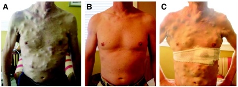
Picture of patient treated with vemurafenib and then developing resistance. Courtesy of the American Society of Clinical Oncology
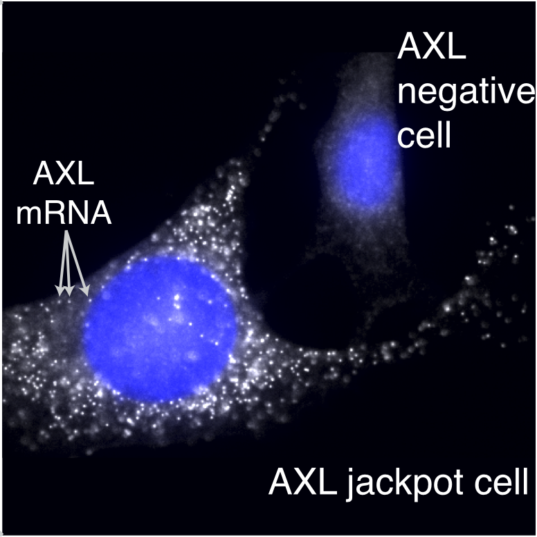
For a long time, everyone assumed that the reason the tumors came back was DNA mistakes on top of the original mistakes, with these new mistakes blocking the activity of the anti-cancer drug. However, new work led by Sydney Shaffer from the Arjun Raj Lab at Penn Bioengineering, published this week in Nature, challenges this view by looking all the way down at individual cancer cells and seeing how they respond to these drugs on a cell-by-cell basis.
Sydney found that in melanoma, contrary to what researchers thought, it need not be a DNA mistake that leads a cell to become resistant to the drug, but rather a change in cellular identity. Just like your body has cells of all different types, like skin cells and brain cells, cancer cells appear to change between different types, but unlike in the body, cancer cells do it in a seemingly random and uncontrolled way, and the cells exploit this variability to allow those rare cells that have changed their type to survive the drug.
Here, we talk with Sydney about the inspiration, triumphs, and challenges she faced in her research.
What was the initial inspiration for looking at drug resistance in melanoma?
For the first two years of working on this project, we actually didn’t have a clear question in mind. I was just trying a bunch of different experiments with melanoma cells, and I noticed something that we found thought-provoking. Whenever we gave the melanoma cells a particular drug, they would become resistant at exactly the same point in time. At first, this may not seem unusual, but for example, if everyone showed up at a restaurant to eat lunch at exactly noon, you would guess this was not happening purely by chance. Maybe classes let out right beforehand? Or a big meeting? For the melanoma cells, we would similarly expect there to be a range of different times for the cells to become resistant, but instead it all happened at once.
This observation helped us figure out that the drug-resistant cells probably already exist before we treat them. It also got us curious about the particular processes that make the cells resistant, and we spent many lab meetings discussing this observation until one postdoc, Gautham Nair, suggested trying some experiments based upon the classical molecular biology experiments of Luria and Delbrück.
Who were Luria and Delbrück, and how did they influence your work?
Max Delbrück and Salvador Luria (below) were scientists who, in the 1940s, performed a clever experiment that demonstrated that bacteria become resistant to viruses through random DNA mutations. According to Wikipedia, Luria actually had the idea for these experiments while watching slot machines!
Delbrück (left) and Luria. Courtesy of the Genetics Society of America.
Their experiment was super simple: it was basically a statistical way to see whether cells “sense and respond” to a challenge, or whether they just passively get a mutation that lets some fraction of them survive the challenge, basically like Darwinian evolution. The idea is that, in the first scenario, there is no history: every cell has an equal chance to respond when challenged.
But in the second scenario, history matters in that if your great-grandparent was a survivor, then all your relatives would be too. If you could redo history over and over, then sometimes maybe your great-great-great-great-grandparent would be a survivor, and so you would get a whole bunch of survivors when the challenge came. Luria and Delbrück’s results showed that this second scenario was what happened with bacteria, providing the first evidence for genetic mutations in bacteria occurring in the absence of selection, and they both went on to win a Nobel prize in 1969.
Arjun actually had just lectured about these experiments in our graduate course on modeling biological systems. We adapted the same strategy and theory as Luria and Delbrück’s experiments for our work but applied it to melanoma and actually found a different result. Our experiments showed that resistance in melanoma does not arise through a heritable DNA mutation.
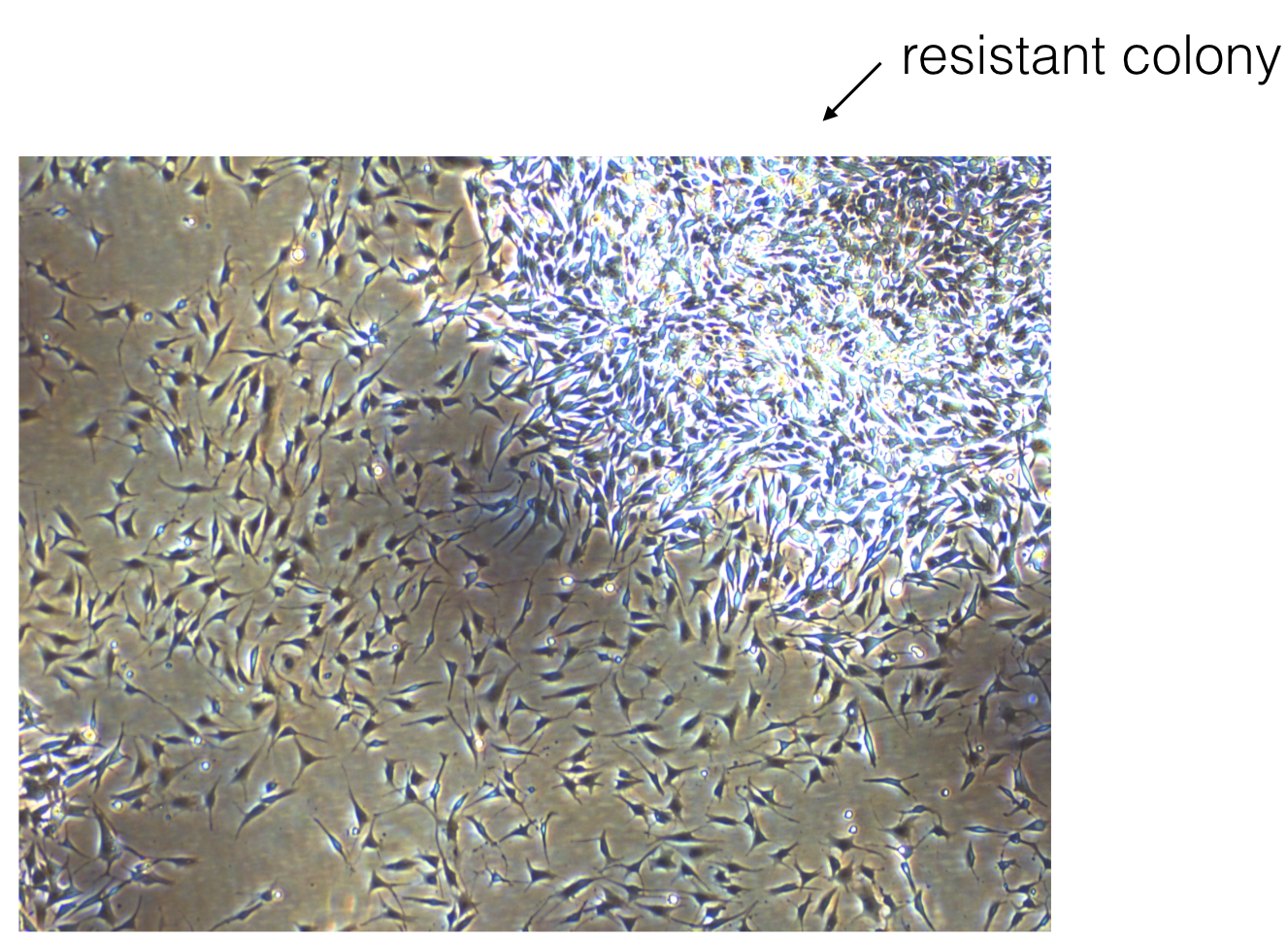
Picture of a resistant colony growing in the Raj lab.
Based upon this work, do you have any ideas for how we might prevent resistance in patients?
Yes. The recommended dosing for many of these drugs is daily. Our work would suggest that something like interval therapy might be more effective, for instance, if you gave the drug for a few days, killed many of the tumor cells, and then stopped the drug. During the time that the drug is stopped, the cells that initially survived the drug (we call these cells pre-resistant) could then transition out of this cell state and back to a sensitive state. Then, when the patient takes the drug again, it would be more effective at killing the remaining tumor cells. Another idea would be to find drugs that are specific to the pre-resistant cells and give these drugs in combination with other targeted therapies.
Were there any “Aha!” moments while working on this project?
One of the most exciting moments of this research was when we first found the pre-resistant cells. Hidden among thousands of pictures of empty cells, we were shocked to actually see the rare cells full of brightly tagged resistance genes (below).
Resistant cells growing in the Raj lab.
What were some low points in working on the project? Do you recall any specific moments that you just felt intellectually and/or emotionally stumped? How did you get through them?
Oh yes, there were definitely low points during this project. One that stands out to me specifically was this one Friday afternoon where I presented at lab meeting. At the time, I only had a little bit of preliminary data. One of the members of the lab asked me a series of questions about resistance: How many different drug doses had I tried? Could I just give a lot and kill them all? What dose of drug is relevant for patients? What about drug resistance? Was I really interested in? All reasonable questions to ask. However, this was really overwhelming to a first-year graduate student because it made me realize that I didn’t have a clearly defined project that I was working on yet. There were just so many different questions that I didn’t know where to start.
Ultimately, with Arjun’s guidance, I came to realize that this was part of the process of figuring out what my thesis project would be, and the vagueness of our ideas at this time was a great thing because it left me open to find a problem that I found really interesting.
At another point in working on this: I remember that we were clearly conceptually stuck. We had identified the rare cells, but it wasn’t clear how to find out if these were the same cells that become resistant to drug. I had an entire lab meeting where we discussed this concern and came to the conclusion that, without some connection between the cells in this state and resistance, the work would be very speculative, which felt unsatisfying to me. Unfortunately, there wasn’t a quick fix to this problem. We just ended up trying a whole bunch of different ideas and eventually one of our strategies worked out.
Were there any funny moments that stand out to you?
Yeah! I was 40 weeks pregnant as we were finishing off our first submission of the paper! As my due date passed, I was really feeling the pressure to finish everything. Each day, I was coming into lab and just hoping I wouldn’t go into labor yet! Actually, the members of our lab had placed bets on when the baby would be born. Fortunately, those who bet on a late arrival ended up winning, and we submitted the paper the day before my daughter, Julien, was born. I was actually still at the hospital when I got the e-mail that the paper went to review.
So even though it might seem like this project is checked off the list with a kick-ass publication, there are probably a bunch of unfinished ideas you have. So,what are you working on next? Will this project ever be “done?”
For sure. The list of unfinished ideas is very long, and some of the questions that came from this work are now being pursued by other people in the lab. Right now, I’m working on ways of measuring the length of time that individual cells remain in these different cell states.
Interested in sharing your research in Penn BE? Contact penngabe@gmail.com for an interview by GABE (Graduate Association of Bioengineers) and let us know!
New research by faculty in the University of Pennsylvania Department of Bioengineering is examining the interplay between cells and their environment and how they impact the cells’ ability to grow and spread, showing that stiffness is not the only factor researchers should consider when studying this process.
The relationship between cellular adhesion and spread is a key factor in cancer metastasis. Better understanding of this dynamic would improve diagnosis of the disease and provide a potential target in combating it; reducing the ability of cells to grip their environment could keep them contained.
Vivek Shenoy (left) and Jason Burdick
The study, published in the Proceedings of the National Academy of Sciences, was led by Vivek Shenoy, professor in the Department of Materials Science and Engineering, co-director of Penn’s Center for Engineering Mechanobiology, and a secondary faculty member in the Department of Bioengineering, along with Xuan Cao and Ehsan Ban, members of his lab. They collaborated with Jason Burdick, professor in the Department of Bioengineering, Boston University’s Christopher Chen, the University of Michigan’s Brendon Baker and the University of Hong Kong’s Yuan Lin.
This collaboration reflects work of The Center for Engineering Mechanobiology, a National Science Foundation-funded Science and Technology Center that supports interdisciplinary research on the way cells exert and are influenced by the physical forces in their environment.
Previous work from Shenoy’s group has shown that the relationship between cancer cells and the extracellular matrix is dynamic, containing feedback mechanisms that can change the ECM’s properties, including overall stiffness. One earlier study investigated how cancer cells attempt to strike a balance in the density of the fibrous netting surrounding them. If there are too few fibers to grip, the cells can’t get enough traction to move. If there are too many, the holes in the net become too small for the cells to pass through.
When someone talks about using “your network” to find a job or answer a question, most people understand that to mean the interconnected web of your friends, family, and acquaintances. But we all have another key network that shapes our life in powerful ways: our brains.
In the brain, impulses whiz from one brain region to another, helping you formulate all of your thoughts and decisions. As science continues to unlock the complexities of the brain, a group of researchers has found evidence that brain networks and social networks actually influence and inform one another.
It found that people who show greater changes in connectivity in their mentalizing system during social exclusion compared to inclusion tend to have a less tightly knit social network — that is, their friends tend not to be friends with one another. By contrast, people with more close-knit social networks, in which many people in the network tend to know one another, showed less change in connectivity in their mentalizing regions.
Six of the students — Zakary Beach, Nicolette Driscoll, Lindsey Fernandez, Jessica Hsu, Jinsu Kim and Ryan Leaphart — are current doctoral students in Bioengineering who earned undergraduate degrees from other top BE programs. Three of the awardees — Lucy Chai, Jake Hsu and Karren Yang — are BE graduating seniors in the Class of 2017. Lucy will spend next year on a Churchill fellowship at Cambridge before starting her NSF fellowship, while Jake has an internship with Genentech‘s Manufacturing Sciences and Technology department, and Karren will attend MIT.
“We are extremely fortunate to attract the very best graduate students in the country,” says David F. Meaney, Solomon R. Pollack Professor and Chair of BE. “This is an external recognition of the high quality of our students across the board.”
The Graduate Research Fellowship Program of the National Science Foundation recognizes and supports outstanding graduate students in NSF-supported science, technology, engineering, and mathematics disciplines who are pursuing research-based master’s and doctoral degrees at accredited United States institutions. For the 2017 competition, the NSF received more than 13,000 applications.