Penn Bioengineers Develop Implantable Living Electrodes

Living Electrode Panels (image courtesy of the Cullen Lab)

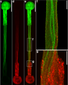
Connecting the human brain to electrical devices is a long-standing goal of neuroscientists, bioengineers, and clinicians, with applications ranging from deep brain stimulation (DBS) to treat Parkinson’s disease to more futuristic endeavors such as Elon Musk’s NeuraLink initiative to record and translate brain activity. However, these approaches currently rely on using implantable metallic electrodes that inherently provoke a lasting immune response due to their non-biological nature, generally complicating the reliability and stability of these interfaces over time. To address these challenges, D. Kacy Cullen, Associate Professor in Neurosurgery and Bioengineering, and Dayo Adewole, a doctoral candidate in Bioengineering, worked with a multi-disciplinary team of collaborators to develop the first “living electrodes” as an implantable, biological bridge between the brain and external devices. In a recent article published in Science Advances, the team demonstrated the fabrication of hair-like microtissue comprised of living neuronal networks and bundled tracts of axons the signal sending fibers of the nervous system protected within soft hydrogel cylinders. They showed that these axon-based living electrodes could be fully controlled and monitored with light thus eliminating the need for electrical contact and are capable of surviving and forming synapses with the brain as demonstrated in an adult rat model. While further advancements are necessary prior to clinical use, the current findings provide the foundation for a new class of “living electrodes” as a biological intermediary between humans and devices capable of leveraging natural mechanisms to potentially provide a stable interface for clinical applications.

Cullen has a primary appointment in the Department of Neurosurgery in the Perelman School of Medicine, with a secondary appointment in the Department of Bioengineering in the School of Engineering and Applied Science, and an appointment in the Corporal Michael J. Crescenz VA Medical Center in Philadelphia.

Ravi Radhakrishnan Adapts Multiscale Modeling Course


Ravi Radhakrishnan, PhD

Ravi Radhakrishnan, Professor and Chair of the Department of Bioengineering and Professor in Chemical and Biomolecular Engineering, is among the many faculty who quickly adapted their courses to an online format in the wake of the COVID-19 pandemic. Now, a recent publication in the American Institute of Chemical Engineers (AIChE) Journal reflects one of these revamped courses. The course BE 559: “Multiscale Modeling of Chemical and Biological Systems” provides theoretical, conceptual, and hands-on modeling experience on three different length and time scales: (1) electronic structure (A, ps); (2) molecular mechanics (100A, ns); and (3) deterministic and stochastic approaches for microscale systems (um, sec). During the course, students gained hands-on experience in running codes on real applications together with the following theoretical formalisms: molecular dynamics, Monte Carlo, free energy methods, deterministic and stochastic modeling. The transition to the online format was greatly facilitated by a grant from the Extreme Science and Engineering Discovery Environment (XSEDE) which provided cloud and supercomputing resources to the students facilitating the computational laboratory experience. Radhakrishnan’s article, “A survey of multiscale modeling: Foundations, historical milestones, current status, and future prospects,” reviews the foundations, historical developments, and current paradigms in multiscale modeling (MSM).

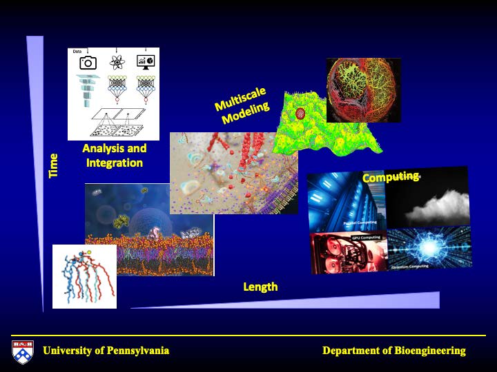
Radhakrishnan aspires to modernize computational science, integrating Multiscale Modeling and Data Science for Biological and Biomedical Science & Engineering. His team does so by integrating multiphysics modeling, computing, data science to tackle applications. The integrative approach is pictorially depicted here in terms of modeling different length and timescales using techniques such as molecular dynamics of atomistic systems, Brownian dynamics of coarse-grained systems, and field equations governing continuum scales of macroscopic systems.

Read the full article in the AIChE Journal: https://doi.org/10.1002/aic.17026

Funding source: National Institutes of Health, Grant/Award Number: CA227550

Penn Engineers Devise Easier Way of Sneaking Antibodies into Cells

Getting a complex protein like an antibody through the membrane of a cell without damaging either is a long-standing challenge in the life sciences. Penn Engineers have found a plug-and-play solution that makes antibodies compatible with the delivery vehicles commonly used to ferry nucleic acids across that barrier.

For almost any conceivable protein, corresponding antibodies can be developed to block it from binding or changing shape, which ultimately prevents it from carrying out its normal function. As such, scientists have looked to antibodies as a way of shutting down proteins inside cells for decades, but there is still no consistent way to get them past the cell membrane in meaningful numbers.

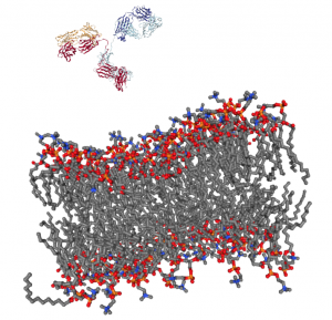
Now, Penn Engineering researchers have figured out a way for antibodies to hitch a ride with transfection agents, positively charged bubbles of fat that biologists routinely use to transport DNA and RNA into cells. These delivery vehicles only accept cargo with a highly negative charge, a quality that nucleic acids have but antibodies lack. By designing a negatively charged amino acid chain that can be attached to any antibody without disrupting its function, they have made antibodies broadly compatible with common transfection agents.

Beyond the technique’s usefulness towards studying intracellular dynamics, the researchers conducted functional experiments with antibodies that highlight the technique’s potential for therapeutic applications. One antibody blocked a protein that decreases the efficacy of certain drugs by prematurely ejecting them from cells. Another blocked a protein involved in the transcription process, which could be an even more fundamental way of knocking out proteins with unwanted effects.

Andrew Tsourkas and Hejia Henry Wang

The study, published in the Proceedings of the National Academy of Sciences, was conducted by Andrew Tsourkas, professor in the Department of Bioengineering, and Hejia Henry Wang, a graduate student in his lab.

Read the full story at the Penn Engineering Medium Blog. Media contact Evan Lerner.

Penn Engineers Solve the Paradox of Why Tissue Gets Stiffer When Compressed

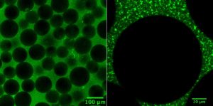
The researchers’ experiments involved making synthetic tissues with artificial “cells.” The fibrin network that surrounds these beads pull on them when compressed; by changing the number of beads in their experimental tissues, the researchers could suss out how cell-fiber interplay contributes to the tissue’s overall properties.

Tissue gets stiffer when it’s compressed. That property can become even more pronounced with injury or disease, which is why doctors palpate tissue as part of a diagnosis, such as when they check for lumps in a cancer screening. That stiffening response is a long-standing biomedical paradox, however: tissue consists of cells within a complex network of fibers, and common sense dictates that when you push the ends of a string together, it loosens tension, rather than increasing it.

Now, in a study published in Nature, University of Pennsylvania’s School of Engineering and Applied Science researchers have solved this mystery by better understanding the mechanical interplay between that fiber network and the cells it contains.

The researchers found that when tissue is compressed, the cells inside expand laterally, pulling on attached fibers and putting more overall tension on the network. Targeting the proteins that connect cells to the surrounding fiber network might therefore be the optimal way of reducing overall tissue stiffness, a goal in medical treatments for everything from cancer to obesity.

Headshots of Paul Janmey and Vivek Shenoy

Paul Janmey and Vivek Shenoy

The study was led by Paul Janmey, Professor in the Perelman School of Medicine’s Department of Physiology and in Penn Engineering’s Department of Bioengineering, and Vivek Shenoy, Eduardo D. Glandt President’s Distinguished Professor in Penn Engineering’s Department of Materials Science and Engineering, Mechanical Engineering and Applied Mechanics, and Bioengineering, along with Anne van Oosten and Xingyu Chen, graduate students in Janmey’s and Shenoy’s labs. Van Oosten is now a postdoctoral fellow at Leiden University in The Netherlands.

Shenoy is Director of Penn’s Center for Engineering Mechanobiology, which studies how physical forces influence the behavior of biological systems; Janmey is the co-director of one of the Center’s working groups, organized around the question, “How do cells adapt to and change their mechanical environment?”

Together, they have been interested in solving the paradox surrounding tissue stiffness.

Read the full story on the Penn Engineering Medium Blog.

Penn BE Undergraduates’ Plate Reader Design Published

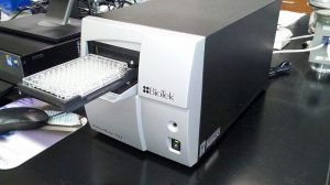
Microplate reader, Wikimedia Commons

In a paper recently published in Biochemistry, a group of University of Pennsylvania Bioengineering students describe the results of their work designing a new, open-source, low-cost microplate reader. Plate readers are instruments designed to measure light absorption and fluorescence emission from molecules useful for clinical biomarker analyses and assays in a diverse array of fields including synthetic biology, optogenetics, and photosensory biology. This new design costs less than $3500, a significantly lower price than other commercially available alternatives. As described in the paper’s abstract, this design is the latest in a growing trend of open-source  hardware to enhance access to equipment for biology labs. The project originated as part of the annual International Genetically Engineering Machine Competition (iGEM), an annual worldwide competition focusing on “push[ing] the boundaries of synthetic biology by tackling everyday issues facing the world” (iGEM website).

The group consists of current junior Andrew Clark (BSE ’20) and recent graduates Karol Szymula (BSE ’18), who works in the lab of Dr. Danielle Bassett, and Michael Patterson (BSE ’18), a Master’s student in Bioengineering and Engineer of Instructional Laboratories. Assistant Professor of Bioengineering Dr. Brian Chow served as their faculty mentor alongside Director of Instructional Labs Sevile Mannickarottu and Michael Magaraci, a Ph.D. candidate in Bioengineering, all of whom serve as co-authors on the published article. The research and design of the project was conducted in the Stephenson Foundation Bioengineering Educational Laboratory here at the University of Pennsylvania’s Department of Bioengineering.