Combined Treatment Takes a Bite Out of Tooth Decay

by Nathi Magubane

Michel Koo of the School of Dental Medicine and David Cormode of the Perelman School of Medicine and the School of Engineering and Applied Science led a team of researchers that uncovered a way to combine two FDA-approved treatments to treat tooth decay that taps into the blend’s bacteria-killing capabilities without disrupting the mouth’s microbiome. (Image: iStock / Alex Sholom)

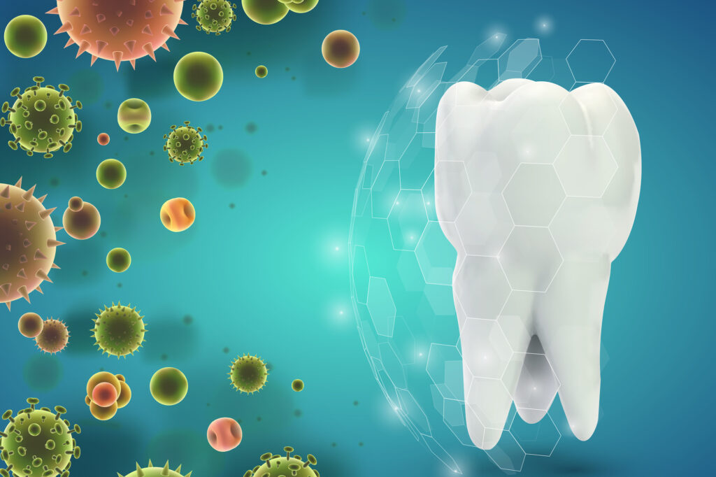
The sting of a toothache or the discovery of a cavity is a universal dread. Dental caries, more commonly known as tooth decay, is an insidious adversary, taking a toll on millions of mouths worldwide. Caries can lead to pain, tooth loss, infection, and, in severe cases, even death.

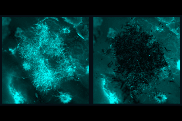
While fluoride-based treatments have long been the gold standard in dentistry, this singular approach is now dated and has limited effect. Current treatments do not sufficiently control biofilm—the main culprit behind dental caries—and prevent enamel demineralization at the same time. This dual dilemma becomes particularly pronounced in high-risk populations where the onset of the disease can be both rapid and severe.

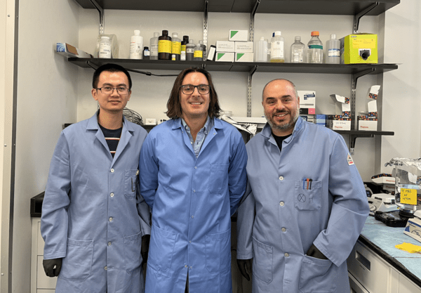
Now, a study from a team of researchers led by Hyun (Michel) Koo of the University of Pennsylvania’s School of Dental Medicine in collaboration with David Cormode of Penn’s Perelman School of Medicine and School of Engineering and Applied Science has unveiled an unexpected synergy in the battle against dental caries. Their research revealed that the combination of ferumoxytol (Fer) and stannous fluoride (SnF2) could point at a potent solution against dental caries. Their findings were published in Nature Communications.

“Traditional treatments often come short in managing the complex biofilm environment in the mouth,” Koo, senior co-author on the study, says. “Our combined treatment not only amplifies the effectiveness of each agent but does so with a lower dosage, hinting at a potentially revolutionary method for caries prevention in high-risk individuals.”

Read the full story in Penn Today.

Hyun (Michel) Koo is a professor in the Department of Orthodontics and in the divisions of Pediatric Dentistry and Community Oral Health and the co-founder of the Center for Innovation & Precision Dentistry in the School of Dental Medicine at the University of Pennsylvania. He is a member of the Penn Bioengineering Graduate Group.

David Cormode is an associate professor of radiology and bioengineering with appointments in Penn’s Perelman School of Medicine and School of Engineering and Applied Science.

Other authors are Yue Huang, Nil Kanatha Pandey, Shrey Shah, and Jessica C. Hsu of Penn’s Perelman School of Medicine; Yuan Liu, Aurea Simon-Soro, Zhi Ren, Zhenting Xiaang, Dongyeop Kim, Tatsuro Ito, Min Jun Oh, and Yong Li of Penn’s School of Dental Medicine; Paul. J Smeets, Sarah Boyer, Xingchen Zhao, and Derk Joester of Northwestern University; and Domenick T. Zero of Indiana University.

The work was supported by the National Institute of Health (grants R01-DE025848 and TL1TR001423 and awards S10OD026871 and R90DE031532) and the National Science Foundation (awards ECCS-2025633 and DMR-1720139).

The Immune Health Future, Today

by Christina Hernandez Sherwood

Breaking the code of the immune system could provide a new fundamental way of understanding, treating, and preventing every type of disease. Penn Medicine is investing in key discoveries about immunity and immune system function, and building infrastructure, to make that bold idea a reality.

Several members of the Penn Bioengineering Graduate Group feature in this story which originally featured in the Penn Medicine Magazine.

Image: Courtesy of Penn Medicine Magazine

This grandfather lives with primary progressive multiple sclerosis (MS), an autoimmune disorder that he controls with a medicine that depletes his body of the type of immune cells that make antibodies. So while he has completed his COVID-19 vaccine course, his immune system function isn’t very strong—and the invitation has arrived at a time when COVID-19 is still spreading rapidly. 

You can imagine the scene as an older gentleman lifts a thick, creamy envelope from his mailbox, seeing his own name written in richly scripted lettering. He beams with pride and gratitude at the sight of his granddaughter’s wedding invitation. Yet his next thought is a sober and serious one. Would he be taking his life in his hands by attending the ceremony?

“In the past, all we could do was [measure] the antibody response,” says Amit Bar-Or, the Melissa and Paul Anderson President’s Distinguished Professor in Neurology at the Perelman School of Medicine, and chief of the Multiple Sclerosis division. “If that person didn’t have a good antibody response, which is likely because of the treatment they’re on, we’d shrug our shoulders and say, ‘Maybe you shouldn’t go because we don’t know if you’re protected.’” 

Today, though, Bar-Or can take a deeper dive into his patients’ individual immune systems to give them far more nuanced recommendations. A clinical test for immune cells produced in response to the COVID-19 vaccine or to the SARS-CoV-2 virus itself—not just antibodies—was one of the first applied clinical initiatives of a major new Immune Health® project at Penn Medicine. Doctors were able to order this test and receive actionable answers through the Penn Medicine electronic health record for patients like the grandfather with MS. 

“With a simple test and an algorithm we can have a very different discussion,” Bar-Or says. A test result showing low T cells, for instance, would tell Bar-Or his patient may get a meaningful jolt in immunity from a vaccine booster, while low antibody levels would suggest passive antibody therapy is more helpful. Or, the test might show his body is already well primed to protect him, making it reasonably safe to attend the wedding.

This COVID-19 immunity test is only the beginning. 

Physicians and scientists at Penn Medicine are imagining a future where patients can get a precise picture of their immune systems’ activity to guide treatment decisions. They are working to bring the idea of Immune Health to life as a new area of medicine. In labs, in complex data models, and in the clinic, they are beginning to make sense out of the depth and breadth of the immune system’s millions of as-yet-undeciphered signals to improve health and treat illnesses of all types. 

Penn Medicine registered the trademark for the term “Immune Health” in recognition of the potential impact of this research area and its likelihood to draw non-academic partners as collaborators in its growth. Today, at the south end of Penn’s medical campus, seven stories of research space are being added atop an office building at 3600 Civic Center Blvd., including three floors dedicated to Immune Health, autoimmunity, and immunology research.

The concept behind the whole project, says E. John Wherry, director of Penn Medicine’s Institute for Immunology and Immune Health (I3H), “is to listen to the immune system, to profile the immune system, and use those individual patient immune fingerprints to diagnose and treat diseases as diverse as immune-related diseases, cancer, cardiovascular disease, Alzheimer’s, and many others.”

The challenge is vast. Each person’s immune system is far more complex than antibodies and T cells alone. The immune system is made of multiple interwoven layers of complex defenders—from our skin and mucous membranes to microscopic memory B cells that never forget a childhood infection—meant to fortify our bodies from germs and disease. It is a sophisticated system that learns and adapts over our lifetimes in numerous ways, and it also falters and fails in some ways we understand and others that remain mysterious. And each person’s intricate internal battlefield is in some way unique.

The immune system is not just a set of defensive barricades, either. It’s also a potential source of deep insight about a person’s physiological functioning and responses to medical treatments.

“The immune system is sensing and keeping track of basically all tissues and all cells in our body all the time,” Wherry says. “It is surveying the body trying to clean up any invaders and restore homeostasis by maintaining good health.”

“Our goal is to essentially break the code of the immune system,” says Jonathan Epstein, executive vice dean of the Perelman School of Medicine and chief scientific officer at Penn Medicine. “By doing so, we believe we will be able to determine your state of health and your response to therapies in essentially every human disease.”

Read the full story in Penn Today.

How the Hippocampus Distinguishes True and False Memories

by Erica Moser

Image: iStock/metamorworks

Let’s say you typically eat eggs for breakfast but were running late and ate cereal. As you crunched on a spoonful of Raisin Bran, other contextual similarities remained: You ate at the same table, at the same time, preparing to go to the same job. When someone asks later what you had for breakfast, you incorrectly remember eating eggs.

This would be a real-world example of a false memory. But what happens in your brain before recalling eggs, compared to what would happen if you correctly recalled cereal?

In a paper published in Proceedings of the National Academy of Sciences, University of Pennsylvania neuroscientists show for the first time that electrical signals in the human hippocampus differ immediately before recollection of true and false memories. They also found that low-frequency activity in the hippocampus decreases as a function of contextual similarity between a falsely recalled word and the target word.

“Whereas prior studies established the role of the hippocampus in event memory, we did not know that electrical signals generated in this region would distinguish the imminent recall of true from false memories,” says psychology professor Michael Jacob Kahana, director of the Computational Memory Lab and the study’s senior author. He says this shows that the hippocampus stores information about an item with the context in which it was presented.

Researchers also found that, relative to correct recalls, the brain exhibited lower theta and high-frequency oscillations and higher alpha/beta oscillations ahead of false memories. The findings came from recording neural activity in epilepsy patients who were already undergoing invasive monitoring to pinpoint the source of their seizures.

Noa Herz, lead author and a postdoctoral fellow in Kahana’s lab at the time of the research, explains that the monitoring was done through intracranial electrodes, the methodology researchers wanted to use for this study. She says that, compared to scalp electrodes, this method “allowed us to more precisely, and directly, measure the neural signals that were generated in deep brain structures, so the activity we are getting is much more localized.”

Read the full story in Penn Today.

Michael Kahana is the Edmund J. and Louise W. Kahn Term Professor of Psychology in the School of Arts & Sciences and director of the Computational Memory Lab at the University of Pennsylvania. He is a member of the Penn Bioengineering Graduate Group.

Center for Innovation & Precision Dentistry Positions Penn as a Leader in Engineering Health

by Devorah Fischler

Kathleen J. Stebe and Michel Koo urge “the academic community to adopt a coordinated approach uniting dental medicine and engineering to support research, training and entrepreneurship to address unmet needs and spur oral health care innovations.” (Image: Min Jun Oh and Seokyoung Yoon)

Penn’s Center for Innovation & Precision Dentistry (CiPD) is the first cross-disciplinary initiative in the nation to unite oral-craniofacial health sciences and engineering.

An institutional partnership formalizing the Center’s dual affiliation between the University of Pennsylvania School of Engineering and Applied Science and School of Dental Medicine makes CiPD unique.

In just two years since CiPD was founded, the outcomes of this newly conceived research partnership have proven its value: microrobots that clean teeth for people with limited mobility, a completely new understanding of bacterial physics in tooth decay, enzymes from plant chloroplasts that degrade plaque, promising futures for lipid nanoparticles in oral cancer treatment and new techniques and materials to restore nerves in facial reconstructive surgery.

In addition, CiPD is training the next generation of dentists, scientists and engineers through an NIH/NIDCR-sponsored postdoctoral training program as well as fellowships from industry.

The center’s Founding Co-Directors, Kathleen J. Stebe, Richer & Elizabeth Goodwin Professor in Chemical and Biomolecular Engineering, and Michel Koo, Professor of Orthodontics in Penn Dental Medicine, published an editorial in the Journal of Dental Research, planting a flag for CiPD’s mission and encouraging others to mirror its method.

The two urge “the academic community to adopt a coordinated approach uniting dental medicine and engineering to support research, training and entrepreneurship to address unmet needs and spur oral health care innovations.”

Read the full story in Penn Engineering Today.

Michel Koo is a member of the Penn Bioengineering Graduate Group.

A Suit of Armor for Cancer-fighting Cells

by Nathi Magubane

Chimeric antigen receptor T cell (CAR T) therapy has delivered promising results, transforming the fight against various forms of cancer, but for many, the therapy comes with severe and potentially lethal side effects. Now, a research team led by Michael Mitchell of the School of Engineering and Applied Science has found a solution that could help CAR T therapies reach their full potential while minimizing severe side effects. (Image: iStock / Meletios Verras)

In recent years, cancer researchers have hailed the arrival of chimeric antigen receptor T cell (CAR T) therapy, which has delivered promising results, transforming the fight against various forms of cancer. The process involves modifying patients’ T-cells to target cancer cells, resulting in remarkable success rates for previously intractable forms of cancer.

Six CAR T cell therapies have secured FDA approval, and several more are in the pipeline. However, these therapies come with severe and potentially lethal side effects, namely cytokine release syndrome (CRS) and neurotoxicity. These drawbacks manifest as a range of symptoms—from high fever and vomiting to multiple organ failure and patient death—posing significant challenges to broader clinical application.

Now, a research team led by Michael Mitchell, associate professor in the School of Engineering and Applied Science at the University of Pennsylvania, has found a solution that could help CAR T therapies reach their full potential while minimizing severe side effects. Their findings are published in the journal Nature Materials.

“Addressing CRS and neurotoxicity without compromising the therapeutic effectiveness of CAR T cells has been a complex challenge,” says Mitchell.

He says that unwanted interactions between CAR T and immune cells called macrophages drive the overactivation of macrophages, which in turn result in the release of toxic cytokines that lead to CRS and neurotoxicity.

“Controlling CAR T-macrophage interactions in vivo is difficult,” Mitchell says. “So, our study introduces a materials engineering-based strategy that involves incorporating a sugar molecule onto the surface of CAR T cells. These sugars are then used as a reactive handle to create a biomaterial coating around these cells directly in the body, which acts as a ‘suit of armor,’ preventing dangerous interactions with macrophages.”

First author Ningqiang Gong, a postdoctoral researcher in the Mitchell Lab, elaborates on the technique, “We attached this sugar molecule to the CAR T cells using metabolic labeling. This modification enables the CAR T cells to attack cancer cells without any hindrance.”

“When symptoms of CRS begin to manifest, we introduce another molecule—polyethylene glycol (PEG)—to create the suit of armor, which effectively blocks dangerous interactions between these engineered T cells, macrophages, and the tumor cells themselves,” Gong says.

Read the full story in Penn Today.

Carl June on the Boundless Potential of CAR T Cell Therapy

by Meagan Raeke

Carl June, at the flash mob celebration of the FDA approval of the CAR T cell therapy he developed, in August 2017. (Image: Courtesy of Penn Medicine Magazine)

For most of modern medicine, cancer drugs have been developed the same way: by designing molecules to treat diseased cells. With the advent of immunotherapy, that changed. For the first time, scientists engineered patients’ own immune systems to recognize and attack diseased cells.

One of the best examples of this pioneering type of medicine is CAR T cell therapy. Invented in the Perelman School of Medicine by Carl June, the Richard W. Vague Professor in Immunotherapy, CAR T cell therapy works by collecting T cells from a patient, modifying those cells in the lab so that they are designed to destroy cancerous cells, and reinfusing them into the patient. June’s research led to the first FDA approval for this type of therapy, in 2017. Six different CAR T cell therapies are now approved to treat various types of blood cancers. Carl June, at the flash mob celebration of the FDA approval of the CAR T cell therapy he developed, in August 2017. (Image: Courtesy of Penn Medicine Magazine)

CAR T cell therapy holds the potential to help millions more patients—if it can be successfully translated to other conditions. June and colleagues, including Daniel Baker, a fourth-year doctoral student in the Cell and Molecular Biology department, discuss this potential in a perspective published in Nature.

In the piece, June and Baker highlight other diseases that CAR T cell therapy could be effective.

“CAR T cell therapy has been remarkably successful for blood cancers like leukemias and lymphomas. There’s a lot of work happening here at Penn and elsewhere to push it to other blood cancers and to earlier stage disease, so patients don’t have to go through chemo first,” June says. “Another big priority is patients with solid tumors because they make up the vast majority of cancer patients. Beyond cancer, we’re seeing early signs that CAR T cell therapy could work in autoimmune diseases, like lupus.”

As for which diseases to pursue as for possible future treatment, June says, “essentially it boils down to two questions: Can we identify a population of cells that are bad? And can we target them specifically? Whether that’s asthma or chronic diseases or lupus, if you can find a bad population of cells and get rid of them, then CAR T cells could be therapeutic in that context.”

“What’s exciting is it’s not just theoretical at this point. There have been clinical reports in other autoimmune diseases, including myasthenia gravis and inflammatory myopathy,” Baker says. “But we are seeing early evidence that CAR T cell therapy will be successful beyond cancer. And it’s really opening the minds of people in the field to think about how else we could use CAR T. For example, there’s some pioneering work at Penn from the Epstein lab for heart failure. The idea is that you could use CAR T cells to get rid of fibrotic tissue after a cardiac injury, and potentially restore the damage following a heart attack.”

Baker adds, “there’s no question that over the last decade, CAR T cell therapy has revolutionized cancer. I’m hoping to play a role in bringing these next generation therapies to patients and make a real impact over the next decade. I think there’s potential for cell therapy to be a new pillar of medicine at large, and not just a new pillar of oncology.”

Read the full story at Penn Medicine Today.

An Improved Delivery System for mRNA Vaccines Provides More Powerful Protection

by Devorah Fischler

(From left to right) Xuexiang Han, Michael Mitchell and Mohamad-Gabriel Alameh

The COVID-19 vaccine swiftly undercut the worst of the pandemic for hundreds of millions around the world. Available sooner than almost anyone expected, these vaccines were a triumph of resourcefulness and skill.

Messenger RNA vaccines, like the ones manufactured by Moderna or Pfizer/BioNTech, owed their speed and success to decades of research reinforcing the safety and effectiveness of their unique immune-instructive technology.

Now, researchers from the University of Pennsylvania School of Engineering and Applied Science and the Perelman School of Medicine are refining the COVID-19 vaccine, creating an innovative delivery system for even more robust protection against the virus.

In addition to outlining a more flexible and effective COVID-19 vaccine, this work has potential to increase the scope of mRNA vaccines writ large, contributing to prevention and treatment for a range of different illnesses.

Michael Mitchell, associate professor in Penn Engineering’s Department of Bioengineering, Xuexiang Han, postdoctoral fellow in Mitchell’s lab, and Mohamad-Gabriel Alameh, postdoctoral fellow in Drew Weissman’s lab at Penn Medicine and incoming assistant professor in the Department of Pathology and Laboratory Medicine at the Perelman School of Medicine, recently published their findings in Nature Nanotechnology.

mRNA, or messenger ribonucleic acid, is the body’s natural go-between. mRNA contains the instructions our cells need to produce proteins that play important roles in our bodies’ health, including mounting immune responses.

The COVID-19 vaccines follow suit, sending a single strand of RNA to teach our cells how to recognize and fight the virus.

Read the full story in Penn Engineering Today.

The Physics of Fat Droplets Reveal DNA Danger

by Devorah Fischler

Fat is a normal and necessary part of the body. Fat cells store and release energy, as well as play significant roles in hormonal regulation and immunity.

Engineers and scientists at the University of Pennsylvania are the first to discover fat-filled lipid droplets’ (pictured above in green) surprising capability to indent and puncture the nucleus, the organelle which contains and regulates a cell’s DNA.

In recent decades, a concerning rise in metabolic illnesses – such as cardiovascular disease, high blood pressure and diabetes – has focused scientific attention on the biology and chemistry of fat, resulting in a wealth of information about how fat cells work.

But fat cells and their metabolic activities are only part of the story.

Fat-filled lipid droplets, tiny spheres of fat many times smaller than fat cells, are a growing subject of scientific interest. Found inside many different cell types, these lipid particles have long been little understood. Studies have begun to illuminate these droplets’ participation in metabolic functions and cellular protection, but we still know next to nothing about the physical nature of fat.

Now, researchers at the University of Pennsylvania School of Engineering and Applied Science have looked beyond biochemistry to publish groundbreaking work on the physics of these droplets, revealing them to be a potential threat to a cell’s nucleus. In the August issue of the Journal of Cell Biology, they are the first to discover fat-filled lipid droplets’ surprising capability to indent and puncture the nucleus, the organelle which contains and regulates a cell’s DNA.

The stakes of their findings are high: a ruptured nucleus can lead to elevated DNA damage that is characteristic of many diseases, including cancer.

The study was led by Dennis E. Discher, Robert D. Bent Professor in the Department of Chemical and Biomolecular Engineering, Bioengineering, and in Mechanical Engineering and Applied Mechanics, Irena Ivanovska, Ph.D. research associate in Penn’s Molecular and Cell Biophysics Lab, and Michael Tobin, Ph.D. candidate in the Department of Bioengineering.

“Intuitively, people think of fat as soft,” says Discher. “And on a cellular level it is. But at this small size of droplet— measuring just a few microns rather than the hundreds of microns of a mature fat cell—it stops being soft. Its shape has a much higher curvature, bending other objects very sharply. This changes its physics in the cell. It can deform. It can damage. It can rupture.”

Read the full story in Penn Engineering Today.

SCALAR: A Microchip Designed to Transform the Production of mRNA Therapeutics and Vaccines

Led by Michael Mitchell and David Issadore of the School of Engineering and Applied Science, a team of researchers has developed a platform that could rapidly accelerate the development of mRNA-based lipid nanoparticle vaccines and therapeutics at both the small and large scale, SCALAR. (Image: iStock / Anatoly Morozov)

Following the global COVID-19 pandemic, the development and rapid deployment of mRNA vaccines highlighted the critical role of lipid nanoparticles (LNPs) in the context of pharmaceuticals. Used as the essential delivery vehicles for fragile RNA-based therapies and vaccines, LNPs protect the RNA from degradation and ensure effective delivery within the body.

Despite their critical importance, the large-scale manufacturing of these LNPs saw numerous bottlenecks during the pandemic, underscoring the need for scalable production techniques that could keep pace with global demand.

Now, in a paper published in the Proceedings of the National Academy of the Sciences, researchers at the University of Pennsylvania describe how the Silicon Scalable Lipid Nanoparticle Generation platform (SCALAR), a reusable silicon- and glass-based platform designed to transform the production landscape of LNPs for RNA therapeutics and vaccines, offers a scalable and efficient solution to the challenges exposed during the COVID-19 crisis.

“We’re excited to create a piece of technology platform that bridges the gap between small-scale discovery and large-scale manufacturing in the realm of RNA lipid nanoparticle vaccines and therapeutics,” says co-author Michael Mitchell, associate professor of bioengineering in the School of Engineering and Applied Science at Penn. “By doing so, we’ve effectively leapfrogged the clunky, time-consuming, and costly barriers that slow down the production ramp-up of promising new RNA medicines and vaccines.”

The intricacies of RNA-based therapies require the RNA to be encased in a delivery system capable of navigating the body’s biological obstacles. LNPs fulfill this role, allowing the RNA to reach the intended cells for maximum therapeutic impact. SCALAR aims to take this a step further, allowing for an unprecedented three orders of magnitude scalability in LNP production rates, addressing the speed and consistency bottlenecks that hinder existing methods.

Sarah Shepherd, the first author of the paper and a recent Ph.D. graduate who worked in the Mitchell Lab, says, “With SCALAR, we’re not just reacting to today’s challenges but proactively preparing for tomorrow’s opportunities and crises. This technology is flexible, uses mixing architectures well-documented in microfluidics, and is scalable enough to meet future demands in real time. That’s an enormous leap forward for the field.”

Shepherd says that SCALAR builds on prior work from the Mitchell lab and is based on a microfluidic chip platform. Akin to a computer chip, wherein a computer’s electrically integrated circuit has numerous little transistors transporting signals as ones or zeroes to produce an output, the SCALAR microchip precisely controls their two key reagents, lipids and RNA, to generate LNPs.

Read the full story in Penn Today.

Penn Engineers Create Low-Cost, Eco-Friendly COVID Test

by Kat Sas

Fabrication steps of the biodegradable BC substrate and the electrochemical devices. (1) Incubation of the bacterium Gluconacetobacter hansenii. (2) BC substrate collected and treated, resulting in a clear sheet. (3) The biodegradable BC sheet is screen-printed, (4) resulting in a device with 3 electrodes, (4) which are cut out using a scissor, (5) resulting in a portable, biodegradable, and inexpensive electrochemical sensor.

The availability of rapid, accessible testing was integral to overcoming the worst surges of the COVID-19 pandemic, and will be necessary to keep up with emerging variants. However, these tests come with unfortunate costs.

Polymerase chain reaction (PCR) tests, the “gold standard” for diagnostic testing, are hampered by waste. They require significant time (results can take up to a day or more) as well as specialized equipment and labor, all of which increase costs. The sophistication of PCR tests makes them harder to tweak, and therefore slower to respond to new variants. They also carry environmental impacts. For example, most biosensor tests developed to date use printed circuit boards, or PCBs, the same materials used in computers. PCBs are difficult to recycle and slow to biodegrade, using large amounts of metal, plastic and non-eco-friendly materials.

In addition, most PCR tests end up in landfills, resulting in material waste and secondary contamination. An analysis by the World Health Organization (WHO) estimated that, as of February 2022, “over 140 million test kits, with a potential to generate 2,600 tonnes of non-infectious waste (mainly plastic) and 731,000 litres of chemical waste (equivalent to one-third of an Olympic-size swimming pool) have been shipped.”

In order to balance the need for fast, affordable and accurate testing while addressing these environmental concerns, César de la Fuente, Presidential Assistant Professor in Bioengineering and Chemical and Biomolecular Engineering in the School of Engineering and Applied Science, with additional primary appointments in Psychiatry and Microbiology within the Perelman School of Medicine, has turned his attention to the urgent need for “green” testing materials.

The de la Fuente lab has been working on creative ways to create faster and cheaper testing for COVID-19 since the outbreak of the pandemic. Utilizing his lab’s focus on machine biology and the treatment of infectious disease, they created RAPID, an aptly named test that generates results in minutes with a high degree of accuracy. An even more cost-effective version, called LEAD, was created using electrodes made from graphite. A third test, called COLOR, was a low-cost optodiagnostic test printed on cotton swabs.

The team’s latest innovation incorporates the speed and cost-effectiveness of previous tests with eco-friendly materials. In a paper published in Cell Reports Physical Science, the group introduces a new test made from Bacterial Cellulose (BC), an organic compound synthesized from several strains of bacteria, as a substitute for PCBs.

Read the full story in Penn Engineering Today.