The bright white spots represent tiny clusters of proteins detected by CluMPS. (Photo by: Thomas Mumford)
Penn Engineers have pioneered a new way to visualize the smallest protein clusters, skirting the physical limitations of light-powered microscopes and opening new avenues for detecting the proteins implicated in diseases like Alzheimer’s and testing new treatments.
In a paper in Cell Systems, Lukasz Bugaj, Assistant Professor in Bioengineering, describes the creation of CluMPS, or Clusters Magnified by Phase Separation, a molecular tool that activates by forming conspicuous blobs in the presence of target protein clusters as small as just a few nanometers. In essence, CluMPS functions like an on/off switch that responds to the presence of clusters of the protein it is programmed to detect.
Normally, says Bugaj, detecting such clusters requires laborious techniques. “With CluMPS, you don’t need anything beyond the standard lab microscope.” The tool fuses with the target protein to form condensates orders of magnitude larger than the protein clusters themselves that resemble the colorful blobs in a lava lamp. “We think the simplicity of the approach is one of its main benefits,” says Bugaj. “You don’t need specialized skills or equipment to quickly see whether there are small clusters in your cells.”
A pair of proteins, YAP and TAZ, has been identified as conductors of bone development in the womb and could provide insight into genetic diseases such as osteogenesis imperfecta, known commonly as “brittle bone disease.” This research, published in Developmental Cell and led by members of the McKay Orthopaedic Research Laboratory of the Perelman School of Medicine, adds understanding to the field of mechanobiology, which studies how mechanical forces influence biology.
“Despite more than a century of study on the mechanobiology of bone development, the cellular and molecular basis largely has remained a mystery,” says the study’s senior author, Joel Boerckel, an associate professor of orthopaedic surgery. “Here, we identify a new population of cells that are key to turning the body’s early cartilage template into bone, guided by the force-activated gene regulating proteins, YAP and TAZ.”
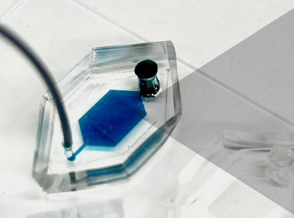
Microwell device with a solution in the reservoir (Image: Courtesy of David E. Reynolds)
Akin to the packages sent from one person to another via an elaborate postal system, cells send tiny parcels that bear contents and packaging material that serve key purposes: To protect the contents from the outside world and to make sure it gets to the right place via a label with an address.
These packages are known as extracellular vesicles (EVs)—lipid-bound molecules that serve a variety of regulatory and maintenance functions throughout the body. They assist in the removal of unwanted materials within the cell, and they transport proteins, aid in DNA and RNA transfer, and promote tumorigeneses in cancerous cells.
Given their myriad roles, EVs have taken center stage for many researchers in the biomedical space as they have the potential to improve current methods of disease detection and treatment. The main challenge, however, is accurately identifying the molecular contents of EVs while also characterizing the EVs, which, unlike other cellular components that are more homogenous, have more heterogeneity.
Now, a team of researchers at the University of Pennsylvania has developed a novel platform, droplet-free double digital assay, for not only profiling individual EVs but also accurately discerning their molecular contents. The researchers took the digital assay, which quantifies the contents of a molecule via binary metric—a 1 corresponds to the presence of a molecule and a zero to the lack thereof—and applies it to the EV. The work is published in Advanced Science.
The team was led by Jina Ko, an assistant professor with appointments in the School of Engineering and Applied Science and Perelman School of Medicine. “Our method allows for highly accurate quantification of the individual molecules inside an EV,” Ko says . “This opens up many doors in the realm of early disease detection and treatment.”
The researchers first compartmentalized individual EVs utilizing a microwell approach to isolate the EVs. Next, they captured individual molecules within the EVs and amplified the signal for clarity. The team then was able to determine the expression levels of pivotal EV biomarkers with remarkable precision via fluorescence.
Jina Ko is an assistant professor in the Department of Pathology and Laboratory Medicine in the Perelman School of Medicine and an assistant professor in the Department of Bioengineering in the School of Engineering and Applied Science at the University of Pennsylvania.
David Reynolds is a Ph.D. candidate in the Department of Bioengineering in Penn Engineering.
Other authors include, Menghan Pan, George Galanis, Yoon Ho Roh, Renee-Tyler T. Morales, Shailesh Senthil Kumar, and Su-Jin Heo of the Department of Bioengineering at Penn Engineering; Jingbo Yang and Xiaowei Xu of the Department of Pathology and Laboratory Medicine at Penn Medicine; and Wei Guo of the Department of Biology in the School of Arts & Sciences at Penn.
The research was supported by the National Institutes of Health: grants R00CA256353, R35 GM141832, and CA174523 (SPORE).
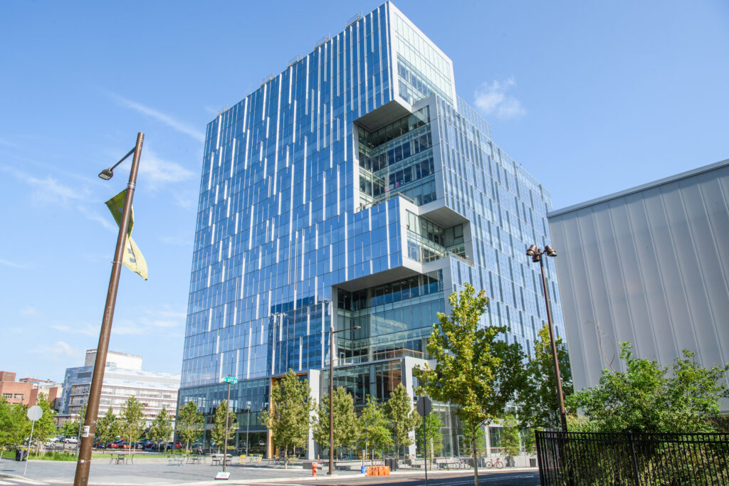
On September 14, Wexford Science & Technology, LLC and the University of Pennsylvania announced that the University has signed a lease for new laboratory space that will usher in a wave of novel vaccine, therapeutics, and engineered diagnostics research to West Philadelphia. Research teams from Penn are poised to move into 115,000 square feet of space at One uCity Square, the 13-story, 400,000 square foot purpose-built lab and office building within the vibrant uCity Square Knowledge Community being developed by Wexford. This is the largest lease in the building, encompassing four floors, and bringing the building to over 90% leased. The building currently includes industry tenants Century Therapeutics (NASDAQ: IPSC), Integral Molecular, Exponent (NASDAQ: EXPO), and Charles River Laboratories (NYSE: CRL).
The new University space will house Penn Medicine’s Institute for RNA Innovation and Penn Engineering’s Center for Precision Engineering for Health, underscoring the University’s commitment to a multi-disciplinary and collaborative approach to research that will attract and retain the best talent and engage partners from across the region. Penn’s decision to locate at One uCity Square reinforces uCity Square’s evolution as a central cluster of academic, clinical, commercial, entrepreneurial, and amenity spaces for the area’s innovation ecosystem, and further cements Philadelphia’s position as a top life sciences market.
Jonathan Epstein, MD, Executive Vice Dean and Chief Scientific Officer of Penn Medicine, shared his anticipation for the opportunities that lie ahead: “Penn Medicine is proud to build on its existing clinical presence in uCity Square and establish an innovative and collaborative research presence at the heart of uCity Square’s multidisciplinary innovation ecosystem. This strategic move underscores our commitment to accelerating advancements in biomedical research, industry collaboration, and equipping our talented teams with the resources they need to shape the future of healthcare.”
Locating the Penn Institute for RNA Innovation in the heart of the uCity Square community brings together researchers across disciplines who are already pursuing new vaccines and treatments, and better ways to deliver them. Their shared work will help to power the next phase of vaccine discovery and development.
Likewise, anchoring the work of Penn Engineering’s Center in the One uCity Square space will allow the School’s multi-disciplinary researchers and their collaborators to advance new clinical and diagnostic methods that will focus on intelligent therapeutics, genome design, diagnostics for discovery of human biology, and engineering the human immune shield.
“Penn Engineering has made a substantial commitment to precision engineering for health, an area that is not only important and relevant to engineering, but also critical to the future of humanity,” said Vijay Kumar, Nemirovsky Family Dean of Penn Engineering. “The space in One uCity Square will add another 30,000 square feet of space for our engineers to develop technologies that will fight future pandemics, cure incurable diseases, and extend healthy life spans around the world.”
Spearheading the Penn Institute for RNA Innovation will be Drew Weissman, MD, PhD, the Roberts Family Professor for Vaccine Research, who along with Katalin Karikó, PhD, adjunct professor of Neurosurgery, discovered foundational mRNA technology that enabled the creation of vital vaccine technology, including the FDA-approved mRNA-based COVID-19 vaccines developed by Pfizer-BioNTech and Moderna.
In this new space at One uCity Square, Weissman and his research team and collaborators will further pursue their groundbreaking research efforts with a goal to develop new therapeutics and vaccines and initiate clinical trials for other devastating diseases.
In addition, two established researchers will join the Institute at One uCity Square: Harvey Friedman, MD, a professor of Infectious Diseases, who leads a team researching various vaccines. He will be joined by Vladimir Muzykantov, MD, PhD, Founders Professor in Nanoparticle Research, who focuses on several projects related to targeting the delivery of drugs, including mRNA, to create more effective, targeted pathways to deliver drugs to the vascular system, treating a wide range of diseases that impact the brain, lung, heart, and blood.
Dan Hammer, Alfred G. and Meta A. Ennis Professor in the Departments of Bioengineering and Chemical and Biomolecular Engineering in Penn Engineering and Director of the Center for Precision Engineering for Health, will oversee the Center’s innovations in diagnostics and delivery, cellular and tissue engineering, and the development of new devices that integrate novel materials with human tissues. The Center will bring together scholars from all departments within Penn Engineering and will help to foster increased collaboration with campus colleagues at Penn’s Perelman School of Medicine and with industry partners.
Joining the Center researchers in One uCity Square are Noor Momin, Sherry Gao, and Michael Mitchell. Noor Momin, who will join Penn Engineering in early 2024 as an assistant professor in Bioengineering, will leverage her lab’s expertise in cardiovascular immunology, protein engineering and pharmacokinetic modeling to develop next-generation treatments and diagnostics for cardiovascular diseases.
Cosette Tomita, a master’s student in Bioengineering, spoke with Penn Engineering Graduate Admissions about her research in cellular therapy and her path to Penn Engineering.
“What were you doing before you came to Penn Engineering?
After college I wanted to get some industry experience before going to graduate school, so I spent a year working for a pharmaceutical company in New Jersey. I learned a lot—but mostly I learned that I wanted to go back into academia. So I was looking for a more research-oriented position to boost my graduate school applications, and I found a position at Penn’s cyclotron facility. Shortly after that, I applied to the master’s program. I’m still working at the cyclotron, so I’m doing the program part time.
How has your experience in the program been so far?
I love the research I’m doing here. I love the collaboration we have and the fact that I’m able to work with whoever I want to. And I can only say good things about my PI, Robert Mach. He’s a very busy man, but he makes time for his people. And he recognizes when somebody has a lot on their plate and he will go to bat for that person.
What’s your research all about?
The focus of my PI’s lab is on neurodegenerative diseases and opiate use, so we’re looking to make imaging agents and antagonists that can help with the opioid crisis.
For my project, I wanted to look at treating neurodegenerative disease from the perspective of cellular therapy. My PI doesn’t have that expertise, so when I came to him with this idea, he said I should talk to Mark Sellmyer in the bioengineering department. He does a lot of cellular therapies, cell engineering, protein engineering and things of that nature. So his lab is more biological.
I don’t have a grant for my research, so my advisors are supporting it out of their own pockets. They could have said, no, you need to work on this project that’s already going on in the lab. But they gave me the intellectual freedom to do what I wanted to do.”
Folding@home is led by Gregory Bowman, a Penn Integrates Knowledge Professor who has appointments in the Departments of Biochemistry and Biophysics in the Perelman School of Medicine and the Department of Bioengineering in the School of Engineering and Applied Science. (Image: Courtesy of Penn Medicine News)
Two heads are better than one. The ethos behind the scientific research project Folding@home is that same idea, multiplied: 50,000 computers are better than one.
Folding@home is a distributed computing project which is used to simulate protein folding, or how protein molecules assemble themselves into 3-D shapes. Research into protein folding allows scientists to better understand how these molecules function or malfunction inside the human body. Often, mutations in proteins influence the progression of many diseases like Alzheimer’s disease, cancer, and even COVID-19.
Penn is home to both the computer brains and human minds behind the Folding@home project which, with its network, forms the largest supercomputer in the world. All of that computing power continually works together to answer scientific questions such as what areas of specific protein implicated in Parkinson’s disease may be susceptible to medication or other treatment.
Using the network hub at Penn, Bowman and his team assign experiments to each individual computer which communicates with other computers and feeds info back to Philly. To date, the network is comprised of more than 50,000 computers spread across the world.
“What we do is like drawing a map,” said Bowman, explaining how the networked computers work together in a type of system that experts call Markov state models. “Each computer is like a driver visiting different places and reporting back info on those locations so we can get a sense of the landscape.”
Individuals can participate by signing up and then installing software to their standard personal desktop or laptop. Participants can direct the software to run in the background and limit it to a certain percentage of processing power or have the software run only when the computer is idle.
When the software is at work, it’s conducting unique experiments designed and assigned by Bowman and his team back at Penn. Users can play scientist and watch the results of simulations and monitor the data in real time, or they can simply let their computer do the work while they go about their lives.
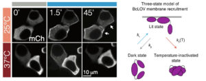
The brighter edges of the cells in the middle and upper right panels show the optogenetic proteins collecting at the membrane after light exposure. At higher temperatures, however, the proteins become rapidly inactivated and thus do not stay at the membrane, resulting in the duller edges seen in the bottom right panel.
Most organisms have proteins that react to light. Even creatures that don’t have eyes or other visual organs use these proteins to regulate many cellular processes, such as transcription, translation, cell growth and cell survival.
The field of optogenetics relies on such proteins to better understand and manipulate these processes. Using lasers and genetically engineered versions of these naturally occurring proteins, known as probes, researchers can precisely activate and deactivate a variety of cellular pathways, just like flipping a switch.
Now, Penn Engineering researchers have described a new type of optogenetic protein that can be controlled not only by light, but also by temperature, allowing for a higher degree of control in the manipulation of cellular pathways. The research will open new horizons for both basic science and translational research.
Lukasz Bugaj, Bomyi Lim, and Brian Chow
Lukasz Bugaj, Assistant Professor in Bioengineering (BE), Bomyi Lim, Assistant Professor in Chemical and Biomolecular Engineering, Brian Chow, Associate Professor in BE, and graduate students William Benman in Bugaj’s lab, Hao Deng in Lim’s lab, and Erin Berlew and Ivan Kuznetsov in Chow’s lab, published their study in Nature Chemical Biology. Arndt Siekmann, Associate Professor of Cell and Developmental Biology at the Perelman School of Medicine, and Caitlyn Parker, a research technician in his lab, also contributed to this research.
The team’s original aim was to develop a single-component probe that would be able to manipulate specific cellular pathways more efficiently. The model for their probe was a protein called BcLOV4, and through further investigation of this protein’s function, they made a fortuitous discovery: that the protein is controlled by both light and temperature.
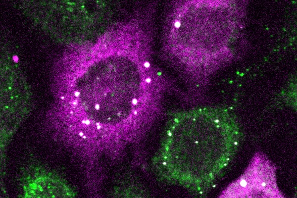
This scProteome-seq array shows separated protein biomarkers (green and magenta spots) from thousands of single cells.
Penn Health-Tech’s Nemirovsky Engineering and Medicine Opportunity (NEMO) Prize awards $80,000 to support early-stage ideas joining engineering and medicine. The goal of the prize is to encourage collaboration between the University of Pennsylvania’s Perelman School of Medicine and the School of Engineering and Applied Science by supporting innovative ideas that might not receive funding from traditional sources.
This year, the NEMO Prize has been awarded to a team of researchers from Penn Engineering’s Department of Bioengineering. Their project aims to develop a technology that can detect multiple cancer biomarkers in single cells from tumor biopsy samples.
As cancer cells grow in the body, one of the characteristics that influences tumor growth and response to treatment is cancer cell state heterogeneity, or differences in cell states. Methods that rapidly catalogue cell heterogeneity may be able to detect rare cells responsible for tumor growth and drug resistance.
Single-cell transcriptomics (scRNA-seq) is the standard method for studying cell states; by amplifying and analyzing the cell’s complement of RNA sequences at a given time, researchers can get a snapshot of what proteins the cell is in the process of making. However, this method does not fully capture the function of the cell. The field of proteomics, which captures the actual protein content of cells along with post-translational modifications, provides a better picture of the cell’s function, but single-cell proteomic methods with the same sensitivity as scRNA-seq do not currently exist.
Alex Hughes, Lukasz Bugaj and Andrew Tsourkas
This collaborative project, which joins Assistant Professors Alex Hughes and Lukasz Bugaj, as well as Professor Andrew Tsourkas, aims to change that by developing multiplexed, sensitive and highly specific single-cell proteomics technologies to advance our understanding of cancer, its detection and its treatment.
This new technology, called scProteome-seq, builds from Hughes’s previous work.
“My specific expertise here is as an inventor of single-cell western blotting, which is the core technology that our team is building on,” says Hughes. “Single-cell proteomics technologies of this type have a track-record of commercial translation for applications in basic science and clinical automation, so our approach has a high potential for real-world impact.”
The current technology from Hughes’ lab separates proteins in cells by their molecular weight and “blots” them on a piece of paper. Improvements to this technology included in this project will remove the limitation of using light-emitting dyes to detect different proteins and instead use DNA barcodes to differentiate them.

 Cosette Tomita, a master’s student in Bioengineering, spoke with Penn Engineering Graduate Admissions about her research in cellular therapy and her path to Penn Engineering.