Penn Bioengineering Postdoc Brittany Taylor Appointed Assistant Professor at University of Florida


Brittany Taylor, PhD

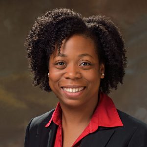
The Department of Bioengineering is proud to congratulate Postdoctoral Researcher Brittany Taylor, PhD on her appointment as a tenure-track Assistant Professor in the J. Crayton Pruitt Family Department of Biomedical Engineering of the Herbert Wertheim College of Engineering at the University of Florida. Taylor’s appointment will begin in January 2021 after four years as a postdoc in Penn Medicine’s McKay Orthopaedic Research Laboratory where she worked under the supervision of Louis Soslowsky, Fairhill Professor in Orthopaedic Surgery and Professor in Bioengineering.

Taylor got her BS in Biomedical Engineering from the University of Virginia where she conducted research under Drs. Cato Laurencin and Edward Botchwey (the latter got his PhD in Penn Bioengineering in 2002). She went on to complete her PhD in Biomedical Engineering in 2016, studying with Dr. Joseph Freeman, in the Musculoskeletal Tissue Regeneration Laboratory at Rutgers University. During her time at Penn, she served as the Co-President of the Biomedical Postdoctoral Council, worked with the Perelman School of Medicine’s PennVIEW program on postdoctoral diversity recruitment, and spearheaded the mentoring circles program, which brings together postdoctoral researchers, graduate students, and undergraduates in informal groups that allow mentorship and learning to flow freely.

The foundation for Taylor’s research interests is a combination of her training in bone tissue engineering, bioactive biomaterials, and tendon injury and repair. Her graduate research focused on a three-dimensional biomimetic pre-vascularized scaffold that simultaneously promoted osteogenic and angiogenic differentiation of human mesenchymal stem cells in vitro and cellular infiltration and neovascularization in vivo without the addition of growth factors of cells. As a postdoctoral fellow, in addition to investigating the role of collagen type V on tendon inflammation and remodeling in a mouse patellar tendon injury model, she also elucidated the biological and mechanical implications of an implantable bilayer delivery system (BiLDS) for controlled and localized release of non-steroidal anti-inflammatory drugs (NSAIDs) to modulate tendon inflammation in a rat rotator cuff injury and repair model. This collection of work exploits the ability of these transformative technologies to provide physical and chemical regenerative cues without the use of exogenous cells; hence avoiding possible complications associated with autologous and allogeneic cell sources and simplifying the regulatory pathway towards clinical application. Taylor’s future research program at the University of Florida will focus on tailored cell-free combinatorial strategies, such as decellularized matrices, tunable delivery systems, and modified extracellular vesicles, to complement and improve the native musculoskeletal tissue regenerative and reparative process.

“Brittany has been an amazing postdoctoral fellow,” says her mentor Louis Soslowsky. “She has learned a lot and contributed to various projects in an exemplary manner. She has been a leader in many arenas here at Penn and I am so proud of what she has done so far. I look forward to following her continued accomplishments at the University of Florida! I know she’ll do great!”

In the course of her pre-faculty career, Taylor achieved an impressive list of accomplishments. She received a Postdoctoral Fellowship for Academic Diversity from the Office of the Vice Provost for Research; a Postdoctoral Enrichment Program (PDEP) award from the Burroughs Wellcome Fund; and a UNCF Bristol-Myers Squibb E.E. Just Postgraduate Fellowship. Additionally, she was named a Rising Star in Cell Mentor’s list of “100 inspiring Black scientists in America” in February 2020 and was given a Rising Star in Biomedical Science Award from MIT in 2019.

“I am grateful for the opportunity to complete my postdoctoral training at Penn,” Taylor says:

“[P]articularly in a lab that is affiliated with the Penn Bioengineering program and the Department of Orthopaedic Surgery, where I had the unique experience of addressing basic science questions using translational animal models, while utilizing my engineering background and having a direct interaction with clinicians. Additionally, I connected with some amazing people here at Penn who had a significant impact on my time at Penn, and will be lifelong friends, colleagues, and mentors.”

Congratulations Dr. Taylor from everyone at Penn Bioengineering!

Students in Penn’s Biomechatronics Course Create Robotic Hands for Their Final Project

by Sophie Burkholder

Andrew Chan (left, M.S.E. in Robotics ‘19) and Omar Abdoun (right, BE M.D./Ph.D. student) present “Cryogripper”

Almost every engineering school in the country offers a course in mechatronics — the overlap of mechanical, electrical, and computer engineering in electromechanical system design — but how many offer a course in biomechatronics? Taught by LeAnn Dourte, Ph.D., a Practice Associate Professor in Bioengineering, Penn Engineering’s Biomechatronics course (BE 570) gives students the chance to think about how the principles of mechatronic design can be used in biological settings involving orthopaedics, cardiovascular systems, and respiration, to name a few.

Throughout the course, students engage in different projects related to circuitry, signal processing, mechanics, motors, and analog controls, eventually applying all of these to biological examples before working on a final culminating project in design teams of two. In a simulation meant to mimic the sort of thinking and design processes that go behind innovations in robotic surgery, students create an electromechanical device that acts as a robotic hand. The catch? The “hand” has to have enough dexterity to pick up a water bead with a slipperiness similar to that of human tissue.

In addition to successfully performing this mechanical task using skills that the students learned throughout the semester, design teams also have to incorporate biological interfaces into the final project, such as using EMG signals to move part of the robotic hand, to give one example. Furthermore, each team needs to have a unique element to their design, whether in the use of a second biological interface, the application of Bluetooth to the system, or even a physical extension of the robotic hand to include the electromechanical equivalents of a shoulder, elbow, or wrist joint.

Carolyn Godone and Mike Furr (both M.S.E. in Bioengineering ‘19) model their design

Students Carolyn Godone and Mike Furr (both M.S.E. in Bioengineering ‘19) created a design inspired by the mechanical iris of a camera lens, using gears to push 3-D printed slices together in a symmetrical pattern to close around an object for pickup. They controlled their unique gripper with a thermal sensing camera that could employ a heat map of the device’s user to rotate, raise, and lower the gripper. Another pair of students, Omar Abdoun (BE M.D./Ph.D. student) and Andrew Chan (M.S.E. in Robotics ‘19), made what they called a “cryogripper”: a tissue moistened with water that freezes on demand when it contacts its target hydrogel. The ice allows the target to be lifted without falling, and the tissue can later be thawed with pumps of warm water to release hydrogel.

After weeks of working on their projects in the George H. Stephenson Foundation Educational Laboratory and Bio-MakerSpace, the class presented their final robotic hands during an open demonstration day (or Demo Day) in the lab. To see all the devices live and in action, watch the Facebook video below!

Week in BioE (March 15, 2019)

by Sophie Burkholder

Synthetic Spinal Discs from a Penn Research Team Might Be the Solution to Chronic Back Pain

Spinal discs, the concentric circles of collagen fiber found between each vertebra of the spine, can be the source of immense back pain when ruptured. Especially for truck and bus drivers, veterans, and cigarette smokers, there is an increased risk in spinal disc rupture due to overuse or deterioration over time. But these patients aren’t alone. In fact, spinal discs erode over time for almost everyone, and are one of the sources of back pain in older patients, especially when the discs erode so much that they allow direct bone-to-bone contact between two or more vertebrae.

Robert Mauck, Ph.D.

Robert Mauck, Ph.D., who is the director of the McKay Orthopaedic Research Laboratory here at Penn and a member of the Bioengineering Graduate Group Faculty, led a research team in creating artificial spinal discs, with an outer layer made from biodegradable polymer and an inner layer made with a sugar-like gel. Their findings appear in Science Translational Medicine. These synthetic discs are also seeded with stem cells that produce collagen over time, meant to replace the polymer as it degrades in vivo over time. Though Mauck and his time are still far from human clinical trials for the discs, they’ve shown some success in goat models so far. If successful, these biodegradable discs could lead to a solution for back pain that integrates itself into the human body over time, potentially eliminating the need of multiple invasive procedures that current solutions require. Mauck’s work was recently featured in Philly.com.

An Untethered, Light-Activated Electrode for Innovations in Neurostimulation

Neurostimulation, a process by which nervous system activity can be purposefully modulated, is a common treatment for patients with some form of paralysis or neurological disorders like Parkinson’s disease. This procedure is typically invasive, and because of the brain’s extreme sensitivity, even the slightest involuntary movement of the cables, electrodes, and other components involved can lead to further brain damage through inflammation and scarring. In an effort to solve this common problem, researchers from the B.I.O.N.I.C. Lab run by Takashi D.Y. Kozai, Ph. D., at the University of Pittsburgh replaced long cables with long wavelength light and a formerly tethered electrode with a smaller, untethered one.

The research team, which includes Pitt senior bioengineering and computer engineering student Kaylene Stocking, centered the device on the principle of the photoelectric effect – a concept first described in a publication by Einstein as the local change in electric potential for an object when hit with a photon. Their design, which includes a 7-8 micron diameter carbon fiber implant, is now patent pending, and Kozai hopes that it will lead to safer and more precise advancements in neurostimulation for patients in the future.

A New Microfluidic Chip Can Detect Cancer in a Drop of Blood

Many forms of cancer cannot be detected until the disease has progressed past the point of optimum treatment time, increasing the risk for patients who receive late diagnoses of these kinds of cancer. But what if the diagnostic process could be simplified and made more efficient so that even a single drop of blood could be enough input to detect the presence of cancer in a patient? Yong Zeng, Ph.D., and his team of researchers at the University of Kansas in Lawrence sought to answer that question.

They designed a self-assembled 3D-nanopatterned microfluidic chip to increase typical microfluidic chip sensitivity so that it can now detect lower levels of tumor-associated exosomes in patient blood plasma. This is in large part due to the nanopatterns in the structure of the chip, which promote mass transfer and increase surface area, which in turn promotes surface-particle interactions in the device. The team applied the device to their studies of ovarian cancer, one of the notoriously more difficult kinds of cancer to detect early on in patients.

A Wearable Respiration Monitor Made from Shrinky Dinks

Michelle Khine, Ph. D., a professor of biomedical engineering at the University of California, Irvine incorporates Shrinky Dinks into her research. After using them once before in a medical device involving microfluidics, her lab recently worked to incorporate them into a wearable respiration monitor – a device that would be useful for patients with asthma, cystic fibrosis, and other chronic pulmonary diseases. The device has the capability to track the rate and volume of its user’s respiration based on measurements of the strain at the locations where the device makes contact with the user’s abdomen.

Paired with Bluetooth technology, this sensor can feed live readings to a smartphone app, giving constant updates to users and doctors, as opposed to the typical pulmonary function test, which only provides information from the time at which the test takes a reading. Though Khine and her team have only tested the device on healthy patients so far, they look forward to testing with patients who have pulmonary disorders, in hopes that the device will provide more comprehensive and accessible data on their respiration.

People and Places

Ashley Kimbel, a high school senior from Grissom High School in Huntsville, Alabama, designed a lightweight prosthetic leg for local Marine, Kendall Bane, after an attack in Afghanistan led him to amputate one of his legs below the knee. Bane, who likes to keep as active as possible, said the new lighter design is more ideal for activities like hiking and mountain biking, especially as any added weight makes balance during these activities more difficult. Kimbel used a CAD-modeling software produced by Siemens called Solid Edge, which the company hopes to continue improving in accessibility so that more students can start projects like Kimbel’s.

This week, we would like to congratulate Angela Belcher, Ph.D., on being named the new head of the Department of Biological Engineering at the Massachusetts Institute of Technology (MIT). With her appointment to this role, now half of the MIT engineering department heads are women. Belcher’s research is in the overlap of materials science and biological engineering, with a particular focus on creating nanostructures based on the evolution of ancient organisms for applications in medical diagnostics, batteries, solar cells, and more.

We would also like to congratulate Eva Dyer, Ph.D., and Chethan Pandarinath, Ph.D., both of whom are faculty members at the Walter H. Coulter Department of Biomedical Engineering at Georgia Tech and Emory University, on receiving research fellowships from the Alfred P. Sloan Foundation. Dr. Dyer, who formerly worked with Penn bioengineering faculty member Dr. Konrad Kording while he was at Northwestern University, leads research in the field of using data analysis methods to quantify neuroanatomy. Dr. Pandarinth leads the Emory and Georgia Tech Systems Neural Engineering Lab, where he works with a team of researchers to use properties of artificial intelligence and machine learning to better understand large neural networks in the brain.