The effectiveness of CAR T cell therapy against a variety of cancers, including solid tumors, could be boosted greatly by using CRISPR-Cas9 technology to knock out the gene for CD5, a protein found on the surface of T cells, according to a preclinical study from investigators at the University of Pennsylvania’s Perelman School of Medicine and Abramson Cancer Center.
CAR T cells are T cells that have been engineered to attack specific targets found on cancer cells. They have had remarkable results in some patients with blood cancers. But they have not performed well against other cancers including solid-tumor cancers, such as pancreatic cancer, prostate cancer, and melanoma. Researchers have been searching for techniques to boost the effectiveness of CAR T cell therapy.
The study, published today in Science Immunology, suggests that knocking out CD5 could be a prime technique. Illuminating the protein’s previously murky role, the researchers found that it works as a powerful immune checkpoint, reining in T cell effectiveness. Removing it, they showed, dramatically enhanced CAR T cell anticancer activity in a variety of preclinical cancer models.
“We’ve discovered in preclinical models that CD5 deletion greatly enhances the function of CAR T cells against multiple cancers,” said senior author Marco Ruella, MD, an assistant professor of Hematology-Oncology, researcher with the Center for Cellular Immunotherapies and the scientific director of Penn Medicine’s Lymphoma Program. “The striking effects we observed across preclinical models suggest that CD5 knockout could be a general strategy for enhancing CAR T cell function.”
The study’s first author is Ruchi Patel, PhD, a recent graduate student from the Ruella Laboratory.
Patients being treated for B-cell non-Hodgkin’s Lymphoma (NHL) who are part of minority populations may not have equal access to cutting-edge CAR T cell therapies, according to a new analysis led by researchers from the Perelman School of Medicine and published in NEJM Evidence.
CAR T cell therapy is a personalized form of cancer therapy that was pioneered at Penn Medicine and has brought hope to thousands of patients who had otherwise run out of treatment options. Six different CAR T cell therapies have been approved since 2017 for a variety of blood cancers, including B-cell NHL that has relapsed or stopped responding to treatment. Image: iStock/PeopleImages
“CAR T cell therapy represents a major leap forward for blood cancer treatment, with many patients living longer than ever before, but its true promise can only be realized if every patient in need has access to these therapies,” says lead author Guido Ghilardi, a postdoctoral fellow in the laboratory of senior author Marco Ruella, an assistant professor of hematology-oncology and scientific director of the Lymphoma Program. “From the scientific perspective, we’re constantly working in the laboratory to make CAR T cell therapy work better, but we also want to make sure that when a groundbreaking treatment like this becomes available, it reaches all patients who might be able to benefit.”
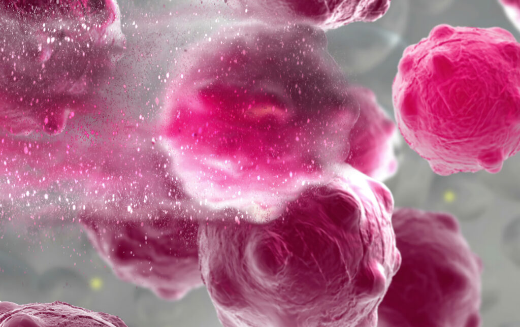
3d illustration of a damaged and disintegrating cancer cell. (Image: iStock/vitanovski)
The development of any type of second cancer following CAR T cell therapy is a rare occurrence, as found in an analysis of more than 400 patients treated at Penn Medicine, researchers from the Perelman School of Medicine at the University of Pennsylvania reported today in Nature Medicine. The team also described a single case of an incidental T cell lymphoma that did not express the CAR gene and was found in the lymph node of a patient who developed a secondary lung tumor following CAR T cell therapy.
CAR T cell therapy, a personalized form of immunotherapy in which each patient’s T cells are modified to target and kill their cancer cells, was pioneered at Penn. More than 30,000 patients with blood cancers in the United States—many of whom had few, if any, remaining treatment options available—have been treated with CAR T cell therapy since the first such therapy was approved in 2017. Some of the earliest patients treated in clinical trials have gone on to experience long-lasting remissions of a decade or more.
Secondary cancers, including T cell lymphomas, are a known, rare risk of several types of cancer treatment, including chemotherapy, radiation, and stem cell transplant. CAR T cell therapy is currently only approved to treat blood cancers that have relapsed or stopped responding to treatment, so patients who receive CAR T cell therapies have already received multiple other types of treatment and are facing dire prognoses.
In November 2023, the FDA announced an investigation into several reported cases of secondary T cell malignancies, including CAR-positive lymphoma, in patients who previously received CAR T cell therapy products. In January 2024, the FDA began requiring drugmakers to add a safety label warning to CAR T cell products. While the FDA review is still ongoing, it remains unclear whether the secondary T cell malignancies were caused by CAR T cell therapy.
As a leader in CAR T cell therapy, Penn has longstanding, clearly established protocols to monitor each patient both during and after treatment – including follow-up for 15 years after infusion – and participates in national reporting requirements and databases that track outcomes data from all cell therapy and bone marrow transplants.
Marco Ruella, M.D.
“When this case was identified, we did a detailed analysis and concluded the T cell lymphoma was not related to the CAR T cell therapy. As the news of other cases came to light, we knew we should go deeper, to comb through our own data to better understand and help define the risk of any type of secondary cancer in patients who have received CAR T cell products,” said senior author Marco Ruella, MD, an assistant professor of Hematology-Oncology and Scientific Director of the Lymphoma Program. “What we found was very encouraging and reinforces the overall safety profile for this type of personalized cell therapy.”
Breaking the code of the immune system could provide a new fundamental way of understanding, treating, and preventing every type of disease. Penn Medicine is investing in key discoveries about immunity and immune system function, and building infrastructure, to make that bold idea a reality.
This grandfather lives with primary progressive multiple sclerosis (MS), an autoimmune disorder that he controls with a medicine that depletes his body of the type of immune cells that make antibodies. So while he has completed his COVID-19 vaccine course, his immune system function isn’t very strong—and the invitation has arrived at a time when COVID-19 is still spreading rapidly.
You can imagine the scene as an older gentleman lifts a thick, creamy envelope from his mailbox, seeing his own name written in richly scripted lettering. He beams with pride and gratitude at the sight of his granddaughter’s wedding invitation. Yet his next thought is a sober and serious one. Would he be taking his life in his hands by attending the ceremony?
“In the past, all we could do was [measure] the antibody response,” says Amit Bar-Or, the Melissa and Paul Anderson President’s Distinguished Professor in Neurology at the Perelman School of Medicine, and chief of the Multiple Sclerosis division. “If that person didn’t have a good antibody response, which is likely because of the treatment they’re on, we’d shrug our shoulders and say, ‘Maybe you shouldn’t go because we don’t know if you’re protected.’”
Today, though, Bar-Or can take a deeper dive into his patients’ individual immune systems to give them far more nuanced recommendations. A clinical test for immune cells produced in response to the COVID-19 vaccine or to the SARS-CoV-2 virus itself—not just antibodies—was one of the first applied clinical initiatives of a major new Immune Health® project at Penn Medicine. Doctors were able to order this test and receive actionable answers through the Penn Medicine electronic health record for patients like the grandfather with MS.
“With a simple test and an algorithm we can have a very different discussion,” Bar-Or says. A test result showing low T cells, for instance, would tell Bar-Or his patient may get a meaningful jolt in immunity from a vaccine booster, while low antibody levels would suggest passive antibody therapy is more helpful. Or, the test might show his body is already well primed to protect him, making it reasonably safe to attend the wedding.
This COVID-19 immunity test is only the beginning.
Physicians and scientists at Penn Medicine are imagining a future where patients can get a precise picture of their immune systems’ activity to guide treatment decisions. They are working to bring the idea of Immune Health to life as a new area of medicine. In labs, in complex data models, and in the clinic, they are beginning to make sense out of the depth and breadth of the immune system’s millions of as-yet-undeciphered signals to improve health and treat illnesses of all types.
Penn Medicine registered the trademark for the term “Immune Health” in recognition of the potential impact of this research area and its likelihood to draw non-academic partners as collaborators in its growth. Today, at the south end of Penn’s medical campus, seven stories of research space are being added atop an office building at 3600 Civic Center Blvd., including three floors dedicated to Immune Health, autoimmunity, and immunology research.
The concept behind the whole project, says E. John Wherry, director of Penn Medicine’s Institute for Immunology and Immune Health (I3H), “is to listen to the immune system, to profile the immune system, and use those individual patient immune fingerprints to diagnose and treat diseases as diverse as immune-related diseases, cancer, cardiovascular disease, Alzheimer’s, and many others.”
The challenge is vast. Each person’s immune system is far more complex than antibodies and T cells alone. The immune system is made of multiple interwoven layers of complex defenders—from our skin and mucous membranes to microscopic memory B cells that never forget a childhood infection—meant to fortify our bodies from germs and disease. It is a sophisticated system that learns and adapts over our lifetimes in numerous ways, and it also falters and fails in some ways we understand and others that remain mysterious. And each person’s intricate internal battlefield is in some way unique.
The immune system is not just a set of defensive barricades, either. It’s also a potential source of deep insight about a person’s physiological functioning and responses to medical treatments.
“The immune system is sensing and keeping track of basically all tissues and all cells in our body all the time,” Wherry says. “It is surveying the body trying to clean up any invaders and restore homeostasis by maintaining good health.”
“Our goal is to essentially break the code of the immune system,” says Jonathan Epstein, executive vice dean of the Perelman School of Medicine and chief scientific officer at Penn Medicine. “By doing so, we believe we will be able to determine your state of health and your response to therapies in essentially every human disease.”
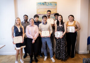
Front, from left to right: Lucy Andersen, Vice Provost for Education Karen Detlefsen, Derek Yang, Ann Ho, and Arianna James. Back, from left to right: Ritesh Isuri, Adiwid (Boom) Devahastin Na Ayudhya, Oualid Merzouga, and Puneeth Guruprasad.
Ten winners of the 2023 Penn Prize for Excellence in Teaching by Graduate Students were announced at a ceremony held April 13 at the Graduate Student Center. The recipients, who represented five of Penn’s 12 schools, were recognized among a pool of 44 Ph.D. candidates and master’s students nominated primarily by undergraduates—a quality unique to and cherished about this Prize.
“It’s a particularly authentic expression of gratitude from undergraduates, and that’s really the pleasure [of presenting these awards],” says Vice Provost for Education Karen Detlefsen, who was present to announce the winners and award them with a certificate. (They also receive a monetary award.) “I’m so proud of our students: Our undergraduates, for taking the time to recognize what it is our graduate students contribute to the student body, and the graduate students who are contributing to the life of the University.
“Students are the lifeblood of the University and without them, we wouldn’t be here.”
The Prize began in the 1999-2000 academic year under former Penn President Judith Rodin. It was spearheaded by then-doctoral-candidate Eric Eisenstein and has been issued every year since. Nominations for the Prize often mention how graduate teaching assistants were able to take a complex subject and make it relatable or craft a course like philosophy or mathematics into an enjoyable—even highly anticipated—experience for students.
“Many nominations show how much students value a TA or a graduate instructor of record who shows that they care for their learning and for them as people, and who makes themself readily available to assist,” says Ian Petrie, director of graduate student programming for the Center for Teaching and Learning, who organizes the selection committee for the Prize. “Typically, however, committee members are also interested in seeing nominations that really point to how a graduate student instructor taught or gave feedback—not just how responsive they were to emails or how many office hours they had.”
He also emphasizes that many winners this year were not just teachers, but mentors—often helping undergraduates or new graduate students navigate not only the course but also Penn as an institution.
Puneeth Guruprasad
One of the winners, Puneeth Guruprasad, hails from Penn Bioengineering. Guruprasad is a fourth-year Ph.D. student in Bioengineering who conducts research in the lab of Marco Ruella, Assistant Professor of Medicine in the Division of Hematology/Oncology in the Perelman School of Medicine. Ruella is also a member of the Center for Cellular Immunotherapies (CCI) and the Penn Bioengineering Graduate Group.
Guruprasad studies mechanisms of resistance to chimeric antigen receptor (CAR) T cell therapy for cancer. He has served as a teaching assistant for five semesters: three for Intro to Biotransport Processes (BE 3500) taught by Alex Hughes, Assistant Professor in Bioengineering, and two for Cellular Engineering (BE 3060), taught by Daniel Hammer, Alfred G. and Meta A. Ennis Professor in Bioengineering and in Chemical and Biomolecular Engineering. Both courses are a part of the core curriculum for undergraduate bioengineering students. His doctoral thesis focuses on how a specific interaction between CAR T cells and tumor cells limits their function across a range of cancers.
“I make myself approachable outside the classroom, and I think that’s one aspect of being a TA: having responsibilities that extend beyond the classroom,” says Guruprasad. “Dozens of times, I’ve spoken to students over coffee, or over some lunch, about what direction they want to take in their life, what they want to do outside of the course, and give them my two cents of advice. I try to individualize.”
This post was adapted from an original story by Brandon Baker in Penn Today. Read the full story and list of 2023 winners here.
A team of researchers led by the School of Arts & Science’s Wei Guo offers new insights into a mechanism that promotes tumor growth. “This information could be used to help clinicians diagnose cancers earlier in the future,” says Guo.
In many instances, the physical manifestation of cancers and the ways they are subsequently diagnosed is via a tumor, tissue masses of mutated cells and structures that grow excessively. One of the major mysteries in understanding what goes awry in cancers relates to the environments within which these structures grow, commonly known as the tumor microenvironment.
These microenvironments play a role in facilitating tumor survival, growth, and spread. Tumors can help generate their own infrastructure in the form of vasculature, immune cells, signaling molecules, and extracellular matrices (ECMs), three-dimensional networks of collagen-rich support scaffolding for a cell. ECMs also help regulate cellular communications, and in the tumor microenvironment ECMs can be a key promoter of tumor growth by providing structural support for cancerous cells and in modulating signaling pathways that promote growth.
Now, new research led by the School of Arts & Science’sWei Guo and published in the journal Nature Cell Biology has bridged the complex structural interactions within the tumor microenvironment to the signals that trigger tumor growth. The researchers studied cancerous liver cells grown on ECMs of varying stiffness and discovered that the stiffening associated with tumor growth can initiate a cascade that increases the production of small lipid-encapsulated vesicles known as exosomes.
“Think of these exosomes as packages that each cell couriers out, and, depending on the address, they get directed to other cells,” says Ravi Radhakrishnan, professor of bioengineering in the School of Engineering and Applied Science and a co-author of the paper.
“By recording the number of packages sent, the addresses on these packages, their contents, and most importantly, how they’re regulated and generated, we can better understand the relationship between a patient’s tumor microenvironment and their unique molecular signaling signatures, hinting at more robust personalized cancer therapies,” Radhakrishnan says.
While studying exosomes in relation to tumor growth and metastasis has been well-documented in recent years, researchers have mostly focused on cataloging their characteristics rather than investigating the many processes that govern the creation and shuttling of exosomes between cells. As members of Penn’s Physical Sciences Oncology Center (PSOC), Guo and Radhakrishnan have long collaborated on projects concerning tissue stiffness. For this paper, they sought to elucidate how stiffening promotes exosome trafficking in cancerous intracellular signaling.
“Our lab previously found that high stiffness promotes the secretion of exosomes,” says Di-Ao Liu, co-first author of the paper and a graduate student in the Guo Lab. “Now, we were able to model the stiffening processes through experiments and identify molecular pathways and protein networks that cause this, which better links ECM stiffening to cancerous signaling.”
Each year, the the Department of Bioengineering seeks exceptional candidates to conduct summer research in bioengineering with the support of two scholarships: the Abraham Noordergraaf Student Summer Bioengineering Research Fund and the Blair Undergraduate Research Fund in the Department of Bioengineering. These scholarships provide a living stipend for students to conduct research on campus in a Penn research lab under the mentorship of a faculty member. The Abraham Noordergraaf Student Summer Bioengineering Research Fund provides financial support for undergraduate or graduate summer research opportunities in bioengineering with a preference for study in the area of cardiovascular systems. Dr. Noordergraaf, who died in 2014, was a founding member and first chair of Penn Bioengineering. The Blair Undergraduate Research Fund in the Department of Bioengineering supports three to five undergraduate research scholars each year with the support of Dr. James C. Blair II. After a competitive round of proposals, the following six scholars were chosen for the Summer 2022 semester. Keep reading below for the research abstracts and bios of the awardees.
The Blair Undergraduate Research Fund in the Department of Bioengineering (Blair Scholars)
Ella Atsavapranee
Student: Ella Atsavapranee (BE Class of 2023)
PI: Michael J. Mitchell, J. Peter and Geri Skirkanich Assistant Professor of Innovation, Bioengineering
“Lipid nanoparticle-mediated delivery of RAS protease to inhibit cancer cell growth”
Mutations in RAS, a family of proteins found in all human cells, drive a third of cancers, including many pancreatic, colorectal, and lung cancers. However, there are still no therapies that can effectively prevent RAS from causing tumor growth. Recently, a protease was engineered to specifically degrade active RAS, offering a promising new tool for treating these cancers. However, many protein-based therapies still cannot be effectively delivered to patients. Lipid nanoparticles (LNPs), which were used in the Pfizer-BioNTech and Moderna COVID-19 vaccines, have emerged as a promising platform for safe and effective delivery of both nucleic acids and proteins. We formulated a library of LNPs using different cationic lipids. We characterized the LNPs by size, charge, and pKa, and tested their ability to deliver fluorescently labeled protease. The LNPs were able to encapsulate and deliver a RAS protease, successfully reducing proliferation of colon cancer cells.
Ella is a senior from Maryland studying bioengineering and chemistry. She works in Dr. Michael Mitchell’s lab, developing lipid nanoparticles to deliver proteins that reduce cancer cell proliferation. She has also conducted research on early-stage cancer detection and therapy monitoring (at Stanford University) and drug delivery across the blood-brain barrier for neurodegenerative diseases (at University of Maryland). She is passionate about translational research, science communication, and promoting diversity in STEM.
Chiadika Eleh
Student: Chiadika Eleh (BE and CIS Class of 2024)
PI: Eric J. Brown, Associate Professor of Cancer Biology, Perelman School of Medicine
“Investigating Viability in ATR and WEE1 Inhibitor Treated Ovarian Cancer Cells”
High-grade serous ovarian cancers (HGSOCs) are an aggressive subtype of ovarian cancer, accounting for up to 80% of all ovarian cancer-related deaths. More than half of HGSOCs are homologous recombination deficient; thus, they lack a favorable response when treated with common chemotherapeutic trials. Therefore, new treatment strategies must be developed to increase the life expectancy and quality of life of HGSOC patients. To address the lack of effective treatment options, the Brown Lab is interested in combining ATR and WEE1 inhibition (ATRi/WEE1i) to target HGSOC cells. It has previously been shown that low-dose ATRi/WEE1i is an effective treatment strategy for CCNE1-amplified ovarian cancer-derived PDX tumors (Xu et al., 2021, Cell Reports Medicine). Therefore, the next step is to characterize the HGSOC-specific response to ATRi/WEE1i treatment. This project aims to characterize the viability phenotype of ovarian cancer (OVCAR3) cells in the presence of ATRi/WEE1i in both single and combination treatments. With further research, Eleh hopes to prove the hypothesis low-dose combination ATRi/WEE1i treatment will result in the synergistic loss of viability in OVCAR3 cells. This goal will be achieved through the treatment of OVCAR3 cells with ranging doses of ATRi and Wee1i over 24 and 48 hour time intervals. We hope that this data will help set a treatment baseline that can be used for all OVCAR30-based viability experiments in the future.
Chiadika Eleh is a Bioengineering and Computer Science junior and a member of Penn Engineering’s Rachleff Scholar program. As a Blair Scholar, she worked in Dr. Eric Brown’s cancer biology lab, where she studied cell cycle checkpoint inhibitors as a form of cancer treatment.
“Tbc1d2b regulates vascular formation during development and tissue repair after ischemia”
The mechanisms behind endothelial cells forming blood vessels remains unknown. We have identified Tbc1d2b as a protein that is integral to the regulation of vascular formation. In order to investigate the role of Tbc1d2b in tubule formation, fibrin gel bead assays will be conducted to evaluate how the presence of Tbc1d2b is required for angiogenesis. Fibrin gel bead assays simulate the extracellular matrix environment to support the in vitro development of vessels from human umbilical vein endothelial cells (HUVEC) coated on cytodex beads. In order to confirm the success of angiogenesis, immunostaining for Phalloidin and CD31 will be conducted. After confirmation that fibrin gel bead assays can produce in vitro tubules, sgRNA CRISPR knockout of Tbc1d2b will be performed on HUVEC cells which will then be used to conduct more fibrin gel bead assays. We hypothesize that HUVEC with the Tbc1d2b knockout phenotype will be unable to form tubules while wild type HUVEC will be able to.
Gloria Lee is a rising senior studying Bioengineering and Physics in the VIPER program from Denver, Colorado. Her research in Dr. Yi Fan’s lab focuses on the role that proteins play in cardiovascular tubule formation.
Abraham Noordergraaf Student Summer Bioengineering Research Fund (Noordergraaf Fellows)
Gary Lin
Student: Gary Lin (Master’s in MEAM Class of 2023)
PI: Michelle J. Johnson, Associate Professor in Physical Medicine and Rehabilitation, Perelman School of Medicine, and in Bioengineering
“Development and Integration of Dynamically Modulating Control Systems in the Rehabilitation Using Community-Based Affordable Robotic Exercise System (Rehab CARES)”
As the number of stroke patients requiring rehabilitative care continues to increase, strain is being put onto the US health infrastructure which already has a shortage of rehabilitation practitioners. To help alleviate this pressure, a cost-effective robotic rehabilitative platform was developed to increase access to rehabilitative care. The haptic TheraDrive, a one-degree of freedom actuated hand crank that can apply assistive and resistive forces, was modified to train pronation and supination at the elbow and pinching of the fingers in addition to flexion and extension of the elbow and shoulder. Two controllers were created including an open-loop force controller and a closed-loop proportional-integral (PI) with adaptive control gains based on subject performance in therapy-game tasks as well as galvanic skin response. Stroke subjects (n=11) with a range of cognitive and motor impairment completed 4 therapy games in both adaptive and non-adaptive versions of the controllers (n=8) while measuring force applied on the TheraDrive handle. Resulting normalized average power versus Upper Extremity Fugl-Meyer (UE-FM) and Montreal Cognitive Assessment (MoCA) correlation analyses showed that power was strongly correlated with UE-FM in 2 of the conditions and moderately correlated with the other 6 while MoCA was moderate correlated to 2 of the conditions and weakly correlated to the rest. Mann-Whitney U-tests between adaptive and non-adaptive versions of each therapy game showed no significant differences with regards to power between controller types (p<0.05).
Gary is a master’s student in the School of Engineering studying Mechanical Engineering and Applied Mechanics with a concentration in Robotic and Mechatronic systems. His research primarily focuses on developing affordable rehabilitation robotics for use in assessment and game-based therapies post neural injury. Many of his interests revolve around the design of mechatronic systems and the algorithms used to control them for use in healthcare spaces.
Priya Shah
Student: Priya Shah (BE Class of 2024)
PI: Alex J. Hughes, Assistant Professor in Bioengineering
“Optogenetic Control of Developing Kidney Cells for Future Treatment of End-Stage Renal Disease”
This project sought to build from prior research in the Hughes Lab on the geometric and mechanical consequences of kidney form on cell and tissue-scale function. While the developmental trajectory of the kidney is well understood, little is currently known about many factors affecting nephron progenitor differentiation rate. Insufficient differentiation of nephron progenitor cells during kidney formation can result in lower nephron number and glomerular density, which is a risk factor for progression to end-stage renal disease later in life. Prior studies indicated that the amount of nephron differentiation – and thus function of the adult kidney – is correlated to the packing of ureteric tubule tips present at the surface of the kidney. Building off of research conducted in the Bugaj Lab, we found that inserting an optogenetic construct into the genome of human embryonic kidney (HEK) cells allowed us to manipulate the contraction of those cells through exposing them to blue light. Manipulating the contraction of the cells allows for the manipulation of the packing of ureteric tubule tips at the kidney surface. We used a lentiviral vector to transduce HEK293 cells with the optogenetic construct and witnessed visible contraction of the cells when they were exposed to blue light. Future work will include using CRISPR-Cas9 to introduce the optogenetic construct into IPS cells.
Priya is a junior studying bioengineering and had the opportunity to work on manipulating developing kidney cells using an optogenetic construct in the Hughes Lab this summer. She is thrilled to continue this research throughout the coming school year. Outside of the lab, Priya is involved with the PENNaach dance team and the Society of Women Engineers, as well as other mentorship roles.
Cosette Tomita
Student: Cosette Tomita (Master’s in MEAM Class of 2023)
“Expression and Characterization of an Anti-Aβ42 scFv”
Background: Amyloid Beta (Aβ42) fibrils contribute to the pathology of Alzheimer’s Disease. Numerous monoclonal antibodies have been developed against Aβ42. In this study we have designed and expressed a short chain variable fragment specific to Aβ42 (Anti-Aβ42 scFv). To characterize our anti-Aβ42 scFv we have performed structural analysis using transmission electron microscopy (TEM) and binding kinetics using microscale thermophoresis (MST) compared to commercially available antibodies 6E10, Aducanumab, and an IgG isotype control. The goal of this study is to determine if labeling densities and binding constants for Aducanumab and anti-Aβ42 scFv are not significantly different.
Method: To characterize Aβ42 fibril associated antibodies we used negative stain TEM. Aβ42 fibrils were stained on a glow discharged copper grid, and incubated with gold conjugated anti-Aβ42 scFv, 6E10—which binds all Aβ species, aducanumab, or IgG isotype control. Labeling densities were calculated as the number of fibril-associated gold particles per 1 μm2 for each image. Next, we used microscale thermophoresis determine the binding kinetics. Antibodies or anti-Aβ42 scFv were labeled with Alexa Fluor-647 and unlabeled Aβ42 was titrated in a serial dilution over 16 capillaries. The average fluorescence intensity was plotted against the antibody or scFv concentration and the curves were analyzed using the GraphPad Prism software to calculate the dissociation constant (KD) values.
Results: We found a significant difference, tested with a one-way ANOVA (P <0.0001), in gold particle associated Aβ fibrils per 1 μm2 between anti-Aβ42 scFv, 6E10, aducanumab, and IgG isotype control. Further analysis of aducanumab and 6CO3 with unpaired student t-test indicates significant differences in fibril associated gold particles between aducanumab vs. 6E10 (P=0.0003), Aducanumab vs. Isotype control (P <0.0001), anti-Aβ42 scFv vs 6E10 (p=0.0072), and anti-Aβ42 scFv vs Isotype Control (P=0.0029) with no significant difference in labeling densities between Aducanumab and anti-Aβ42 scFv. The expected KD values from MST were 1.8μM for Aducanumab and anti-Aβ42 scFv, 10.3nM for 6E10 and no expected binding for the isotype control. The experimental KD values for anti-Aβ42 scFv and 6E10 are 0.1132μM and 1.467μM respectively. The KD value for Isotype control was undetermined, as expected, however, the KD for Aducanumab was undetermined due to suboptimal assay conditions. Due to confounding variables in the experimental set up such as the use of Aβ1-16 compared to Aβ42 and the use of different fluorophores—5-TAMRA, Alexa Fluor 647 or FITC— the experimental KD values were off by several orders of magnitude.
Conclusion: We have illustrated similar labeling densities between Aducanumab and our anti-Aβ42 scFv. In the future, we will further optimize the MST assay conditions and compare the KD values obtained by MST with other techniques such as surface plasma resonance.
Cosette was born and raised in Chicago land area. Go Sox! She attended University of Missouri where she majored in Chemistry and Biology. She synthesized sigma-2 radiotracers and developed advanced skills in biochemical techniques in Dr. Susan Lever’s lab. After graduation, she moved to NJ to work at Lantheus, a radiopharmaceutical company. She missed academia and the independence of program and project development, so she came to work at the Penn Cyclotron facility before entering the Bioengineering master’s program.
Buffone got his Ph.D. in Chemical Engineering from SUNY Buffalo in Buffalo, NY in 2012, working with advisor Sriram Neelamegham, Professor of Chemical and Biological Engineering. Buffone completed previous postdoctoral studies at Roswell Park Comprehensive Cancer Center with Joseph T.Y. Lau, Distinguished Professor of Oncology in the department of Cellular and Molecular Biology. Upon coming to Penn in 2015, Buffone has worked in the Hammer Lab under advisor Daniel A. Hammer, Alfred G. and Meta A. Ennis Professor in Bioengineering and in Chemical and Biomolecular Engineering, first as a postdoc and later a research associate. Buffone also spent a year as a Visiting Scholar in the Center for Bioengineering and Tissue Regeneration, directed by Valerie M. Weaver, Professor at the University of California, San Francisco in 2019.
While at Penn, Buffone was a co-investigator on an R21 grant through the National Institutes of Health (NIH) which supported his time as a research associate. Buffone is excited to start his own laboratory where he plans to train a diverse set of trainees.
Buffone’s research area lies at the intersection of genetic engineering, immunology, and glycobiology and addresses how to specifically tailor the trafficking and response of immune cells to inflammation and various diseases. The work seeks to identify and subsequently modify critical cell surface and intracellular signaling molecules governing the recruitment of various blood cell types to distal sites. The ultimate goal of his research is to tailor and personalize the innate and adaptive immune response to specific diseases on demand.
“None of this would have been possible without the unwavering support of all of my mentors, past and present, and most especially Dan Hammer,” Buffone says. “His support in helping me transition into an independent scientist and his understanding of my outside responsibilities as a dad with two young children is truly the reason why I am standing here today. It’s a testament to Dan as both a person and a mentor.”
The U.S. Food and Drug Administration has expanded its approval for Kymriah, a personalized cellular therapy developed at the Abramson Cancer Center, this time for the treatment of adults with relapsed/refractory follicular lymphoma who have received at least two lines of systemic therapy. “Patients with follicular lymphoma who relapse or don’t respond to treatment have a poor prognosis and may face a series of treatment options without a meaningful, lasting response,” said Stephen J. Schuster, the Robert and Margarita Louis-Dreyfus Professor in Chronic Lymphocytic Leukemia and Lymphoma in the Division of Hematology Oncology. It’s the third FDAapproval for the “living drug,” which was the first of its kind to be approved, in 2017, and remains the only CAR T cell therapy approved for both adult and pediatric patients.
“In just over a decade, we have moved from treating the very first patients with CAR T cell therapy and seeing them live healthy lives beyond cancer to having three FDA-approved uses of these living drugs which have helped thousands of patients across the globe,” said Carl June, MD, the Richard W. Vague Professor in Immunotherapy in the department of Pathology and Laboratory Medicine in Penn’s Perelman School of Medicine and director of the Center for Cellular Immunotherapies in the Abramson Cancer Center and director of the Parker Institute for Cancer Immunotherapy at Penn. “Today’s news is new fuel for our work to define the future of cell therapy and set new standards in harnessing the immune system to treat cancer.”
Research from June, a member of the Penn Bioengineering Graduate Group, led to the initial FDA approval for the CAR T therapy (sold by Novartis as Kymriah) for treating acute lymphoblastic leukemia (ALL), one of the most common childhood cancers.
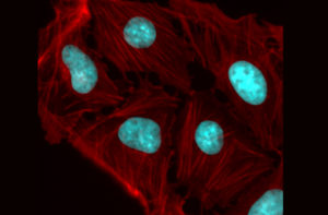
A colorized microscope image of an osteosarcoma shows how cellular fibers can transfer physical force between neighboring nuclei, influencing genes. The Penn Anti-Cancer Engineering Center will study such forces, looking for mechanisms that could lead to new treatments or preventative therapies.
Advances in cell and molecular technologies are revolutionizing the treatment of cancer, with faster detection, targeted therapies and, in some cases, the ability to permanently retrain a patient’s own immune system to destroy malignant cells.
However, there are fundamental forces and associated challenges that determine how cancer grows and spreads. The pathological genes that give rise to tumors are regulated in part by a cell’s microenvironment, meaning that the physical push and pull of neighboring cells play a role alongside the chemical signals passed within and between them.
The Penn Anti-Cancer Engineering Center (PACE) will bring diverse research groups from the School of Engineering and Applied Science together with labs in the School of Arts & Sciences and the Perelman School of Medicine to understand these physical forces, leveraging their insights to develop new types of treatments and preventative therapies.
The Center’s founding members are Dennis Discher, Robert D. Bent Professor with appointments in the Departments of Chemical and Biomolecular Engineering (CBE), Bioengineering (BE) and Mechanical Engineering and Applied Mechanics (MEAM), and Ravi Radhakrishnan, Professor and chair of BE with an appointment in CBE.
Discher, an expert in mechanobiology and in delivery of cells and nanoparticles to solid tumors, and Radhakrishnan, an expert on modeling physical forces that influence binding events, have long collaborated within the Physical Sciences in Oncology Network. This large network of physical scientists and engineers focuses on cancer mechanisms and develops new tools and trainee opportunities shared across the U.S. and around the world.
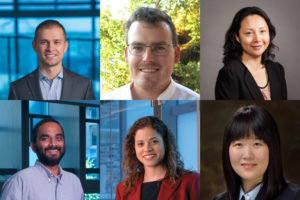
Lukasz Bugaj, Alex Hughes, Jenny Jiang, Bomyi Lim, Jennifer Lukes and Vivek Shenoy (Clockwise from upper left).
Among the PACE Center’s initial research efforts are studies of the genetic and immune mechanisms associated with whether a tumor is solid or liquid and investigations into how physical stresses influence cell signaling.