Week in BioE (May 10, 2018)

Advances in Cancer Detection
glioblastoma
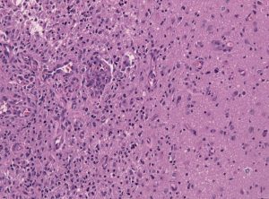
Tumor-brain-interface in a glioblastoma biopsy specimen.

Among the deadliest and most difficult to treat types of cancer is glioblastoma, an especially aggressive form of brain cancer. Widely available imaging techniques can diagnose the tumor, but often the diagnosis is too late to treat the cancer effectively. Although blood-based cancer biomarkers can provide for earlier detection of cancer, these markers face the difficult task of crossing the blood-brain barrier (BBB), which prevents all but the tiniest molecules from moving from the brain to the bloodstream.

A study recently published in Scientific Reports, coauthored by Hong Chen, PhD, Assistant Professor of Biomedical Engineering at Washington University in St. Louis (WUSTL), reports of successful deployment of a strategy consisting of focused ultrasound (FUS), enhanced green fluorescent protein (eGFP), and systemically injected microbubbles to see if the BBB could be opened temporarily to allow biomarkers to pass from the brain into the bloodstream. The authors used eGFP-activated mouse models of glioblastoma, injecting the microbubbles into the mice and then exposing the mice to varying acoustic pressures of FUS. They found that circulating blood levels of eGFP were several thousand times higher in the FUS-treated mice compared to non-treated mice, which would significantly facilitate the detection of the marker in blood tests.

The method has some way to go before it can be used in humans. For one thing, the pressures used in the Scientific Reports study would damage blood vessels, so it must be determined whether lower pressures would still provide detectable transmission of proteins across the BBB. In addition, the authors must exclude the possibility of FUS unexpectedly enhancing tumor growth.

In other body areas, with easier access from tissue to the bloodstream, engineers have developed a disease-screening pill that, when ingested and activated by infrared light, can indicate tumor locations on optical tomography. The scientists, led by Greg M. Thurber, PhD, Assistant Professor of Biomedical and Chemical Engineering at the University of Michigan, reported their findings in Molecular Pharmaceutics.

The authors of the study used negatively charged sulfate groups to facilitate absorption by the digestive system of molecular imaging agents. They tested a pill consisting of a combination of these agents and found that their model tumors were visible. The next steps will include optimizing the imaging agent dosage loaded into the pill to optimize visibility. The authors believe their approach could eventually replace uncomfortable procedures like mammograms and invasive diagnostic procedures.

Liquid Assembly Line to Produce Drug Microparticles

Pharmaceuticals owe their effects mostly to their chemical composition, but the packaging of these drugs into must be done precisely. Many drugs are encapsulated in solid microparticles, and engineering consistent size and drug loading in these particles is key. However, common drug manufacturing techniques, such as spray drying and ball milling, produce uneven results. 

University of Pennsylvania engineers developed a microfluidic system in which more than ten thousand of these devices run in parallel, all on a silicon-and-glass chip that can fit into a shirt pocket, to produce a paradigm shift in microparticle manufacturing. The team, led by David Issadore, Assistant Professor in the Department of Bioengineering, outlined the design of their system in the journal Nature Communications.

The Penn team first tested their system by making simple oil-in-water droplets, at a rate of more than 1 trillion droplets per hour. Using materials common to current drug manufacturing processes, they manufactured polycapralactone  microparticles at a rate of ‘only’ 328 billion particles per hour. Further testing backed by pharma company GlaxoSmithKline will follow.

Preventing Fungal Infections of Dental Prostheses

Dental prostheses are medical devices that many people require, particularly as they age. One of the chief complications with prostheses is fungal infections, with an alarming rate of two-thirds among people wearing dentures. These infections can cause a variety of problems, spreading to other parts of the digestive system and affecting nutrition and overall well-being. Fungal infections can be controlled in part by mouthwashes, microwave treatments, and light therapies, but none of them have high efficacy.

To address this issue, Praveen Arany, DDS, PhD, Assistant Professor, Department of Oral Biology and Biomedical Engineering at SUNY Buffalo, combined 3D printing technology and polycaprolactone microspheres containing amphotericin-B, an antifungal agent. Initial fabrication of the prostheses is described in an article in Materials Today Communications, along with successful in vitro testing with fungal biofilm. If further testing proves effective, these prostheses could be used in dental patients in whom the current treatments are either ineffective or contraindicated.

People and Places

West Virginia University has announced that it will launch Master’s and doctoral programs in Biomedical Engineering. The programs will begin enrolling students in the fall. The graduate tracks augment a Bachelor’s degree program begun in 2014.

Week in BioE (February 20, 2018)

Modeling Hemostasis With Microfluidics

hemostasis
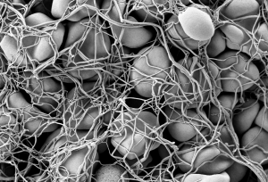
Electronic scanning microscopic image of red blood cells forming a clot

Hemostasis is the process by which blood stops flowing from damaged blood vessels. It is a complex process involving multiple molecules and forces, and our current understanding is limited by our inability to test these factors simultaneously in the laboratory. Some tests, for instance, can tell us much about clotting — a part of hemostasis — but little about the other elements at play. In particular, the role in hemostasis of the endothelium, which is the cell layer that lines the blood vessels, has generally been omitted from previous studies.

However, a new article in Nature Communications details the use of microfluidics technology, which is often used to model organ systems outside the body, to engineer a more complete model of hemostasis. Led by Wilbur A. Lam, M.D., Ph.D., Assistant Professor in the Wallace H. Coulter Department of Biomedical Engineering at Georgia Tech, the study authors fabricated microfluidics devices and then seeded vascular channels in the devices with human aortic and umbilical vein cells to simulate the endothelium.

Using the device, the authors were able to visualize hemostatic plug formation with whole blood and with blood from subjects with hemophilia. Although the authors concede that their model best represents capillary bleeding, rather than bleeding from larger vessels, they are confident that their model reliably represents the interaction of the endothelium with multiple varieties of blood cells.

Shedding Light on Cancer Response and Resistance

Penn’s founder, Benjamin Franklin, has a famous axiom: “an ounce of prevention is worth a pound of cure.” If Franklin were alive today, he would likely agree with two common axioms in cancer treatment: 1) if you can’t see it, you can’t treat it; and 2) if you treat it, treat all of it. Recent publications from investigators at Columbia and the University of Maryland reveal how imaging technologies can help predict to  outcomes and how nanotechnology is offering a new therapeutic tools for fighting cancer.

Using diffuse optical tomography (DOT), which employs near-infrared spectroscopy to obtain three-dimensional images, scientists at Columbia have shown in an article in Radiology that treatment response could be predicted as early as two weeks after the start of therapy. The authors, led by Andreas H. Hielscher, PhD, Professor of Biomedical Engineering, Electrical Engineering, and Radiology at Columbia, applied DOT in 40 women with breast cancer undergoing chemotherapy. They found that DOT imaging features were associated, some very strongly, with treatment outcomes at 5 months.  Given their positive findings, the authors intend to continue testing DOT in a larger cohort prospective study.

Another major issue in cancer chemotherapy is multidrug resistance (MDR),  a highly frustrating complication resulting from lengthy treatment. In MDR, cancer types can find ways to overcome the effects of chemotherapy, resulting in relapse, often with deadly consequences. Therefore, among the challenges that oncologists face is the need to predict MDR, preferably before treatment even begins.

Based on the knowledge that adenosine triphosphate (ATP), a common organic molecule in energy generation, is involved in MDR, scientists at the University of Maryland engineered nanoparticles that could target cancer cells and, when exposed to near-infrared laser irradiation, reduce the amount of ATP in the cells . The scientists, led by Xiaoming Shawn He, Ph.D., Professor of Bioengineering at Maryland, published their findings in Nature Communications.

Dr. He’s team tested their nanoparticles in vitro and subsequently in mice and found decreased tumor sizes in mice treated with the particles, as well as more deaths of cancer cells. In addition, two of seven mice treated with the nanoparticles plus light experienced complete tumor eradication. The findings offer hope that MDR could be overcome with direct delivery of targeted treatment to resistant tumors.

Preserving the Tooth

A frustrating problem often encountered by dentists is the growth of new cavities around existing fillings. Microbes are often critical catalysts for these new cavities. Using antimicrobial agents at cavity-repair sites could make a real difference. However, mesoporous silica has proved suboptimal for this purpose.

However, help might be on the way. A study in a recent issue of Scientific Reports, written by a trio of authors led by Benjamin D. Hatton, Ph.D., Assistant Professor at the Institute of Biomaterials & Biomedical Engineering of the University of Toronto, reports the successful synthesis of 500-nm nanocomposite spheres combining silica with octenidine dihydrochloride, a common antiseptic. The newly synthesized nanospheres outperformed earlier attempts with mesoporous silica. The authors will continue to develop these nanoparticles to deliver other drugs for longer periods of time.

Week in BioE (February 2, 2018)

Broccoli + Yogurt = Cancer Prevention?

broccoliGeorge H.W. Bush refused to eat it, but maybe he should start. It turns out that broccoli, combined with bioengineered yogurt, could provide effect cancer prevention. We’ve known for some time that compounds in certain fresh vegetables can increase chemoprevention, but the levels are usually too low to be effective, or they can’t be assimilated optimally by the body.  However, scientists in Singapore found that engineered bacteria, when ingested by mice with colorectal cancer, had anticancer effects. The bacteria caused the secretion of an enzyme by the cancer cells that transformed glucosinolates — compounds found in vegetables — into molecules with anticancer efficacy. The scientists report their findings in Nature Biomedical Engineering.

The authors programmed an E. coli cell line to bind to heparan sulfate proteoglycan, a cell surface protein that occurs in colorectal cancer cells. Once the engineered bacteria bound to the cancer cells, the bacteria secreted myrosinase, an enzyme that commonly occurs in many plants to defend them against aphids. In the cell model employed by the authors, myrosinase caused the conversion of glucosinolates into sulforaphane, which in turn could inhibit cancer cell growth.

The scientists then applied their system in a mouse model of colorectal cancer, feeding the mice yogurt infused with the engineered bacteria. They found that the mice fed broccoli plus the yogurt developed fewer and smaller tumors than mice fed broccoli alone. Additional testing is necessary, of course, but the study authors believe that their engineered bacteria could be used both as a preventive tool in high-risk patients and as a supplement for cancer patients after surgery to remove their tumors.

The Gates of CRISPR

About two years ago, software giant Microsoft unveiled Azimuth, a gene-editing tool for CRISPR/Casa9 that it had developed in collaboration with scientists at the Broad Institute. Now, in response to concerns that CRIPR may edit more of the genome than a bioengineer wants, the team has introduced a tool called Elevation. A new article in Nature Biomedical Engineering discusses the new tool.

In the article, the team, co-led by John C. Doench, Ph.D., Institute Scientist at the Broad Institute, describes how it developed Azimuth and Elevation, both of which are machine learning models, and deployed the tools to compare their ability to predict off-target editing with the ability of other approaches. The Elevation model outperformed the other methods. In addition, the team has implemented a cloud-based service for end-to-end RNA design, which should alleviate some of the time and resource handicaps that scientists face in using CRISPR.

Reducing Infant Mortality With an App

Among the challenges still faced in the developing world with regard to health care is high infant mortality, with the most common cause being perinatal asphyxia, or lack of oxygen reaching the infant during delivery. In response, Nigerian graduate student Charles C. Onu, a Master’s student in the computer science lab of Doina Precup, Ph.D., at McGill University in Montreal, founded a company called Ubenwa, an Igbo word that means “baby’s cry.”

With Ubenwa and scientists from McGill, Onu developed a smartphone app and a wearable that apply machine learning to instantly diagnose birth asphyxia based on the sound of a baby’s cry. In initial testing, the device performed well, with sensitivity of more than 86% and specificity of more than 89%. You can read more about the development and testing of Ubenwa at Arxiv.

People and Places

Several universities have announced that they are introducing new centers for research in bioengineering. Purdue University secured $27 million in funding from Semiconductor Research Corp. for its Center for Brain-inspired Computing Enabling Autonomous Intelligence, or C-BRIC, which opened last month. The center will develop, among other technologies, robotics that can operate without human intervention.

In Atlanta, Emory University received a $400 million pledge from the Robert W. Woodruff Foundation for two new centers — the Winship Cancer Institute Tower and a new Health Sciences Research Building. The latter will host five research teams, including one specializing in biomedical engineering. Further north in Richmond, Virginia Commonwealth University announced that it will begin construction on a new $92 million Engineering Research Building in the fall.  The uppermost floors of the new building will include labs for the college’s Department of Biomedical Engineering.

Finally, North Carolina’s Elon College will introduce a bachelor’s degree program in engineering in the fall. The program will offer concentrations in biomedical engineering and computer engineering. Sirena Hargrove-Leak, Ph.D., is director of the program.

Oncology/Engineering Review Published

oncology
Mike Mitchell, Ph.D.

Michael Mitchell, Ph.D., who will arrive in the Spring 2018 semester as assistant professor in the Department of Bioengineering, is the first author on a new review published in Nature Reviews Cancer on the topic of engineering and the physical sciences and their contributions to oncology. The review was authored with Rakesh K. Jain, Ph.D., who is Andrew Werk Cook Professor of Radiation Oncology (Tumor Biology) at Harvard Medical School, and Robert Langer, Sc.D., who is Institute Professor in Chemical Engineering at the David H. Koch Institute for Integrative Cancer Research at MIT. Dr. Mitchell is currently in his final semester as a postdoctoral fellow at the Koch Institute and is a member of Dr. Langer’s lab at MIT.

The review focuses on four key areas of development for oncology in recent years: the physical microenvironment of the tumor; technological advances in drug delivery; cellular and molecular imaging; and microfluidics and microfabrication. Asked about the review, Dr. Mitchell said, “We’ve seen exponential growth at the interface of engineering and physical sciences over the last decade, specifically through these advances. These novel tools and technologies have not only advanced our fundamental understanding of the basic biology of cancer but also have accelerated the discovery and translation of new cancer therapeutics.”

Week in BioE (August 25, 2017)

Beyond Sunscreen

skin cancer
The sun

Excessive exposure to the sun remains a leading cause of skin cancers. The common methods of protection, including sunscreens and clothing, are the main ways in which people practice prevention. Amazingly, new research shows that what we eat could affect our cancer risk from sun exposure as well.  Joseph S. Takahashi, Ph.D., who is chair of the Department of Neuroscience at the University of Texas Southwestern Medical Center’s Peter O’Donnell Jr. Brain Institute, was one of a team of scientists who recently published a paper in Cell Reports that found that by restricting the times when animals ate, their relative risk from exposure to ultraviolet light could change dramatically.

We tend to think of circadian rhythms as being among the reasons why we get sleepy at night, but the skin has a circadian clock as well, and this clock regulates the expression of certain genes by the epidermis, the visible outermost layer of the skin. The Cell Reports study found that food intake also affected these changes in gene expression. Restricting the eating to time windows throughout a 24h cycle, rather than providing food all the time, led to reduced levels of a skin enzyme that repairs damaged DNA — the underlying cause of sun-induced skin cancer. The study was conducted in mice, so no firm conclusions about the effects in humans can be drawn yet, but avoiding midnight snacks could be beneficial to more than your weight.

Let’s Get Small

Nanotechnology is one of the most common buzzwords nowadays in engineering, and the possible applications in health are enormous. For example, using tiny particles to interfere with the cancer signaling could give us a tool to stop cancer progression far earlier than what is possible today. One of the most recent approaches is the use of star-shaped gold particles — gold nanostars — in combination with an antibody-based therapy to treat cancer.

The study authors, led by Tuan Vo-Dinh, Ph.D., the R. Eugene and Susie E. Goodson Professor of Biomedical Engineering at Duke, combined the gold nanostars with anti-PD-L1 antibodies. The antibodies target a protein that is expressed in a variety of cancer types. Focusing a laser on the gold nanostars heats up the particles, destroying the cancer cells bound to the nanoparticles. Unlike past nanoparticle designs, the star shape concentrate the energy from the laser at their tips, thus requiring less exposure to the laser. Studies using the nanostar technology in mice showed a significant improvement in the cure rate from primary and metastatic tumors, and a resistance to cancer when it was reintroduced months later.

Nanotechnology is not the only new frontier for cancer therapies. One very interesting area is using plant viruses as a platform to attack cancers. Plant viruses stimulate a natural response to fight tumor progression, and these are viewed by some as ‘nature’s nanoparticles’. The viruses are complex structures, and offer the possibility of genetic manipulation to make them even more effective in the future. At Case Western Reserve University, scientists led by Nicole Steinmetz, Ph.D., associate professor of biomedical engineering, used a virus that normally affects potatoes to deliver cancer drugs in mice. Reporting their findings in Nano Letters, the authors used potato virus X (PVX) to form nanoparticles that they injected into the tumors of mice with melanoma, alongside a widely used chemotherapy drug, doxorubicin. Tumor progression was halted. Most importantly, the co-administration of drug and virus was more effective than packing the drug in the virus before injection.  This co-administration approach is different than past studies that focus on packaging the drug into the nanoparticle first, and represents an important shift in the field.

Educating Engineers “Humanely”

Engineering curricula are nothing if not rigorous, and that level of rigor doesn’t leave much room for education in the humanities and social sciences. However, at Wake Forest University, an initiative led by founding dean of engineering Olga Pierrakos, Ph.D., will have 50 undergraduate engineering students enrolled in a new program at the college’s Downtown campus in Winston-Salem, N.C. The new curriculum plans for an equal distribution of general education/free electives relative to engineering coursework, with the expectation that the expansion of the liberal arts into and engineering degree will develop students with a broader perspective on how engineering can shape society.

People in the News

At the University of Illinois, Urbana-Champaign, Rashid Bashir, Ph.D., Grainger Distinguished Chair in Engineering and professor in the Department of Bioengineering, has been elevated to the position of executive associate dean and chief diversity officer at UIUC’s new Carle Illinois College of Medicine. The position began last week. Professor Michael Insana, Ph.D., replaces Dr. Bashir as department chair.

At the University of Virginia, Jeffrey W. Holmes, Ph.D., professor of biomedical engineering and medicine, will serve as the director of a new Center for Engineering in Medicine (CEM). The center is to be built using $10 million in funding over the next five years. The goal of the center is to increase the collaborations among engineers, physicians, nursing professionals, and biomedical scientists.

Tsourkas Joint Venture Featured in “Inquirer”

Tsourkas
Andrew Tsourkas, Ph.D.

Andrew Tsourkas, Ph.D., who is an associate professor in the Department of Bioengineering, cofounded PolyAurum LLC, a company using gold particles to develop technologies to improve cancer therapies, in 2015. Dr. Tsourkas founded the company with two faculty members from the Perelman School of Medicine: Jay Dorsey, M.D., Ph.D., and Dave Cormode, Ph.D., the latter of whom is also a secondary factory member in BE. The name PolyAurum combines the word polymer with aurum, the Latin word for “gold.” Gold has been found to be able to enhance the effects of radiation therapy in cancer without damaging healthy tissue.

Dr. Tsourkas’s work with his colleagues at PolyAurum was featured recently in the The Philadelphia Inquirer. Debra Travers, the CEO of PolyAurum and herself a cancer survivor, was interviewed by the newspaper for its business section.

According to the article, Drs. Tsourkas and Cormode

have worked to make gold more biocompatible, resulting in PolyAurum’s current technology, Dorsey said. The gold nanocrystals are contained in a biodegradable polymer that allows enough metal to collect in a tumor. The polymer then breaks down, releasing the gold for excretion from the body so that it does not build up in key organs.

Read more at the Inquirer Web site.