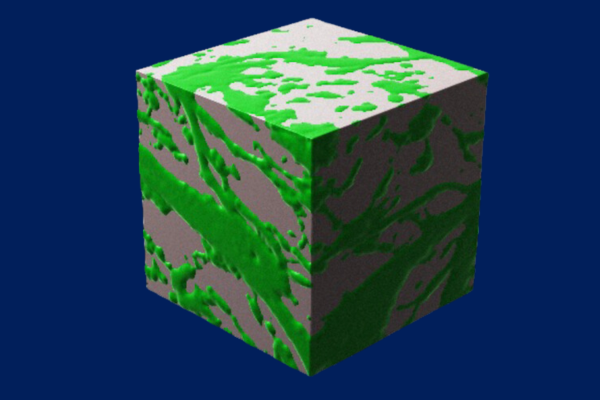
Bicontinuous materials, like this representation of a cube of gelatin and hyaluronic acid, have greater internal surface area, allowing cells to travel faster between two points. (Credit: Karen Xu)
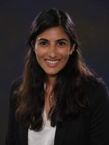
One of the most important but least understood aspects of healing is cell migration, or the process of cells moving from one part of the body to another. “If you are an ambulance out in the woods,” says Karen Xu, an M.D/Ph.D. student in Medicine and Bioengineering, “and there are no paths for you to move forward, it will be a lot harder for you to get to a site that needs you.”
Earlier this year, Xu co-authored a paper in Nature Communications describing a new cue to help cells get to where they need to go: a material made chiefly of hyaluronic acid and gelatin, two gooey substances commonly found outside cells in joints and connective tissue.
“Hundreds of thousands of people tear their meniscus every year,” says Robert Mauck, Mary Black Ralston Professor in Orthopaedic Surgery in Penn Medicine and Professor in Bioengineering at Penn Engineering and one of Xu’s advisors, as well as a senior author on the paper. “This material could potentially speed up their recovery.”
What makes the material — known as a hydrogel due to its blend of gelatinous matter and water — unique is that the combination of hyaluronic acid and gelatin forms a complex network of paths, providing cells many different ways to travel between two points.
This property is known as bicontinuity, and is exemplified by two discrete continuous phases that are each connected throughout the entire volume of the material (for example with a sponge, with phases of cellulose and air; in the hydrogel, this is comprised of gelatin and hyaluronic acid) resulting in a dizzying array of patterns that dramatically increase the surface area inside the material.
To test the hydrogel’s efficacy, Xu and her collaborators — including co-advisor Jason Burdick, formerly the Robert D. Bent Professor in Bioengineering at Penn Engineering and now the Bowman Endowed Professor at the University of Colorado Boulder, and the paper’s other senior author — first created several different versions of the hydrogel to find the sweet spot at which the constituents formed the bicontinuous structure and had the highest internal surface area. “We found that a precise combination of the various hydrogel components and control over their mixing was needed to form the bicontinuous structure,” says Burdick.
Karen Xu, a 2024 doctoral graduate in Bioengineering at the University of Pennsylvania, is one of 100 doctoral students in the U. S. and Canada selected to receive a $25,000 Scholar Award from the P.E.O. Sisterhood.
The P.E.O. Scholar Awards were established in 1991 to provide substantial merit-based awards for women of the United States and Canada who are pursuing a doctoral-level degree at an accredited college or university. Scholar Awards recipients are a select group of women chosen for their high level of academic achievement and their potential for having a positive impact on society.
The P.E.O., founded January 21, 1869, at Iowa Wesleyan College, Mount Pleasant, Iowa, is a philanthropic educational organization dedicated to supporting higher education for women. There are approximately 6,000 local chapters in the United States and Canada with nearly a quarter of a million active members.
Xu graduated summa cum laude with a B.S.E. in Biomedical Engineering from Duke University in 2018, after which she joined the M.D.-Ph.D. program at the University of Pennsylvania. She completed her Ph.D. in Bioengineering in spring 2024, funded by an NIH NRSA F30 fellowship, and is set to earn her M.D. in 2026. Under the mentorship of Jason Burdick, Bowman Endowed Professor in Chemical and Biological Engineering at the University of Colorado Boulder and Adjunct Professor in Bioengineering in Penn Engineering, and Robert Mauck, Mary Black Ralston Professor in Orthopaedic Surgery in the Perelman School of Medicine and in Bioengineering in Penn Engineering, her doctoral research has focused on engineering disease models to facilitate therapeutic discoveries. Her doctoral thesis involved the fabrication of hydrogels as tissue mimics to investigate how extracellular environments affect cell behaviors, thereby informing repair of dense connective tissues.
Beyond her research, Xu has taught with the Educational Pipeline Program at the Netter Center and the Perelman School of Medicine, where she hopes to inspire and support the next generation of healthcare workers and scientists.
Congratulations to the fourteen Bioengineering students to receive 2023 National Science Foundation Graduate Research Fellowship Program (NSF GRFP) fellowships. The prestigious NSF GRFP program recognizes and supports outstanding graduate students in NSF-supported fields. The recipients honorees were selected from a highly-competitive, nationwide pool. Further information about the program can be found on the NSF website.
Carlos Armando Aguila, Ph.D. student in Bioengineering, is a member of the Center of Neuroengineering and Therapeutics, advised by Erin Conrad, Assistant Professor in Neurology, and Brian Litt, Professor in Bioengineering and Neurology. His research focuses on analyzing electroencephalogram (EEG) signals to better understand epilepsy.
Joseph Lance Victoria Casila is a Ph.D. student in Bioengineering in the lab of Riccardo Gottardi, Assistant Professor in Pediatrics and Bioengineering. His research focuses on probing environmental factors that influence stem cell differentiation towards chondrogenesis for cartilage engineering and regeneration.
Trevor Chan is a Ph.D. student in Bioengineering in the lab of Felix Wehrli, Professor of Radiologic Science. His research is in developing computational methods for medical image refinement and analysis. Two ongoing projects are: self-supervised methods for CT super-resolution and assessment of osteoporosis, and semi-supervised segmentation of 3D and 4D echocardiograms for surgical correction of congenital heart-valve defects.
Rakan El-Mayta is an incoming Ph.D. student in the lab of Drew Weissman, Roberts Family Professor in Vaccine Research. Rakan studies messenger RNA-lipid nanoparticle vaccines for the treatment and prevention of infectious diseases. Prior to starting in the Bioengineering graduate program, he worked as a Research Assistant in Weissman lab and in the lab of Michael Mitchell, Associate Professor in Bioengineering.
Austin Jenk is a Ph.D. student in the lab of Robert Mauck, Mary Black Ralston Professor in Orthopaedic Surgery and Bioengineering. Austin aims to develop early intervention, intra-articular therapeutics to combat the onset of post-traumatic osteoarthritis following acute joint injuries. His work focuses on developing a therapeutic that can be employed not only in conventional healthcare settings, but also emergency and battlefield medicine.
Jiageng Liu is a Ph.D. student in the lab of Alex Hughes, Assistant Professor in Bioengineering. His work aims to precisely control the bio-physical/chemical properties of iPSC-derived organoids with advanced synthetic biology approaches to create functional replacement renal tissues.
Alexandra Neeser is a Ph.D. student in the lab of Leyuan Ma, Assistant Professor of Pathology and Laboratory Medicine. Her research focuses on solid tumor microenvironment delivery of therapeutics.
William Karl Selboe Ojemann, a Ph.D. Student in Bioengineering, is a member of the Center for Neuroengineering and Therapeutics directed by Brian Litt, Professor in Bioengineering and Neurology. His research is focused on developing improved neurostimulation therapies for epilepsy and other neurological disorders.
Savan Patel (BSE Class of 2023) conducted research in the lab of Michael Mitchell, Associate Professor in Bioengineering, where he worked to develop lipid nanoparticle formulations for immunotherapy and extrahepatic delivery of mRNA. He will be joining the Harvard-MIT HST MEMP Ph.D. program in the fall of 2023.
David E. Reynolds, a Ph.D. student in Bioengineering, is a member of the lab of Jina Ko, Assistant Professor in Bioengineering and Pathology and Laboratory Medicine. His research focuses on developing novel and translatable technologies to address currently intractable diagnostic challenges for precision medicine.
Andre Roots is a Ph.D. student in the lab of Christopher Madl, Assistant Professor in Materials Science and Engineering. His research focuses on the use of protein engineering techniques and an optimized 3D human skeletal muscle microtissue platform to study the effects of biophysical material properties on cells.
Emily Sharp, a second year Ph.D. student in Bioengineering, is a member of the lab of Robert Mauck, Mary Black Ralston Professor in Orthopaedic Surgery and Bioengineering, part of the McKay Orthopaedic Research Laboratories. Her research focuses on designing multi-functional biomaterials to enhance tissue repair, specifically intervertebral disc repair following herniation and discectomy.
Nat Thurlow is a Ph.D. student in the lab of Louis J. Soslowsky, Fairhill Professor in Orthopedic Surgery and Bioengineering. Their current work focuses on delineating the roles of collagens V and XI in tendon mechanics, fibril structure, and gene expression during tendon development and healing.
Maggie Wagner, Ph.D. student in Bioengineering, is a member in the labs of Josh Baxter, Assistant Professor of Orthopaedic Surgery, and Flavia Vitale, Assistant Professor in Neurology and Bioengineering. Her research focuses on the development of novel sensors to record and monitor muscle neuromechanics.
The Solomon R. Pollack Award for Excellence in Graduate Bioengineering Research is given annually to the most deserving Bioengineering graduate students who have successfully completed research that is original and recognized as being at the forefront of their field. This year, the Department of Bioengineering at the University of Pennsylvania recognizes the stellar work of four graduate students in Bioengineering.
Margaret Billingsley
Dissertation: “Ionizable Lipid Nanoparticles for mRNA CAR T Cell Engineering”
Maggie Billingsley
Margaret earned a bachelor’s degree in Biomedical Engineering from the University of Delaware where she conducted research in the Day Lab on the use of antibody-coated gold nanoparticles for the detection of circulating tumor cells. She conducted doctoral research in the lab of Michael J. Mitchell, J. and Peter Skirkanich Assistant Professor in Bioengineering. After defending her thesis at Penn in 2022, Margaret began postdoctoral training at the Massachusetts Institute of Technology (MIT) in the Hammond Lab where she is investigating the design and application of polymeric nanoparticles for combination therapies in ovarian cancer. She plans to use these experiences to continue a research career focused on drug delivery systems.
“Maggie was an absolutely prolific Ph.D. student in my lab, who pioneered the development of new mRNA lipid nanoparticle technology to engineer the immune system to target and kill tumor cells,” says Mitchell. “Maggie is incredibly well deserving of this honor, and I am so excited to see what she accomplishes next as a Postdoctoral Fellow at MIT and ultimately as a professor running her own independent laboratory at a top academic institution.”
Victoria Muir
Dissertation: “Designing Hyaluronic Acid Granular Hydrogels for Biomaterials Applications”
Victoria Muir
Victoria is currently a Princeton University Presidential Postdoctoral Research Fellow in the lab of Sujit S. Datta, where she studies microbial community behavior in 3D environments. She obtained her Ph.D. in 2022 as an NSF Graduate Research Fellow at Penn Bioengineering under the advisement of Jason A. Burdick, Adjunct Professor in Bioengineering at Penn and Bowman Endowed Professor in Chemical and Biological Engineering at the University of Colorado, Boulder. She received a B.ChE. in Chemical Engineering from the University of Delaware in 2018 as a Eugene DuPont Scholar. Outside of research, Victoria is highly active in volunteer and leadership roles within the American Institute of Chemical Engineers (AIChE), currently serving as Past Chair of the Young Professionals Community and a member of the Career and Education Operating Council (CEOC). Victoria’s career aspiration is to become a professor of chemical engineering and to lead a research program at the interaction of biomaterials, soft matter, and microbiology.
“Victoria was a fantastic Ph.D. student,” says Burdick. “She worked on important projects related to granular materials from the fundamentals to applications in tissue repair. She was also a leader in outreach activities, a great mentor to numerous undergraduates, and is already interviewing towards an independent academic position.”
Sadhana Ravikumar
Dissertation: “Characterizing Medial Temporal Lobe Neurodegeneration Due to Tau Pathology in Alzheimer’s Disease Using Postmortem Imaging”
Sadhana Ravikumar
Sadhana completed her B.S. in Electrical Engineering at the University of Cape Town, South Africa in 2014 and her M.S. in Biomedical Engineering from Carnegie Mellon University in 2017. Outside of the lab, she enjoys spending time in nature and exploring restaurants in Philadelphia with friends. She focused her doctoral work on the development of computational image analysis techniques applied to ex vivo human brain imaging data in the Penn Image Computing and Science Laboratory of Paul Yushkevich, Professor of Radiology at the Perelman School of Medicine and member of the Penn Bioengineering Graduate Group. She hopes to continue working at the intersection of machine learning and biomedical imaging to advance personalized healthcare and drug development.
“Dr. Sadhana Ravikumar’s Ph.D. work is a tour de force that combines novel methodological contributions crafted to address the challenge of anatomical variability in ultra-high resolution ex vivo human brain MRI with new clinical knowledge on the contributions of molecular pathology to neurodegeneration in Alzheimer’s disease,” says Yushkevich. “I am thrilled that this excellent contribution, as well as Sadhana’s professionalism and commitment to mentorship, have been recognized through the Sol Pollack award.”
Hannah Zlotnick
Dissertation: “Remote Force Guided Assembly of Complex Orthopaedic Tissues”
Hannah Zlotnick
Hannah was a Ph.D. candidate in the lab of Robert Mauck, Mary Black Ralston Professor in Orthopaedic Surgery and in Bioengineering. She successfully defended her thesis and graduated in August 2022. During her Ph.D., Hannah advanced the state-of-the-art in articular cartilage repair by harnessing remote fields, such as magnetism and gravity. Using these non-invasive forces, she was able to control cell positioning within engineered tissues, similar to the cell patterns within native cartilage, and enhance the integration between cartilage and bone. Her work could be used in many tissue engineering applications to recreate complex tissues and tissue interfaces. Hannah earned a B.S. in Biological Engineering from the Massachusetts Institute of Technology (MIT) in 2017 during which time she was also a member of the women’s varsity soccer team. At Penn, Hannah was also involved in the Graduate Association of Bioengineers (GABE) intramurals & leadership, and helped jumpstart the McKay DEI committee. Since completing her Ph.D., Hannah has begun her postdoctoral research as a Schmidt Science Fellow in Jason Burdick’s lab at the University of Colorado Boulder where she looks to improve in vitro disease models for osteoarthritis.
“Hannah was an outstanding graduate student, embodying all that is amazing about Penn BE – smart, driven, inventive and outstanding in every way,” says Mauck. “ I can’t wait to see where she goes and what she accomplishes!”
Congratulations to our four amazing 2023 Sol Pollack Award winners!
She joins 28 early-career scientists from around the world in this year’s cohort, with each receiving support for one to two years, $100,000 in salary support per year, individualized mentoring, and a series of professional development sessions as they pivot to the next stages of their research agendas.
The fellowship is a program of Schmidt Futures, the philanthropic initiative of Eric and Wendy Schmidt that aims to tackle society’s toughest challenges by supporting interdisciplinary researchers at the start of their careers.
“Our latest group of Schmidt Science Fellows embodies our vision for this Program at its inception five years ago,” says Eric Schmidt, co-founder of Schmidt Futures and former CEO and Chairman of Google. “We find the most talented next-generation leaders from around the world and back these impressive young adults with the resources and networks they need to realize their full potential while addressing some of the big scientific questions facing the world. Congratulations to the 2022 Schmidt Science Fellows, I am excited to see where your science takes you and what you will achieve.”
Working at the intersection of materials science, biology, and applied clinical research, Zlotnick’s postdoctoral work will involve developing advanced bioprinting techniques for regenerative medicine. Such advances are necessary to recreate the multi-cellular composition of orthopedic tissues, such as those found in the knee joint. Lab-grown tissue models can then be used to broaden our understanding of how degenerative diseases progress after injury, limit the need for animal models, and serve as a platform for therapeutic discovery.
As a child, Sonal Mahindroo would go to her orthopaedics appointments with her family, slowly becoming more and more fascinated by the workings and conditions of the musculoskeletal system. While being treated for scoliosis, she would receive children’s books from her doctor that helped provide clear and simplified explanations of orthopaedic topics, which supported her interest.
Nearly a decade later, Mahindroo is still interested in expanding her orthopaedic knowledge, and a Penn Medicine program is helping fuel that expansion. Now a senior at St. Bonaventure University in New York, Mahindroo spends her time at the university’s lab. But in addition to that, this year, she was able to take part in more learning opportunities with Penn Medicine’s support, via the McKay Orthopaedic Research Lab’s Diversity, Equity, and Inclusion (DEI) committee’s conference grant program.
McKay’s DEI committee — consisting of faculty, post-docs, graduate students, and staff — offers a welcoming environment and resources that support people of all identities, empowering them to bring forward unique perspectives to orthopaedic research.
“Our goal is to improve diversity and culture both within McKay and in the orthopaedic research community outside of Penn,” said Sarah Gullbrand, PhD, a research assistant professor at the McKay Lab. “We wanted to provide an opportunity for students to attend a conference and make connections to help them pursue their interest in orthopaedic research.”
The McKay conference grant supports undergraduate students who have been unable to get hands-on research experience. Participants are provided with the opportunity to network with leaders in the field of orthopaedic research, listen to cutting-edge research presentations, and learn about ways to get involved in orthopaedic research themselves.
“When launching the conference grant program earlier this year, I was motivated by my own experience attending a conference as an undergraduate. That experience really increased my interest in attending graduate school and taught me a lot about the breadth of research in orthopaedics,” said Hannah Zlotnick, a PhD student at the McKay Lab and member of the DEI committee. Through the McKay Conference Grants, the committee has supported two cohorts of students. “So far, we’ve been able to fund 11 undergraduate students from around the country to virtually attend orthopaedics conferences and receive early exposure to careers in STEM.”
Along with the conference grant, the McKay Lab holds workshops, book clubs, and other programs focused on DEI-related topics. As part of their efforts for promoting gender diversity in the field, the McKay Lab has previously partnered with the Perry Initiative to offer direct orthopaedic experiences for girls in high school, where they can learn how to suture, and perform mock fracture fixation surgeries on sawbones.
As a primarily male-populated field, orthopaedics could benefit greatly from diversity efforts. While women comprise approximately 50 percent of medical school graduates in the United States, they represent only 14 percent of orthopaedic surgery residents.
“The only women on staff at my orthopaedist’s office were receptionists. There were no female physicians or engineers to make my scoliosis brace,” Mahindroo said. “It was really cool coming to the McKay Lab and seeing how much the field has progressed since then.”
N.B. Hannah Zlotnick is a PhD student in Bioengineering studying in the lab of Robert Mauck, Mary Black Ralston Professor in Bioengineering and Orthopaedic Surgery.
The Department of Bioengineering is proud to congratulate Claudia Loebel, M.D., Ph.D. on her appointment as Assistant Professor in the Department of Materials Science and Engineering at the University of Michigan. Loebel is part of the University of Michigan’s Biological Sciences Scholar program, which recruits junior instructional faculty in major areas of biomedical investigation. Loebel’s appointment will begin in Fall 2021.
Loebel got her M.D. in 2011 from Martin-Luther University in Halle-Wittenberg, Germany and her Ph.D. in Health Sciences and Technology from ETH Zurich, Switzerland in 2016. There she worked under her advisors Professors Marcy Zenobi-Wong from ETH Zurich and David Eglin from AO Research Institute Davos. At Penn, she conducted postdoctoral research in the Polymeric Biomaterials Laboratory of Jason Burdick, Robert D. Bent Professor in Bioengineering, and as a Visiting Research Scholar in the Mauck Laboratory of the McKay Orthopaedic Research Laboratory in the Perelman School of Medicine.
Loebel was awarded a K99/R00 Pathway to Independence Award through the National Institutes of Health (NIH), which supports her remaining time as a postdoc as well as her time as an independent investigator at the University of Michigan. Loebel is excited about training the next generation of scientists and engineers and being part of their journey in becoming independent and diverse thinkers.
Loebel’s research area is inspired by the interface between material science and regenerative engineering and how it can address specific problems related to tissue development, repair, and regeneration. By developing mechanically and strucatally dynamic biomaterials, microfabrication, and matrix manipulation techniques her works aim to recreate complex cell-matrix interactions and model tissue morphogenesis and disease. The ultimate goal of her research is to use these engineered systems to develop and translate more effective therapeutic treatments for diseases such as fibrotic, inflammatory, and congenital disorders. Her lab’s work will initially focus on developing engineering lung alveolar organoids, aiming to build models of acute and chronic pulmonary diseases and for personalized medicine.
Loebel says, “I am grateful to all my Ph.D. and postdoc mentors for their continuous support and especially Jason who, over the last few years, has trained me in becoming an independent scientist and mentor. This transition would not have been possible without such a great mentor team behind me.”
Congratulations Dr. Loebel from everyone at Penn Bioengineering!
New research from Robert Mauck, Mary Black Ralston Professor in Orthopaedic Surgery and Bioengineering and Director of Penn Medicine’s McKay Orthopaedic Research Laboratory, announces a “new biosealant therapy may help to stabilize injuries that cause cartilage to break down, paving the way for a future fix or – even better – begin working right away with new cells to enhance healing.” Their research was published in Advanced Healthcare Materials. The study’s lead author was Jay Patel, a former postdoctoral fellow in the McKay Lab and now Assistant Professor at Emory University and was contributed to by Claudia Loebel, a postdoctoral research in the Burdick lab and who will begin an appointment as Assistant Professor at the University of Michigan in Fall 2021. In addition, the technology detailed in this publication is at the heart of a new company (Forsagen LLC) spun out of Penn with support from the Penn Center for Innovation (PCI) Ventures Program, which will attempt to spearhead the system’s entry into the clinic. It is co-founded by both Mauck and Patel, along with study co-author Jason Burdick, Professor in Bioengineering, and Ana Peredo, a PhD student in Bioengineering.
Next up in the Penn Bioengineering student spotlight series is Sonia Bansal. Sonia got her B.S. in Biomedical Engineering at Columbia University in 2014. She then came to Penn, where she recently got her Ph.D. in September of 2020 in Bioengineering under the advisement of Robert Mauck, Mary Black Ralston Professor of Orthopaedic Surgery and Professor of Bioengineering. Her dissertation is entitled “Functional and Structural Remodeling of the Meniscus with Growth and Injury” and focuses on the ways the knee meniscus changes while being actively loaded (growth) and under aberrant loading (injurious) conditions. She has presented her work internationally and has first authored four papers, with two more in preparation. She is passionate about K-12 STEM outreach and teaching at the collegiate level. She has been on the teaching team for six classes in the department, and is the first recipient of the Graduate Fellowship for Teaching Excellence from the Bioengineering department.
What drew you to the field of Bioengineering?
I first got interested in Bioengineering when I realized that it would let me merge my interests in biology and the human body with my desire to solve big questions by building and creating solutions. I applied to college knowing it was what I wanted to study.
What kind of research do you conduct, and what is the focus of your thesis?
My research is focused on the knee meniscus, specifically the impacts of its complex extracellular matrix and how that matrix changes during growth and after meniscal injury. My interests are largely translational, and in the future, I’d like to think about how we can use preclinical animal models to create effective therapeutics and drive clinical decision making in the orthopedic space.
What did you study for your undergraduate degree? How does it pair with the work you’re doing now, and what advice would you give to your undergraduate self?
I studied Biomedical Engineering during my undergraduate education and worked in cartilage tissue engineering. These experiences helped guide me to my Ph.D. work here at Penn. The two pieces of advice I’d give my undergraduate self is to ask for help and that it’s important to get more than five hours of sleep a night.
What’s your favorite thing to do on Penn’s campus or in Philly?
My favorite thing to do on campus was to read papers/write lectures/work on grants at a local coffee shop. I used to go to HubBub when it still existed, Saxby’s, and United By Blue.
Have you done or learned anything new or interesting during quarantine?
I have embarked on a journey in culinary fermentation (variety of pickles and sourdough, of course), and recently started homebrewing!
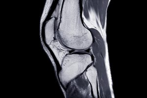
MRI Knee joint or Magnetic resonance imaging sagittal view for detect tear or sprain of the anterior cruciate ligament (ACL).
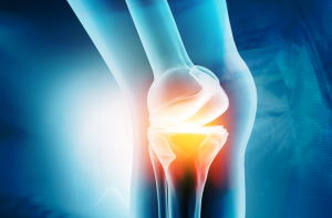
Using a magnetic field and hydrogels, a team of researchers in the Perelman School of Medicine have demonstrated a new possible way to rebuild complex body tissues, which could result in more lasting fixes to common injuries, such as cartilage degeneration. This research was published in Advanced Materials.
“We found that we were able to arrange objects, such as cells, in ways that could generate new, complex tissues without having to alter the cells themselves,” says the study’s first author, Hannah Zlotnick, a graduate student in bioengineering who works in the McKay Orthopaedic Research Laboratory at Penn Medicine. “Others have had to add magnetic particles to the cells so that they respond to a magnetic field, but that approach can have unwanted long-term effects on cell health. Instead, we manipulated the magnetic character of the environment surrounding the cells, allowing us to arrange the objects with magnets.”
In humans, tissues like cartilage can often break down, causing joint instability or pain. Often, the breakdown isn’t in total, but covers an area, forming a hole. Current fixes are to fill those holes in with synthetic or biologic materials, which can work but often wear away because they are not the same exact material as what was there before. It’s similar to fixing a pothole in a road by filling it with gravel and making a tar patch: The hole will be smoothed out but eventually wear away with use because it’s not the same material and can’t bond the same way.
What complicates fixing cartilage or other similar tissues is that their makeup is complex.
“There is a natural gradient from the top of cartilage to the bottom, where it contacts the bone,” Zlotnick explains. “Superficially, or at the surface, cartilage has a high cellularity, meaning there is a higher number of cells. But where cartilage attaches to the bone, deeper inside, its cellularity is low.”
So the researchers, which included senior author Robert Mauck, PhD, director of the McKay Lab and a professor of Orthopaedic Surgery and Bioengineering, sought to find a way to fix the potholes by repaving them instead of filling them in. With that in mind, the research team found that if they added a magnetic liquid to a three-dimensional hydrogel solution, cells, and other non-magnetic objects including drug delivery microcapsules, could be arranged into specific patterns that mimicked natural tissue through the use of an external magnetic field.

 Carlos Armando Aguila, Ph.D. student in Bioengineering, is a member of the Center of Neuroengineering and Therapeutics, advised by
  Joseph Lance Victoria Casila is a Ph.D. student in Bioengineering in the lab of
  Trevor Chan is a Ph.D. student in Bioengineering in the lab of
  Rakan El-Mayta is an incoming Ph.D. student in the lab of
  Austin Jenk is a Ph.D. student in the lab of
  Jiageng Liu is a Ph.D. student in the lab of
  Alexandra Neeser is a Ph.D. student in the lab of
  William Karl Selboe Ojemann, a Ph.D. Student in Bioengineering, is a member of the Center for Neuroengineering and Therapeutics directed by
  Savan Patel (BSE Class of 2023) conducted research in the lab of
  David E. Reynolds, a Ph.D. student in Bioengineering, is a member of the lab of
  Andre Roots is a Ph.D. student in the lab of
  Emily Sharp, a second year Ph.D. student in Bioengineering, is a member of the lab of
  Nat Thurlow is a Ph.D. student in the lab of
  Maggie Wagner, Ph.D. student in Bioengineering, is a member in the labs of


 New research from