How HIV Infection Shrinks the Brain’s White Matter

by Katherine Unger Baillie

Researchers from Penn and CHOP detail the mechanism by which HIV infection blocks the maturation process of brain cells that produce myelin, a fatty substance that insulates neurons.

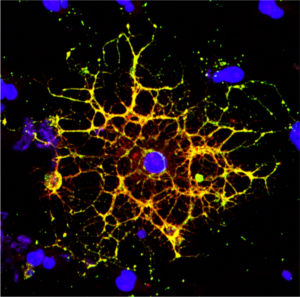
A confocal microscope image shows an oligodendrocyte in cell culture, labeled to show the cell nucleus in blue and myelin proteins in red, green, and yellow. Researchers from Penn and CHOP have shown that HIV infection prevents oligodendrocytes from maturing, leading to a reduction in white matter in the brain. (Image: Raj Putatunda)

It’s long been known that people living with HIV experience a loss of white matter in their brains. As opposed to gray matter, which is composed of the cell bodies of neurons, white matter is made up of a fatty substance called myelin that coats neurons, offering protection and helping them transmit signals quickly and efficiently. A reduction in white matter is associated with motor and cognitive impairment.

Earlier work by a team from the University of Pennsylvania and Children’s Hospital of Philadelphia (CHOP) found that antiretroviral therapy (ART)—the lifesaving suite of drugs that many people with HIV use daily—can reduce white matter, but it wasn’t clear how the virus itself contributed to this loss.

In a new study using both human and rodent cells, the team has hammered out a detailed mechanism, revealing how HIV prevents the myelin-making brain cells called oligodendrocytes from maturing, thus putting a wrench in white matter production. When the researchers applied a compound blocking this process, the cells were once again able to mature.

The work is published in the journal Glia.

“Even when people with HIV have their disease well-controlled by antiretrovirals, they still have the virus present in their bodies, so this study came out of our interest in understanding how HIV infection itself affects white matter,” says Kelly Jordan-Sciutto, a professor in Penn’s School of Dental Medicine and co-senior author on the study. “By understanding those mechanisms, we can take the next step to protect people with HIV infection from these impacts.”

“When people think about the brain, they think of neurons, but they often don’t think about white matter, as important as it is,” says Judith Grinspan, a research scientist at CHOP and the study’s other co-senior author. “But it’s clear that myelination is playing key roles in various stages of life: in infancy, in adolescence, and likely during learning in adulthood too. The more we find out about this biology, the more we can do to prevent white matter loss and the harms that can cause.”

Jordan-Sciutto and Grinspan have been collaborating for several years to elucidate how ART and HIV affect the brain, and specifically oligodendrocytes, a focus of Grinspan’s research. Their previous work on antiretrovirals had shown that commonly used drugs disrupted the function of oligodendrocytes, reducing myelin formation.

In the current study, they aimed to isolate the effect of HIV on this process. Led by Lindsay Roth, who recently earned her doctoral degree within the Biomedical Graduate Studies group at Penn and completed a postdoctoral fellowship working with Jordan-Sciutto and Grinspan, the investigation began by looking at human macrophages, one of the major cell types that HIV infects.

Read the full story in Penn Today.

Kelly Jordan-Sciutto is vice chair and professor in the University of Pennsylvania School of Dental Medicine’s Department of Basic & Translational Sciences and is director of Biomedical Graduate Studies. She is a member of the Penn Bioengineering Graduate Group.

Week in BioE (February 21, 2019)

by Sophie Burkholder

Detecting Infectious Diseases with Paper-Based Devices

Dr. Linnes’ paper device. Image used courtesy of Erin Easterling, Purdue College of Engineering.

Despite great advancements in diagnostics technology over the past few decades, patient accessibility to these technologies remains one of the biggest challenges of the field today. Particularly in low-resource areas, even simple processes can end up taking weeks or months to return results from tests that are normally completed in days. But what if these tests could be simplified to smaller, at-home tests based on properties of microfluidics – something like a pregnancy test but for infectious diseases like HIV?

Jacqueline Linnes, Ph.D., and her team of researchers at Purdue University are working towards finding a way to do just that by creating paper-based devices that use microfluidics to help carry out the necessary diagnostic tests. Specifically, her lab designed such a paper-based system that can detect HIV nucleic acids within 90 minutes of receiving a drop of patient blood. The success of this design shows promise for producing devices for diseases whose diagnostics process involve similar pathways of pathogen detection, opening the door to more applications of at-home tests based in the properties of paper microfluidics.

Here at Penn, undergraduate bioengineering students enrolled in the two-semester laboratory course Bioengineering Modeling, Analysis, and Design (BE 309 & BE 310) have the chance to create their own models of paper microfluidics delivery systems based on given time constraints in a multi-step process. Though the students’ challenge only involves water as a substrate, Linnes’ research demonstrates the later implications of studying fluid flow through a medium as cheap and accessible as paper.

Watch the video below demonstrating Dr. Linnes’ device:

Funding for Cancer Research in Tumor Mimicry and Imaging

Two of the deadliest forms of cancer today are breast cancer and pancreatic cancer, with the latter having a five-year survival rate of only about 8%. Because cancer treatments are often adjusted according to a unique patient-to-patient basis, learning how to improve predictions of tumor behavior could help determine proper therapies sooner.

Chien-Chi Lin, Ph.D., an associate professor of biomedical engineering at Indiana University – Purdue University Indianapolis, recently received a grant from the National Institute of Health to advance his research in pancreatic cancer treatment. His project under the grant involves the development of bio-inspired, responsive, and viscoelastic (BRAVE) cell-laden hydrogels to help understand cell interactions in pancreatic ductal adenocarcinoma, which is the most common form of malignancy in the pancreas. These hydrogels mimic tumor tissue, as well as model tumor development over time, helping to eventually find better ways of treating pancreatic cancer.

In other news surrounding cancer-related research, a team of researchers led by Kenneth Tichauer, Ph.D., at the Illinois Institute of Technology won the university’s Nayar Prize for their development of the Agent-Dependent Early Photon Tomography (ADEPT) Cancer Imager, a machine designed to find early tumor development in the lymph nodes of breast cancer patients. Through the use of a special dyeing process that now dyes the entire lymph node, providing a sharper image that allows for a quicker discovery of smaller tumors.

Penn’s Women in Computer Science (WiCS) Hosts FemmeHacks

Penn President Amy Gutmann and Penn Engineering Dean Vijay Kumar stopped by FemmeHacks at the Pennovation Center Feb. 9. The annual event is a beginner-friendly collegiate hackathon for women-identifying people with an interest in computer programming, and featured a day of all-levels workshops Feb. 8. The event is sponsored by Penn’s Women in Computer Science student organization.

Though the event is not specifically tailored towards applications in bioengineering, skills relating to coding and software development are increasingly important for those interested in pursuing a career in medical device design. In fact, in the evaluation of new medical devices, the FDA often focuses more on software over hardware, as the former is associated with more security liabilities, due to its relative novelty.

Read the full story and see pictures on FemmeHacks on Penn Today.

People & Places

In December, the department of biomedical engineering at the University of Alabama at Birmingham received the Ruth L. Kirschstein National Research Service Awards Institutional Research Training Grant, which will support predoctoral students enrolled in the university’s biomedical engineering graduate programs. The department plans to use the grant for research in cardiovascular tissue engineering.

Case Western Reserve University and Cleveland Clinic announced the launch of an alliance last year with the goal of creating better synergy across the two renowned institutions, hoping to provide more opportunities for students with interest in medicine at all levels, from high school to postdoctoral education. Though researchers from both institutions frequently partner on projects, this new alliance will create a more structured platform for future collaborations.

We would like to commend Steven George, M.D./Ph.D., on his new position as the chair of the Department of Biomedical Engineering at the University of California at Davis. His research involves the development of “organ-on-a-chip” technologies using stem cells and microfluidics to mimic human organ functions of vascularized cardiac, tumor, and pancreatic tissues.

Finally, we want to congratulate Paul Yock, M.D., on his being chosen to receive the National Academy of Engineering’s 2019 Fritz J. and Dolores H. Russ Prize. The prize honors two of Dr. Yock’s inventions from his research in interventional cardiology, one of which is Rapid Exchange, which is a kind of stenting and balloon angioplasty system. Dr. Yock is the Martha Meier Weiland Professor in the School of Medicine and Professor of Bioengineering.