“The proposed studies lay the foundation to make a major scientific impact in the childhood leukemia field and ultimately improve outcomes for children,” says Vining.
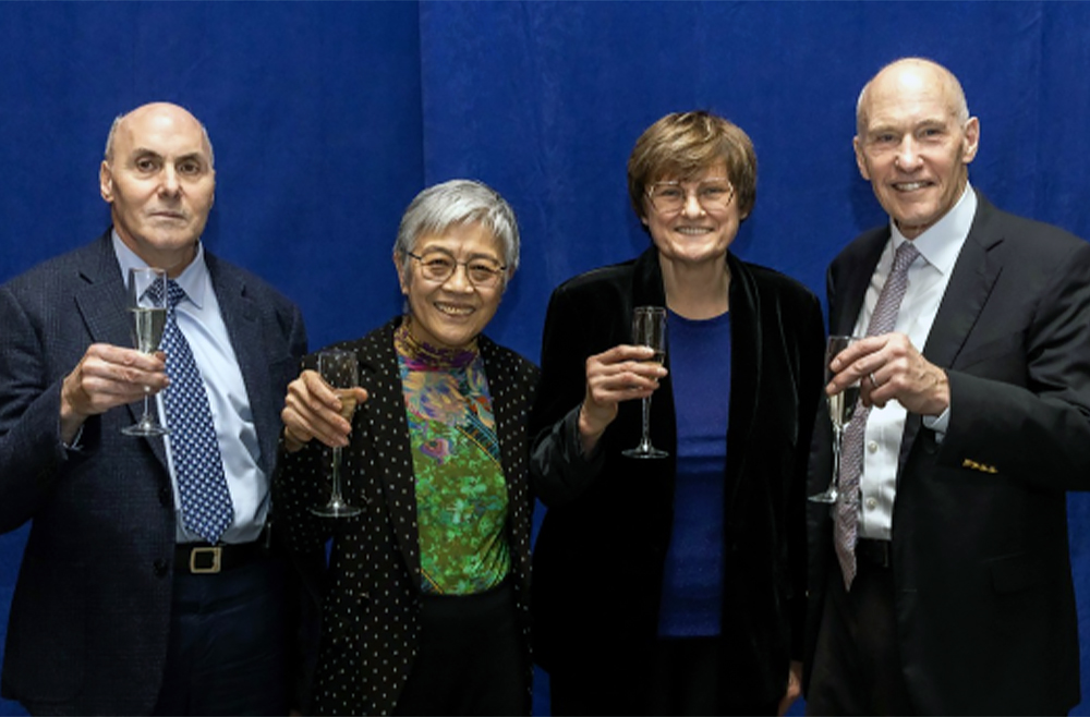
(From left to right) Breakthrough Prize recipients Drew Weissman, Virginia M-Y Lee, Katalin Karikó, and Carl June at a reception on Feb. 13. (Image: Courtesy of Penn Medicine News)
In popular culture, scientific discovery is often portrayed in “Eureka!” moments of sudden realization: a lightbulb moment, coming sometimes by accident. But in real life—and in Penn Medicine’s rich history as a scientific innovator for more than 250 years—scientific breakthroughs can never truly be distilled down to a single, “ah-ha” moment. They’re the result of years of hard work, perseverance, and determination to keep going, despite repeated, often discouraging, barriers and setbacks.
“Research is [like taking], four, or six, or eight steps back, and then a little stumble forward,” said Drew Weissman, MD, PhD, the Roberts Family Professor of Vaccine Research. “You keep doing that over and over and somehow, rarely, you can get to the top of the step.”
For Weissman and his research partner, Katalin Karikó, PhD, an adjunct professor of Neurosurgery, that persistence—documented in thousands of news stories across the globe—led to the mRNA technology that enabled two lifesaving COVID-19 vaccines, earning the duo numerous accolades, including the highest scientific honor, the 2023 Nobel Prize in Medicine.
Weissman and Karikó were also the 2022 recipients of the Breakthrough Prize in Life Sciences, the world’s largest science awards, popularly known as the “Oscars of Science.” Founded in 2012 by a group of web and tech luminaries including Google co-founder Sergey Brin and Meta CEO Mark Zuckerberg, the Breakthrough Prizes recognize “the world’s top scientists working in the fundamental sciences—the disciplines that ask the biggest questions and find the deepest explanations.” With six total winners, including four from the Perelman School of Medicine (PSOM), Penn stands alongside Harvard and MIT as the institutions whose researchers have been honored with the most Breakthrough Prizes.
Virginia M.–Y. Lee, PhD, the John H. Ware 3rd Professor in Alzheimer’s Research, was awarded the Prize in 2020 for discovering how different forms of misfolded proteins can move from cell to cell and lead to neurodegenerative disease progression. Carl June, MD, the Richard W. Vague Professor in Immunotherapy, is the most recent recipient and will be recognized at a star-studded red-carpet event in April for pioneering the development of CAR T cell therapy, which programs patients’ own immune cells to fight their cancer.
The four PSOM Breakthrough Prize recipients were honored on Tuesday, Feb. 13, 2024, when a new large-scale installation was unveiled in the lobby of the Biomedical Research Building to celebrate each laurate and their life-changing discoveries. During a light-hearted panel discussion, the honorees shared how a clear purpose, dogged determination, and a good sense of humor enabled their momentum forward.
Weissman presented the Department of Bioengineering’s 2022 Herman P. Schwan Distinguished Lecture: “Nucleoside-modified mRNA-LNP therapeutics.” Read more stories featuring Weissman in the BE Blog here.
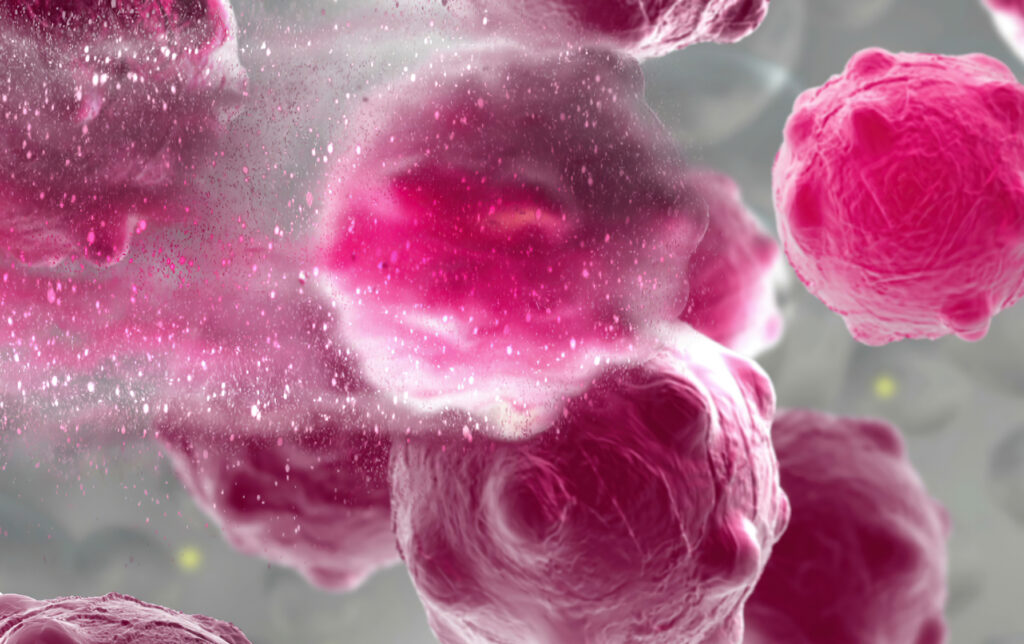
3d illustration of a damaged and disintegrating cancer cell. (Image: iStock/vitanovski)
The development of any type of second cancer following CAR T cell therapy is a rare occurrence, as found in an analysis of more than 400 patients treated at Penn Medicine, researchers from the Perelman School of Medicine at the University of Pennsylvania reported today in Nature Medicine. The team also described a single case of an incidental T cell lymphoma that did not express the CAR gene and was found in the lymph node of a patient who developed a secondary lung tumor following CAR T cell therapy.
CAR T cell therapy, a personalized form of immunotherapy in which each patient’s T cells are modified to target and kill their cancer cells, was pioneered at Penn. More than 30,000 patients with blood cancers in the United States—many of whom had few, if any, remaining treatment options available—have been treated with CAR T cell therapy since the first such therapy was approved in 2017. Some of the earliest patients treated in clinical trials have gone on to experience long-lasting remissions of a decade or more.
Secondary cancers, including T cell lymphomas, are a known, rare risk of several types of cancer treatment, including chemotherapy, radiation, and stem cell transplant. CAR T cell therapy is currently only approved to treat blood cancers that have relapsed or stopped responding to treatment, so patients who receive CAR T cell therapies have already received multiple other types of treatment and are facing dire prognoses.
In November 2023, the FDA announced an investigation into several reported cases of secondary T cell malignancies, including CAR-positive lymphoma, in patients who previously received CAR T cell therapy products. In January 2024, the FDA began requiring drugmakers to add a safety label warning to CAR T cell products. While the FDA review is still ongoing, it remains unclear whether the secondary T cell malignancies were caused by CAR T cell therapy.
As a leader in CAR T cell therapy, Penn has longstanding, clearly established protocols to monitor each patient both during and after treatment – including follow-up for 15 years after infusion – and participates in national reporting requirements and databases that track outcomes data from all cell therapy and bone marrow transplants.
Marco Ruella, M.D.
“When this case was identified, we did a detailed analysis and concluded the T cell lymphoma was not related to the CAR T cell therapy. As the news of other cases came to light, we knew we should go deeper, to comb through our own data to better understand and help define the risk of any type of secondary cancer in patients who have received CAR T cell products,” said senior author Marco Ruella, MD, an assistant professor of Hematology-Oncology and Scientific Director of the Lymphoma Program. “What we found was very encouraging and reinforces the overall safety profile for this type of personalized cell therapy.”
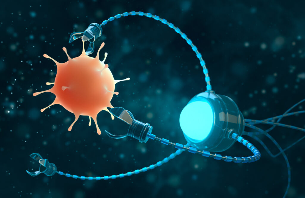
Biofilms—structured communities of microorganisms that create a protective matrix shielding them from external threats, including antibiotics—are responsible for about 80% of human infections and present a significant challenge in medical treatments, often resisting conventional methods.
In a Q&A with Penn Today, Hyun (Michel) Koo of the School of Dental Medicine and Edward Steager of the School of Engineering and Applied Science at Penn discuss an innovative approach they’ve partnered on: the use of small-scale robotics, microrobots, to offer a promising solution to tackle these persistent infections, bringing new capabilities and precision to the field of biomedical engineering.
Q: What is the motivation behind opting for tiny robots to tackle infections?
Koo: Treating biofilms is a broad yet unresolved biomedical problem, and conversely, the strategies for tackling biofilms are limited for a number of reasons. For instance, biofilms typically occur on surfaces that can be tricky to reach, like between the teeth in the oral cavity, the respiratory tract, or even within catheters and implants, so treatments for these are usually restricted to antibiotics (or antimicrobials) and other physical methods reliant on mechanical disruption. However, this touches on the problem of antimicrobial resistance: targeting specific microorganisms present in these structures is difficult, so antibiotics often fail to reach and penetrate the biofilm’s protective layers, leading to persistent infections and increased risk of antibiotic resistance.
We needed a way to circumvent these constraints, so Ed and I teamed up in 2017 to develop new, more precise and effective approaches that leverage the engineers’ ability to generate solutions that we, the clinicians and life science researchers, identify.
Hyun (Michel) Koo is a professor in the Department of Orthodontics and in the divisions of Pediatric Dentistry and Community Oral Health and the co-founder of the Center for Innovation & Precision Dentistry in the School of Dental Medicine at the University of Pennsylvania. He is a member of the Penn Bioengineering Graduate Group.
Edward Steager is a senior research investigator in Penn’s School of Engineering and Applied Science.
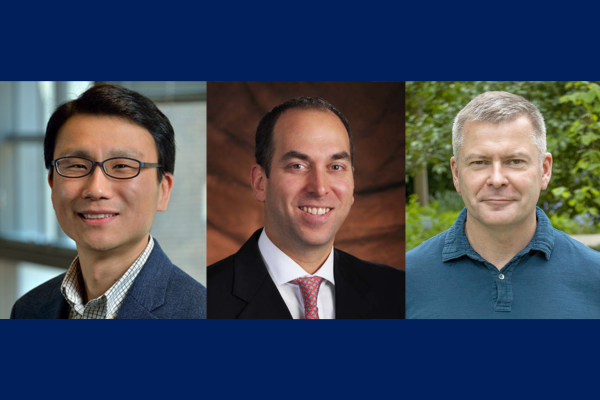
Daeyeon Lee (left), Oren Friedman (center) and Sergei Vinogradov (right)
Each year, the Nemirovsky Engineering and Medicine Opportunity (NEMO) Prize, funded by Penn Health-Tech, awards $80,000 to a collaborative team of researchers from the University of Pennsylvania’s Perelman School of Medicine and the School of Engineering and Applied Science for early-stage, interdisciplinary ideas.
This year, the NEMO Prize has been awarded to Penn Engineering’s Daeyeon Lee, Russel Pearce and Elizabeth Crimian Heuer Professor in Chemical and Biomolecular Engineering, Oren Friedman, Associate Professor of Clinical Otorhinolaryngology in the Perelman School of Medicine, and Sergei Vinogradov, Professor in the Department of Biochemistry and Biophysics in the Perelman School of Medicine and the Department of Chemistry in the School of Arts & Sciences. Together, they are developing a new therapy that improves the survival and success of soft-tissue grafts used in reconstructive surgery.
More than one million people receive soft-tissue reconstructive surgery for reasons such as tissue trauma, cancer or birth defects. Autologous tissue transplants are those where cells and tissue such as fat, skin or cartilage are moved from one part of a patient’s body to another. As the tissue comes from the patient, there is little risk of transplant rejection. However, nearly one in four autologous transplants fail due to tissue hypoxia, or lack of oxygen. When transplants fail the only corrective option is more surgery. Many techniques have been proposed and even carried out to help oxygenate soft tissue before it is transplanted to avoid failures, but current solutions are time consuming and expensive. Some even have negative side effects. A new therapy to help oxygenate tissue quickly, safely and cost-effectively would not only increase successful outcomes of reconstructive surgery, but could be widely applied to other medical challenges.
The therapy proposed by this year’s NEMO Prize recipients is a conglomerate or polymer of microparticles that can encapsulate oxygen and disperse it in sustainable and controlled doses to specific locations over periods of time up to 72 hours. This gradual release of oxygen into the tissue from the time it is transplanted to the time it functionally reconnects to the body’s vascular system is essential to keeping the tissue alive.
“The microparticle design consists of an oxygenated core encapsulated in a polymer shell that enables the sustained release of oxygen from the particle,” says Lee. “The polymer composition and thickness can be controlled to optimize the release rate, making it adaptable to the needs of the hypoxic tissue.”
These life-saving particles are designed to be integrated into the tissue before transplantation. However, because they exist on the microscale, they can also be applied as a topical cream or injected into tissue after transplantation.
“Because the microparticles are applied directly into tissues topically or by interstitial injection (rather than being administered intravenously), they surpass the need for vascular channels to reach the hypoxic tissue,” says Friedman. “Their micron-scale size combined with their interstitial administration, minimizes the probability of diffusion away from the injury site or uptake into the circulatory system. The polymers we plan to use are FDA approved for sustained-release drug delivery, biocompatible and biodegrade within weeks in the body, presenting minimal risk of side effects.”
The research team is currently testing their technology in fat cells. Fat is an ideal first application because it is minimally invasive as an injectable filler, making it versatile in remodeling scars and healing injury sites. It is also the soft tissue type most prone to hypoxia during transplant surgeries, increasing the urgency for oxygenation therapy in this particular tissue type.
Congratulations to the members of the Penn Bioengineering community who were awarded 2023 Accelerating from Lab to Market Pre-Seed Grants from the University of Pennsylvania Office of the Vice Provost for Research (OVPR).
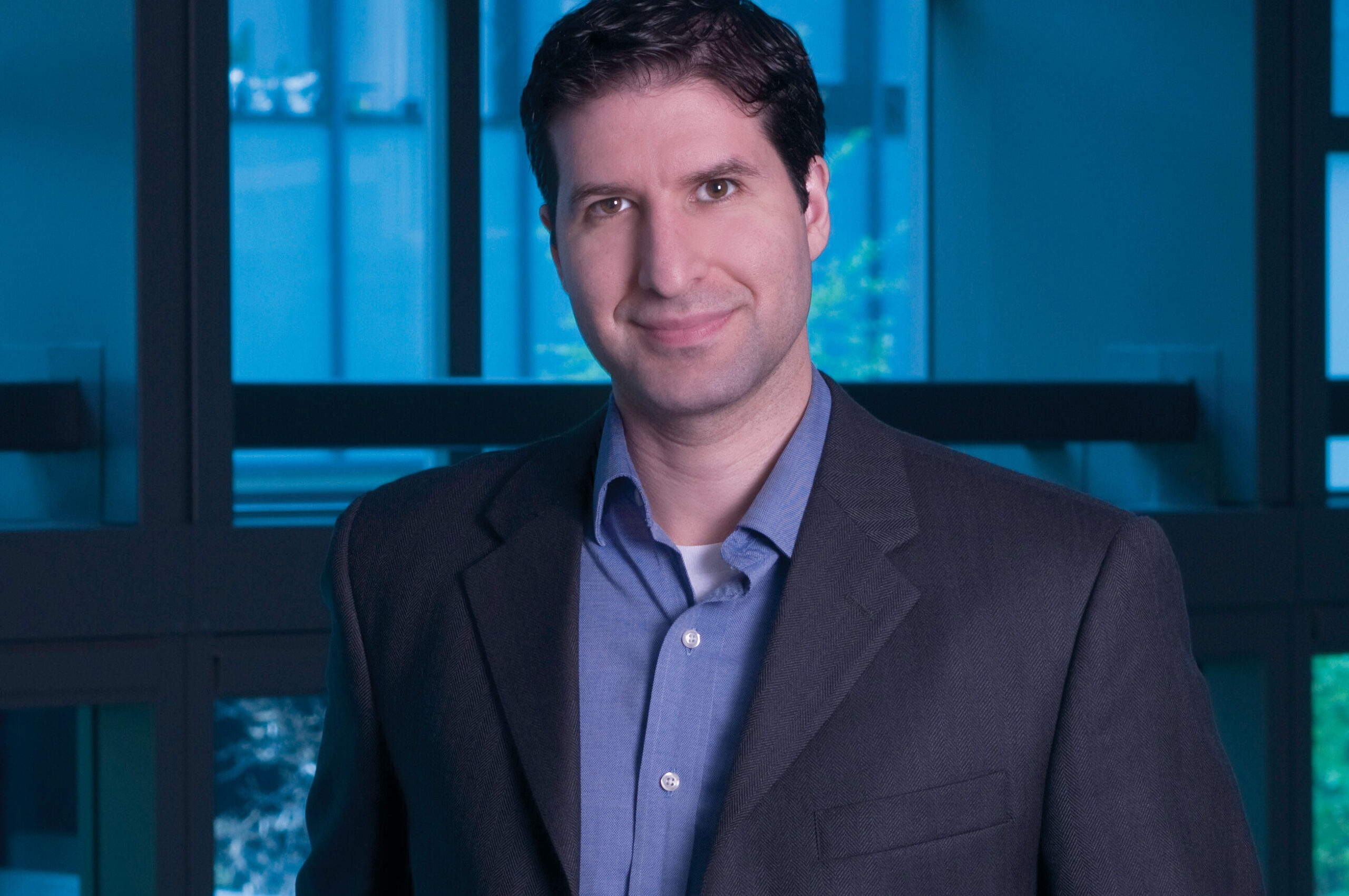
Andrew Tsourkas, Ph.D.
Three faculty affiliated with Bioengineering were included among the four winners. Andrew Tsourkas, Professor in Bioengineering and Co-Director of the Center for Targeted Therapeutics and Translational Nanomedicine (CT3N), was awarded for his project titled “Precise labeling of protein scaffolds with fluorescent dyes for use in biomedical applications.” Tsourkas’s team created protein scaffold that can better control the location and orientation of fluorescent dyes, commonly used for a variety of biomedical applications, such as labeling antibodies or fluorescence-guided surgery. The Tsourkas Lab specializes in “creating novel targeted imaging and therapeutic agents for the detection and/or treatment of diverse diseases.”
Also awarded were Penn Bioengineering Graduate Group members Mark Anthony Sellmeyer, Assistant Professor in Radiology in the Perelman School of Medicine, and Rahul M. Kohli, Associate Professor of Medicine in the Division of Infectious Diseases in the Perelman School of Medicine.
From the OVPR website:
“Penn makes significant commitments to academic research as one of its core missions, including investment in faculty research programs. In some disciplines, the path by which discovery makes an impact on society is through commercialization. Pre-seed grants are often the limiting step for new ideas to cross the ‘valley of death’ between federal research funding and commercial success. Accelerating from Lab to Market Pre-Seed Grant program aims to help to bridge this gap.”
Read the full list of winning projects and abstracts at the OVPR website.
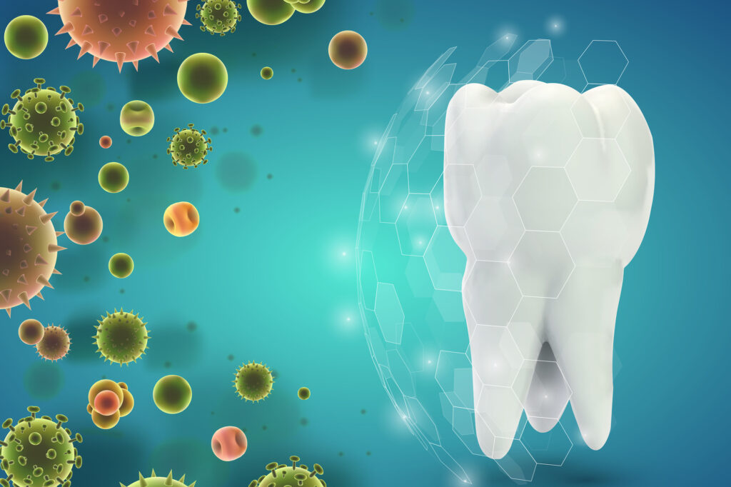
Michel Koo of the School of Dental Medicine and David Cormode of the Perelman School of Medicine and the School of Engineering and Applied Science led a team of researchers that uncovered a way to combine two FDA-approved treatments to treat tooth decay that taps into the blend’s bacteria-killing capabilities without disrupting the mouth’s microbiome. (Image: iStock / Alex Sholom)
The sting of a toothache or the discovery of a cavity is a universal dread. Dental caries, more commonly known as tooth decay, is an insidious adversary, taking a toll on millions of mouths worldwide. Caries can lead to pain, tooth loss, infection, and, in severe cases, even death.
While fluoride-based treatments have long been the gold standard in dentistry, this singular approach is now dated and has limited effect. Current treatments do not sufficiently control biofilm—the main culprit behind dental caries—and prevent enamel demineralization at the same time. This dual dilemma becomes particularly pronounced in high-risk populations where the onset of the disease can be both rapid and severe.
“Traditional treatments often come short in managing the complex biofilm environment in the mouth,” Koo, senior co-author on the study, says. “Our combined treatment not only amplifies the effectiveness of each agent but does so with a lower dosage, hinting at a potentially revolutionary method for caries prevention in high-risk individuals.”
David Cormode is an associate professor of radiology and bioengineering with appointments in Penn’s Perelman School of Medicine and School of Engineering and Applied Science.
Other authors are Yue Huang, Nil Kanatha Pandey, Shrey Shah, and Jessica C. Hsu of Penn’s Perelman School of Medicine; Yuan Liu, Aurea Simon-Soro, Zhi Ren, Zhenting Xiaang, Dongyeop Kim, Tatsuro Ito, Min Jun Oh, and Yong Li of Penn’s School of Dental Medicine; Paul. J Smeets, Sarah Boyer, Xingchen Zhao, and Derk Joester of Northwestern University; and Domenick T. Zero of Indiana University.
The work was supported by the National Institute of Health (grants R01-DE025848 and TL1TR001423 and awards S10OD026871 and R90DE031532) and the National Science Foundation (awards ECCS-2025633 and DMR-1720139).
Breaking the code of the immune system could provide a new fundamental way of understanding, treating, and preventing every type of disease. Penn Medicine is investing in key discoveries about immunity and immune system function, and building infrastructure, to make that bold idea a reality.
This grandfather lives with primary progressive multiple sclerosis (MS), an autoimmune disorder that he controls with a medicine that depletes his body of the type of immune cells that make antibodies. So while he has completed his COVID-19 vaccine course, his immune system function isn’t very strong—and the invitation has arrived at a time when COVID-19 is still spreading rapidly.
You can imagine the scene as an older gentleman lifts a thick, creamy envelope from his mailbox, seeing his own name written in richly scripted lettering. He beams with pride and gratitude at the sight of his granddaughter’s wedding invitation. Yet his next thought is a sober and serious one. Would he be taking his life in his hands by attending the ceremony?
“In the past, all we could do was [measure] the antibody response,” says Amit Bar-Or, the Melissa and Paul Anderson President’s Distinguished Professor in Neurology at the Perelman School of Medicine, and chief of the Multiple Sclerosis division. “If that person didn’t have a good antibody response, which is likely because of the treatment they’re on, we’d shrug our shoulders and say, ‘Maybe you shouldn’t go because we don’t know if you’re protected.’”
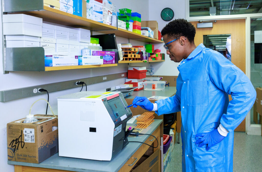
Today, though, Bar-Or can take a deeper dive into his patients’ individual immune systems to give them far more nuanced recommendations. A clinical test for immune cells produced in response to the COVID-19 vaccine or to the SARS-CoV-2 virus itself—not just antibodies—was one of the first applied clinical initiatives of a major new Immune Health® project at Penn Medicine. Doctors were able to order this test and receive actionable answers through the Penn Medicine electronic health record for patients like the grandfather with MS.
“With a simple test and an algorithm we can have a very different discussion,” Bar-Or says. A test result showing low T cells, for instance, would tell Bar-Or his patient may get a meaningful jolt in immunity from a vaccine booster, while low antibody levels would suggest passive antibody therapy is more helpful. Or, the test might show his body is already well primed to protect him, making it reasonably safe to attend the wedding.
This COVID-19 immunity test is only the beginning.
Physicians and scientists at Penn Medicine are imagining a future where patients can get a precise picture of their immune systems’ activity to guide treatment decisions. They are working to bring the idea of Immune Health to life as a new area of medicine. In labs, in complex data models, and in the clinic, they are beginning to make sense out of the depth and breadth of the immune system’s millions of as-yet-undeciphered signals to improve health and treat illnesses of all types.
Penn Medicine registered the trademark for the term “Immune Health” in recognition of the potential impact of this research area and its likelihood to draw non-academic partners as collaborators in its growth. Today, at the south end of Penn’s medical campus, seven stories of research space are being added atop an office building at 3600 Civic Center Blvd., including three floors dedicated to Immune Health, autoimmunity, and immunology research.
The concept behind the whole project, says E. John Wherry, director of Penn Medicine’s Institute for Immunology and Immune Health (I3H), “is to listen to the immune system, to profile the immune system, and use those individual patient immune fingerprints to diagnose and treat diseases as diverse as immune-related diseases, cancer, cardiovascular disease, Alzheimer’s, and many others.”
The challenge is vast. Each person’s immune system is far more complex than antibodies and T cells alone. The immune system is made of multiple interwoven layers of complex defenders—from our skin and mucous membranes to microscopic memory B cells that never forget a childhood infection—meant to fortify our bodies from germs and disease. It is a sophisticated system that learns and adapts over our lifetimes in numerous ways, and it also falters and fails in some ways we understand and others that remain mysterious. And each person’s intricate internal battlefield is in some way unique.
The immune system is not just a set of defensive barricades, either. It’s also a potential source of deep insight about a person’s physiological functioning and responses to medical treatments.
“The immune system is sensing and keeping track of basically all tissues and all cells in our body all the time,” Wherry says. “It is surveying the body trying to clean up any invaders and restore homeostasis by maintaining good health.”
“Our goal is to essentially break the code of the immune system,” says Jonathan Epstein, executive vice dean of the Perelman School of Medicine and chief scientific officer at Penn Medicine. “By doing so, we believe we will be able to determine your state of health and your response to therapies in essentially every human disease.”
Let’s say you typically eat eggs for breakfast but were running late and ate cereal. As you crunched on a spoonful of Raisin Bran, other contextual similarities remained: You ate at the same table, at the same time, preparing to go to the same job. When someone asks later what you had for breakfast, you incorrectly remember eating eggs.
This would be a real-world example of a false memory. But what happens in your brain before recalling eggs, compared to what would happen if you correctly recalled cereal?
In a paper published in Proceedings of the National Academy of Sciences, University of Pennsylvania neuroscientists show for the first time that electrical signals in the human hippocampus differ immediately before recollection of true and false memories. They also found that low-frequency activity in the hippocampus decreases as a function of contextual similarity between a falsely recalled word and the target word.
“Whereas prior studies established the role of the hippocampus in event memory, we did not know that electrical signals generated in this region would distinguish the imminent recall of true from false memories,” says psychology professor Michael Jacob Kahana, director of the Computational Memory Lab and the study’s senior author. He says this shows that the hippocampus stores information about an item with the context in which it was presented.
Researchers also found that, relative to correct recalls, the brain exhibited lower theta and high-frequency oscillations and higher alpha/beta oscillations ahead of false memories. The findings came from recording neural activity in epilepsy patients who were already undergoing invasive monitoring to pinpoint the source of their seizures.
Noa Herz, lead author and a postdoctoral fellow in Kahana’s lab at the time of the research, explains that the monitoring was done through intracranial electrodes, the methodology researchers wanted to use for this study. She says that, compared to scalp electrodes, this method “allowed us to more precisely, and directly, measure the neural signals that were generated in deep brain structures, so the activity we are getting is much more localized.”
Michael Kahana is the Edmund J. and Louise W. Kahn Term Professor of Psychology in the School of Arts & Sciences and director of the Computational Memory Lab at the University of Pennsylvania. He is a member of the Penn Bioengineering Graduate Group.
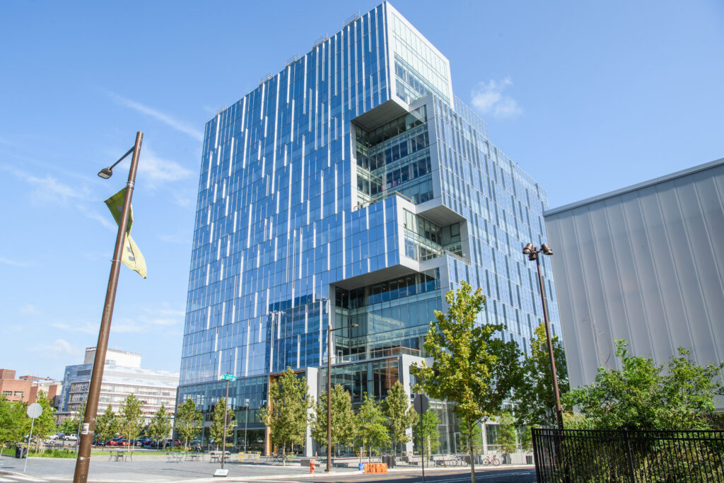
On September 14, Wexford Science & Technology, LLC and the University of Pennsylvania announced that the University has signed a lease for new laboratory space that will usher in a wave of novel vaccine, therapeutics, and engineered diagnostics research to West Philadelphia. Research teams from Penn are poised to move into 115,000 square feet of space at One uCity Square, the 13-story, 400,000 square foot purpose-built lab and office building within the vibrant uCity Square Knowledge Community being developed by Wexford. This is the largest lease in the building, encompassing four floors, and bringing the building to over 90% leased. The building currently includes industry tenants Century Therapeutics (NASDAQ: IPSC), Integral Molecular, Exponent (NASDAQ: EXPO), and Charles River Laboratories (NYSE: CRL).
The new University space will house Penn Medicine’s Institute for RNA Innovation and Penn Engineering’s Center for Precision Engineering for Health, underscoring the University’s commitment to a multi-disciplinary and collaborative approach to research that will attract and retain the best talent and engage partners from across the region. Penn’s decision to locate at One uCity Square reinforces uCity Square’s evolution as a central cluster of academic, clinical, commercial, entrepreneurial, and amenity spaces for the area’s innovation ecosystem, and further cements Philadelphia’s position as a top life sciences market.
Jonathan Epstein, MD, Executive Vice Dean and Chief Scientific Officer of Penn Medicine, shared his anticipation for the opportunities that lie ahead: “Penn Medicine is proud to build on its existing clinical presence in uCity Square and establish an innovative and collaborative research presence at the heart of uCity Square’s multidisciplinary innovation ecosystem. This strategic move underscores our commitment to accelerating advancements in biomedical research, industry collaboration, and equipping our talented teams with the resources they need to shape the future of healthcare.”
Locating the Penn Institute for RNA Innovation in the heart of the uCity Square community brings together researchers across disciplines who are already pursuing new vaccines and treatments, and better ways to deliver them. Their shared work will help to power the next phase of vaccine discovery and development.
Likewise, anchoring the work of Penn Engineering’s Center in the One uCity Square space will allow the School’s multi-disciplinary researchers and their collaborators to advance new clinical and diagnostic methods that will focus on intelligent therapeutics, genome design, diagnostics for discovery of human biology, and engineering the human immune shield.
“Penn Engineering has made a substantial commitment to precision engineering for health, an area that is not only important and relevant to engineering, but also critical to the future of humanity,” said Vijay Kumar, Nemirovsky Family Dean of Penn Engineering. “The space in One uCity Square will add another 30,000 square feet of space for our engineers to develop technologies that will fight future pandemics, cure incurable diseases, and extend healthy life spans around the world.”
Spearheading the Penn Institute for RNA Innovation will be Drew Weissman, MD, PhD, the Roberts Family Professor for Vaccine Research, who along with Katalin Karikó, PhD, adjunct professor of Neurosurgery, discovered foundational mRNA technology that enabled the creation of vital vaccine technology, including the FDA-approved mRNA-based COVID-19 vaccines developed by Pfizer-BioNTech and Moderna.
In this new space at One uCity Square, Weissman and his research team and collaborators will further pursue their groundbreaking research efforts with a goal to develop new therapeutics and vaccines and initiate clinical trials for other devastating diseases.
In addition, two established researchers will join the Institute at One uCity Square: Harvey Friedman, MD, a professor of Infectious Diseases, who leads a team researching various vaccines. He will be joined by Vladimir Muzykantov, MD, PhD, Founders Professor in Nanoparticle Research, who focuses on several projects related to targeting the delivery of drugs, including mRNA, to create more effective, targeted pathways to deliver drugs to the vascular system, treating a wide range of diseases that impact the brain, lung, heart, and blood.
Dan Hammer, Alfred G. and Meta A. Ennis Professor in the Departments of Bioengineering and Chemical and Biomolecular Engineering in Penn Engineering and Director of the Center for Precision Engineering for Health, will oversee the Center’s innovations in diagnostics and delivery, cellular and tissue engineering, and the development of new devices that integrate novel materials with human tissues. The Center will bring together scholars from all departments within Penn Engineering and will help to foster increased collaboration with campus colleagues at Penn’s Perelman School of Medicine and with industry partners.
Joining the Center researchers in One uCity Square are Noor Momin, Sherry Gao, and Michael Mitchell. Noor Momin, who will join Penn Engineering in early 2024 as an assistant professor in Bioengineering, will leverage her lab’s expertise in cardiovascular immunology, protein engineering and pharmacokinetic modeling to develop next-generation treatments and diagnostics for cardiovascular diseases.